Influenza A H1N1 Nucleoprotein Antibody - (A/WSN/1933) - BSA Free
Novus Biologicals | Catalog # NBP2-16965
![Immunoprecipitation: Influenza A H1N1 Nucleoprotein Antibody - (A/WSN/1933) [NBP2-16965]](https://resources.rndsystems.com/images/products/Influenza-A-nucleoprotein-Antibody-Immunoprecipitation-NBP2-16965-img0008.jpg "Immunoprecipitation: Influenza A H1N1 Nucleoprotein Antibody - (A/WSN/1933) [NBP2-16965]")
Loading...
Key Product Details
Species Reactivity
Validated:
Virus
Cited:
Virus
Applications
Validated:
Immunohistochemistry, Immunohistochemistry-Paraffin, Western Blot, Sandwich ELISA, Immunocytochemistry/ Immunofluorescence, Immunoprecipitation
Cited:
Western Blot
Label
Unconjugated
Antibody Source
Polyclonal Rabbit IgG
Format
BSA Free
Loading...
Product Specifications
Immunogen
Recombinant protein of Influenza A H1N1 Nucleoprotein (A/WSN/1933(H1N1). The exact sequence is proprietary.
Specificity
Influenza A H1N1 Nucleoprotein. (A/WSN/1933)
Clonality
Polyclonal
Host
Rabbit
Isotype
IgG
Scientific Data Images for Influenza A H1N1 Nucleoprotein Antibody - (A/WSN/1933) - BSA Free
Immunoprecipitation: Influenza A H1N1 Nucleoprotein Antibody - (A/WSN/1933) [NBP2-16965]
Immunoprecipitation: Influenza A H1N1 Nucleoprotein Antibody [NBP2-16965] - IP of Influenza A H1N1 Nucleoprotein from transfected 293T cell lysates. IP image submitted by a verified customer review.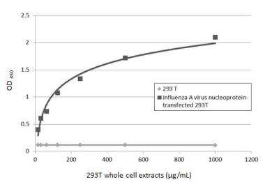
Sandwich ELISA: Influenza A H1N1 Nucleoprotein Antibody [NBP2-16965] - Detection of non-transfected and Influenza A virus nucleoprotein transfected 293T whole cell extracts using Influenza A H1N1 Nucleoprotein antibody [1236] as capture antibody at concentration of 5 ug/mL and Influenza A H1N1 Nucleoprotein antibody as detection antibody at concentration of 1 ug/mL. Rabbit IgG antibody (HRP) (NBP2-19301) was diluted at 1:10000 and used to detect the primary antibody.
 [NBP2-16965] -")
ELISA: Influenza A H1N1 Nucleoprotein Antibody - (A/WSN/1933) [NBP2-16965] -
ELISA: Influenza A H1N1 Nucleoprotein Antibody - (A/WSN/1933) [NBP2-16965] - Sandwich ELISA detection of recombinant full-length Influenza A virus NP (nucleoprotein) protein, DDDDK tag using Influenza A virus NP (nucleoprotein) antibody [GT1236] as capture antibody at concentration of 5 ug/mL and Influenza A virus NP (nucleoprotein) antibody (NBP2-16965) as detection antibody at concentration of 1 ug/mL. Rabbit IgG antibody (HRP) was diluted at 1:10000 and used to detect the primary antibody. [NBP2-16965] -")
Immunocytochemistry/ Immunofluorescence: Influenza A H1N1 Nucleoprotein Antibody - (A/WSN/1933) [NBP2-16965] -
Immunocytochemistry/ Immunofluorescence: Influenza A H1N1 Nucleoprotein Antibody - (A/WSN/1933) [NBP2-16965] - Immunofluorescent analysis of Influenza virus infected cells using Influenza A H1N1 Nucleoprotein antibody antibody (NBP2-16965).Sample: Influenza A and B virus infected cells slide.
Green: Influenza A H1N1 Nucleoprotein antibody antibody (NBP2-16965) diluted at 1:100.
Blue: Fluoroshield with DAPI.
 [NBP2-16965] -")
Western Blot: Influenza A H1N1 Nucleoprotein Antibody - (A/WSN/1933) [NBP2-16965] -
Western Blot: Influenza A H1N1 Nucleoprotein Antibody - (A/WSN/1933) [NBP2-16965] - Influenza A virus lysate (H1N1) infected 293T whole cell extract was separated by 12% SDS-PAGE, and the membrane was blotted with Influenza A virus NP (nucleoprotein) antibody (NBP2-16965) diluted at 1:50000. The HRP-conjugated anti-rabbit IgG antibody was used to detect the primary antibody. [NBP2-16965] -")
Western Blot: Influenza A H1N1 Nucleoprotein Antibody - (A/WSN/1933) [NBP2-16965] -
Western Blot: Influenza A H1N1 Nucleoprotein Antibody - (A/WSN/1933) [NBP2-16965] - Influenza A viral lysate (1 ug & 1ng ug) were separated by 10% SDS-PAGE, and the membrane was blotted with Influenza A virus NP (nucleoprotein) antibody (NBP2-16965) diluted at 1:5000. The HRP-conjugated anti-rabbit IgG antibody was used to detect the primary antibody. [NBP2-16965] -")
ELISA: Influenza A H1N1 Nucleoprotein Antibody - (A/WSN/1933) [NBP2-16965] -
Sandwich ELISA detection of recombinant full-length Influenza A virus NP (nucleoprotein) protein, DDDDK tag using Influenza A virus NP (nucleoprotein) antibody (NBP2-16965) as capture antibody at concentration of 5 ug/mL and Influenza A virus NP (nucleoprotein) antibody [GT1236] as detection antibody at concentration of 1 ug/mL. Mouse IgG antibody (HRP) was diluted at 1:10000 and used to detect the primary antibody. [NBP2-16965] -")
Western Blot: Influenza A H1N1 Nucleoprotein Antibody - (A/WSN/1933) [NBP2-16965] -
Non-infected (-) and infected (+) H1299 whole cell extracts (5 ug) were separated by 10% SDS-PAGE, and the membrane was blotted with Influenza A virus Nucleoprotein antibody (NBP2-16965) diluted at 1:10000. The HRP-conjugated anti-rabbit IgG antibody was used to detect the primary antibody. [NBP2-16965] -")
Western Blot: Influenza A H1N1 Nucleoprotein Antibody - (A/WSN/1933) [NBP2-16965] -
Influenza A (H1N1) viral lysate (0.1 ug) was separated by 10% SDS-PAGE, and the membrane was blotted with Influenza A virus Nucleoprotein antibody (NBP2-16965) diluted at 1:50000. The HRP-conjugated anti-rabbit IgG antibody was used to detect the primary antibody. [NBP2-16965] -")
Western Blot: Influenza A H1N1 Nucleoprotein Antibody - (A/WSN/1933) [NBP2-16965] -
Non-infected (-) and infected (+) DF1 whole cell extracts (5 ug) were separated by 10% SDS-PAGE, and the membrane was blotted with Influenza A virus NP (nucleoprotein) antibody (NBP2-16965) diluted at 1:10000. The HRP-conjugated anti-rabbit IgG antibody was used to detect the primary antibody. [NBP2-16965] -")
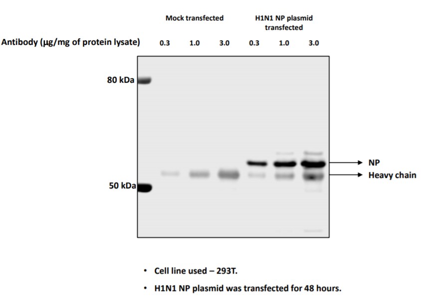
Western Blot: Influenza A H1N1 Nucleoprotein Antibody - (A/WSN/1933) [NBP2-16965] -
Non-transfected (-) and transfected (+) 293T whole cell extracts (30 ug) were separated by 10% SDS-PAGE, and the membrane was blotted with Influenza A virus Nucleoprotein antibody (NBP2-16965) diluted at 1:50000. The HRP-conjugated anti-rabbit IgG antibody was used to detect the primary antibody.Applications for Influenza A H1N1 Nucleoprotein Antibody - (A/WSN/1933) - BSA Free
Application
Recommended Usage
Immunocytochemistry/ Immunofluorescence
1:100 - 1:1000
Immunohistochemistry-Paraffin
Assay dependent
Immunoprecipitation
Assay dependent
Sandwich ELISA
Assay dependent
Western Blot
1:5000 - 1:20000
Reviewed Applications
Read 1 review rated 5 using NBP2-16965 in the following applications:
Formulation, Preparation, and Storage
Purification
Antigen Affinity-purified
Formulation
PBS, 20% Glycerol
Format
BSA Free
Preservative
0.025% Proclin 300
Concentration
Concentrations vary lot to lot. See vial label for concentration. If unlisted please contact technical services.
Shipping
The product is shipped with polar packs. Upon receipt, store it immediately at the temperature recommended below.
Stability & Storage
Aliquot and store at -20C or -80C. Avoid freeze-thaw cycles.
Product Documents for Influenza A H1N1 Nucleoprotein Antibody - (A/WSN/1933) - BSA Free
Certificate of Analysis
To download a Certificate of Analysis, please enter a lot or batch number in the search box below.
Product Specific Notices for Influenza A H1N1 Nucleoprotein Antibody - (A/WSN/1933) - BSA Free
This product is for research use only and is not approved for use in humans or in clinical diagnosis. Primary Antibodies are guaranteed for 1 year from date of receipt.
Citations for Influenza A H1N1 Nucleoprotein Antibody - (A/WSN/1933) - BSA Free
 Powered by Bioz
Customer Reviews for Influenza A H1N1 Nucleoprotein Antibody - (A/WSN/1933) - BSA Free (1)
5 out of 5
1 Customer Rating
Have you used Influenza A H1N1 Nucleoprotein Antibody - (A/WSN/1933) - BSA Free?
Submit a review and receive an Amazon gift card!
$25/€18/£15/$25CAN/¥2500 Yen for a review with an image
$10/€7/£6/$10CAN/¥1110 Yen for a review without an image
Submit a review
Customer Images
Showing
1
-
1 的
1 review
Showing All
Filter By:
-
Application: ImmunoprecipitationSample Tested: 293T cell lysateSpecies: HumanVerified Customer | Posted 11/26/2020IP of H1N1 NP protein from transfected lysates.

There are no reviews that match your criteria.
Protocols
Find general support by application which include: protocols, troubleshooting, illustrated assays, videos and webinars.
- Antigen Retrieval Protocol (PIER)
- Antigen Retrieval for Frozen Sections Protocol
- Appropriate Fixation of IHC/ICC Samples
- Cellular Response to Hypoxia Protocols
- Chromogenic IHC Staining of Formalin-Fixed Paraffin-Embedded (FFPE) Tissue Protocol
- Chromogenic Immunohistochemistry Staining of Frozen Tissue
- ClariTSA™ Fluorophore Kits
- Detection & Visualization of Antibody Binding
- ELISA Sample Preparation & Collection Guide
- ELISA Troubleshooting Guide
- Fluorescent IHC Staining of Frozen Tissue Protocol
- Graphic Protocol for Heat-induced Epitope Retrieval
- Graphic Protocol for the Preparation and Fluorescent IHC Staining of Frozen Tissue Sections
- Graphic Protocol for the Preparation and Fluorescent IHC Staining of Paraffin-embedded Tissue Sections
- Graphic Protocol for the Preparation of Gelatin-coated Slides for Histological Tissue Sections
- How to Run an R&D Systems DuoSet ELISA
- How to Run an R&D Systems Quantikine ELISA
- How to Run an R&D Systems Quantikine™ QuicKit™ ELISA
- ICC Cell Smear Protocol for Suspension Cells
- ICC Immunocytochemistry Protocol Videos
- ICC for Adherent Cells
- IHC Sample Preparation (Frozen sections vs Paraffin)
- Immunocytochemistry (ICC) Protocol
- Immunocytochemistry Troubleshooting
- Immunofluorescence of Organoids Embedded in Cultrex Basement Membrane Extract
- Immunofluorescent IHC Staining of Formalin-Fixed Paraffin-Embedded (FFPE) Tissue Protocol
- Immunohistochemistry (IHC) and Immunocytochemistry (ICC) Protocols
- Immunohistochemistry Frozen Troubleshooting
- Immunohistochemistry Paraffin Troubleshooting
- Immunoprecipitation Protocol
- Preparing Samples for IHC/ICC Experiments
- Preventing Non-Specific Staining (Non-Specific Binding)
- Primary Antibody Selection & Optimization
- Protocol for Heat-Induced Epitope Retrieval (HIER)
- Protocol for Making a 4% Formaldehyde Solution in PBS
- Protocol for VisUCyte™ HRP Polymer Detection Reagent
- Protocol for the Fluorescent ICC Staining of Cell Smears - Graphic
- Protocol for the Fluorescent ICC Staining of Cultured Cells on Coverslips - Graphic
- Protocol for the Preparation & Fixation of Cells on Coverslips
- Protocol for the Preparation and Chromogenic IHC Staining of Frozen Tissue Sections
- Protocol for the Preparation and Chromogenic IHC Staining of Frozen Tissue Sections - Graphic
- Protocol for the Preparation and Chromogenic IHC Staining of Paraffin-embedded Tissue Sections
- Protocol for the Preparation and Chromogenic IHC Staining of Paraffin-embedded Tissue Sections - Graphic
- Protocol for the Preparation and Fluorescent ICC Staining of Cells on Coverslips
- Protocol for the Preparation and Fluorescent ICC Staining of Non-adherent Cells
- Protocol for the Preparation and Fluorescent ICC Staining of Stem Cells on Coverslips
- Protocol for the Preparation and Fluorescent IHC Staining of Frozen Tissue Sections
- Protocol for the Preparation and Fluorescent IHC Staining of Paraffin-embedded Tissue Sections
- Protocol for the Preparation of Gelatin-coated Slides for Histological Tissue Sections
- Protocol for the Preparation of a Cell Smear for Non-adherent Cell ICC - Graphic
- Quantikine HS ELISA Kit Assay Principle, Alkaline Phosphatase
- Quantikine HS ELISA Kit Principle, Streptavidin-HRP Polymer
- R&D Systems Quality Control Western Blot Protocol
- Sandwich ELISA (Colorimetric) – Biotin/Streptavidin Detection Protocol
- Sandwich ELISA (Colorimetric) – Direct Detection Protocol
- TUNEL and Active Caspase-3 Detection by IHC/ICC Protocol
- The Importance of IHC/ICC Controls
- Troubleshooting Guide: ELISA
- Troubleshooting Guide: Immunohistochemistry
- Troubleshooting Guide: Western Blot Figures
- Western Blot Conditions
- Western Blot Protocol
- Western Blot Protocol for Cell Lysates
- Western Blot Troubleshooting
- Western Blot Troubleshooting Guide
- View all Protocols, Troubleshooting, Illustrated assays and Webinars
Loading...