INSL5 Antibody - BSA Free
Novus Biologicals | Catalog # NBP1-86343
![Immunohistochemistry-Paraffin: INSL5 Antibody [NBP1-86343]](https://resources.rndsystems.com/images/products/INSL5-Antibody-Immunohistochemistry-Paraffin-NBP1-86343-img0006.jpg "Immunohistochemistry-Paraffin: INSL5 Antibody [NBP1-86343]")
Loading...
Key Product Details
Validated by
Orthogonal Validation
Species Reactivity
Human
Applications
Immunohistochemistry, Immunohistochemistry-Paraffin, Western Blot
Label
Unconjugated
Antibody Source
Polyclonal Rabbit IgG
Format
BSA Free
Loading...
Product Specifications
Immunogen
This antibody was developed against Recombinant Protein corresponding to amino acids: LEYIRTVIYICASSRWRRHLEGIPQAQQAETGNSFQLPHKREFSEENPAQNLPKVDASGEDRLWGGQMPTEELWKSKKHSVMSR
Clonality
Polyclonal
Host
Rabbit
Isotype
IgG
Scientific Data Images for INSL5 Antibody - BSA Free
![Immunohistochemistry-Paraffin: INSL5 Antibody [NBP1-86343]](https://resources.rndsystems.com/images/products/INSL5-Antibody-Immunohistochemistry-Paraffin-NBP1-86343-img0005.jpg "Immunohistochemistry-Paraffin: INSL5 Antibody [NBP1-86343]")
Immunohistochemistry-Paraffin: INSL5 Antibody [NBP1-86343]
Immunohistochemistry-Paraffin: INSL5 Antibody [NBP1-86343] - Staining of human liver shows low expression as expected.![Immunohistochemistry-Paraffin: INSL5 Antibody [NBP1-86343]](https://resources.rndsystems.com/images/products/INSL5-Antibody-Immunohistochemistry-Paraffin-NBP1-86343-img0004.jpg "Immunohistochemistry-Paraffin: INSL5 Antibody [NBP1-86343]")
Immunohistochemistry-Paraffin: INSL5 Antibody [NBP1-86343]
Immunohistochemistry-Paraffin: INSL5 Antibody [NBP1-86343] - Staining of human rectum shows high expression.![INSL5 Antibody - BSA Free Western Blot: INSL5 Antibody - BSA Free [NBP1-86343]](https://resources.rndsystems.com/images/products/nbp1-86343_rabbit-polyclonal-insl5-antibody-30420251343625.jpg "Western Blot: INSL5 Antibody - BSA Free [NBP1-86343]")
Western Blot: INSL5 Antibody - BSA Free [NBP1-86343]
Analysis in control (vector only transfected HEK293T lysate) and INSL5 over-expression lysate (Co-expressed with a C-terminal myc-DDK tag (~3.1 kDa) in mammalian HEK293T cells).
Western Blot: INSL5 Antibody - BSA Free [NBP1-86343] -
INSL5 induces glucose metabolism to aerobic glycolysis reprogramming in NPC cellsAAnalysis of glycolytic gene expression by qRT–PCR in INSL5 overexpressed CNE1 cell lines.BAnalysis of glycolytic gene expression by immunoblot in INSL5 overexpressing cells with or without GPCR142 knockdown.C–EThe metabolomics identified increased glycolytic intermediate metabolites in INSL5 overexpressed CNE1 cells (C), CNE2 cells (D), and HK1 cells (E). CNE1 and HK1 were from five independent samples, and CNE2 were from seven independent samples.FSchematic diagram of aerobic glycolysis pathway and TCA pathway. Red: INSL5 upregulated glycolytic genes and metabolites. Green: INSL5 downregulated TCA intermediates.GThe extracellular acidification rate (ECAR) was measured in cells with or without INSL5 overexpression using a Seahorse XF96 Extracellular Flux analyzer.HThe oxygen consumption rate (OCR) was measured in cells with or without INSL5 overexpression using a Seahorse XF96 Extracellular Flux analyzer.I–LGlucose uptake (I), HK2 enzyme activity (J), ATP concentration (K), and lactate production (L) in CNE2 and HK1 stable cells.MGlucose uptake in CNE2 wide‐type or GPCR142 knockdown cells stimulated with INSL5 peptide (50 ng/ml) for 24 h.NATP concentration, HK2 enzyme activity, and lactate production in INSL5 wide‐type or knockdown CNE2‐EBV cells.Data information: In (G–I, M and N), data are presented as mean +/- SEM, in (C–E and J–L), data are presented as mean +/- SD, from three different experiments, and P‐values were determined by unpaired t‐test. *P < 0.05, **P < 0.01, ***P < 0.001, ns, no significance. Exact P‐values are specified in Appendix Table S4.Source data are available online for this figure. Image collected and cropped by CiteAb from the following open publication (https://pubmed.ncbi.nlm.nih.gov/32657028), licensed under a CC-BY license. Not internally tested by Novus Biologicals.
Flow Cytometry: INSL5 Antibody - BSA Free [NBP1-86343] -
Overexpression of INSL5 promotes cell cycle progression and suppresses cell apoptosisAThe cell cycle of vector control or INSL5 overexpressing CNE1, CNE2, and HK1 cells were analyzed by flow cytometry assay.BStatistical analysis of cell percentage in each cell cycle phase.CCyclin D, cyclin E, cyclin B, and p27 expression levels were detected by Western blotting in cells with or without INSL5 overexpression.DWestern blotting for c‐myc, BCL2, and BCL‐xL in CNE1, CNE2, and HK1 with or without INSL5 overexpression.ECNE1 stable cell line was treated with DDP or 5‐FU and stained with annexin V/propidium iodide (PI), and measured by flow cytometry. Data shown are representative of three independent experiments.FStatistical analysis of the effects of INSL5 overexpression on cell apoptosis under DDP or 5‐FU treatment.GWestern blotting for apoptosis pathway in CNE1, CNE2, and HK1 with or without INSL5 overexpression under 5‐FU treatment.Data information: In (F), data are presented as mean +/- SEM, from three different experiments, and P‐values were determined by unpaired t‐test. *P < 0.05, **P < 0.01, ***P < 0.001, ns, no significance. Exact P‐values are specified in Appendix Table S4.Source data are available online for this figure. Image collected and cropped by CiteAb from the following open publication (https://pubmed.ncbi.nlm.nih.gov/32657028), licensed under a CC-BY license. Not internally tested by Novus Biologicals.
Immunohistochemistry: INSL5 Antibody - BSA Free [NBP1-86343] -
INSL5 promotes the progression of NPC via accelerating cell proliferation and invasion depending on GPCR142AExogenous expression of INSL5 in NPC cells. Representative immunoblotting (upper panel) and immunofluorescent staining (lower panel) showed stable exogenous expression of INSL5 in both CNE1, CNE2, and HK1 NPC cell lines. Scale bars represent 20 μm.BMTT assay of vector control or INSL5 overexpressing CNE1, CNE2, and HK1 NPC cell lines (upper panel) either transfected with control siRNA (NC) or GPCR142 siRNA (#1 and #2) (lower panel). n = 4 biological replicates for CNE1 and CNE2 cell line, n = 6 biological replicates for HK1.C–HColony formation (C and D), Brdu incorporation (E and F), and migration assays (G and H) of vector control or INSL5 overexpressing CNE1, CNE2, and HK1 NPC cell lines either transfected with control siRNA (NC) or GPCR142 siRNA (#1 and #2). Representative images are shown in (C), (E), and (G) for colony formation, Brdu incorporation, and migration assays, respectively. Number of colonies, the percentage of Brdu positive cells, and migrated cells per field of view were plotted in (D, F, and H), respectively. The results are from three different experiments. Scale bars represent 20 μm in (E) and 100 μm in (G)I, JXenograft tumor growth of INSL5 overexpression NPC HK1 stable cell lines in nude mice. Tumor size (I) and tumor weight (J) of two groups. n = 11 mice per group.Data information: In (B, I, and J), data are presented as mean +/- SD, in (D, F, and H), data are presented as mean +/- SEM, from three different experiments, and P‐values were determined by unpaired t‐test. *P < 0.05, **P < 0.01, ***P < 0.001, ns, no significance. Exact P‐values are specified in Appendix Table S4.Source data are available online for this figure. Image collected and cropped by CiteAb from the following open publication (https://pubmed.ncbi.nlm.nih.gov/32657028), licensed under a CC-BY license. Not internally tested by Novus Biologicals.
Immunocytochemistry/ Immunofluorescence: INSL5 Antibody - BSA Free [NBP1-86343] -
INSL5 promotes the progression of NPC via accelerating cell proliferation and invasion depending on GPCR142AExogenous expression of INSL5 in NPC cells. Representative immunoblotting (upper panel) and immunofluorescent staining (lower panel) showed stable exogenous expression of INSL5 in both CNE1, CNE2, and HK1 NPC cell lines. Scale bars represent 20 μm.BMTT assay of vector control or INSL5 overexpressing CNE1, CNE2, and HK1 NPC cell lines (upper panel) either transfected with control siRNA (NC) or GPCR142 siRNA (#1 and #2) (lower panel). n = 4 biological replicates for CNE1 and CNE2 cell line, n = 6 biological replicates for HK1.C–HColony formation (C and D), Brdu incorporation (E and F), and migration assays (G and H) of vector control or INSL5 overexpressing CNE1, CNE2, and HK1 NPC cell lines either transfected with control siRNA (NC) or GPCR142 siRNA (#1 and #2). Representative images are shown in (C), (E), and (G) for colony formation, Brdu incorporation, and migration assays, respectively. Number of colonies, the percentage of Brdu positive cells, and migrated cells per field of view were plotted in (D, F, and H), respectively. The results are from three different experiments. Scale bars represent 20 μm in (E) and 100 μm in (G)I, JXenograft tumor growth of INSL5 overexpression NPC HK1 stable cell lines in nude mice. Tumor size (I) and tumor weight (J) of two groups. n = 11 mice per group.Data information: In (B, I, and J), data are presented as mean +/- SD, in (D, F, and H), data are presented as mean +/- SEM, from three different experiments, and P‐values were determined by unpaired t‐test. *P < 0.05, **P < 0.01, ***P < 0.001, ns, no significance. Exact P‐values are specified in Appendix Table S4.Source data are available online for this figure. Image collected and cropped by CiteAb from the following open publication (https://pubmed.ncbi.nlm.nih.gov/32657028), licensed under a CC-BY license. Not internally tested by Novus Biologicals.
Immunohistochemistry: INSL5 Antibody - BSA Free [NBP1-86343] -
INSL5 promotes the progression of NPC via accelerating cell proliferation and invasion depending on GPCR142AExogenous expression of INSL5 in NPC cells. Representative immunoblotting (upper panel) and immunofluorescent staining (lower panel) showed stable exogenous expression of INSL5 in both CNE1, CNE2, and HK1 NPC cell lines. Scale bars represent 20 μm.BMTT assay of vector control or INSL5 overexpressing CNE1, CNE2, and HK1 NPC cell lines (upper panel) either transfected with control siRNA (NC) or GPCR142 siRNA (#1 and #2) (lower panel). n = 4 biological replicates for CNE1 and CNE2 cell line, n = 6 biological replicates for HK1.C–HColony formation (C and D), Brdu incorporation (E and F), and migration assays (G and H) of vector control or INSL5 overexpressing CNE1, CNE2, and HK1 NPC cell lines either transfected with control siRNA (NC) or GPCR142 siRNA (#1 and #2). Representative images are shown in (C), (E), and (G) for colony formation, Brdu incorporation, and migration assays, respectively. Number of colonies, the percentage of Brdu positive cells, and migrated cells per field of view were plotted in (D, F, and H), respectively. The results are from three different experiments. Scale bars represent 20 μm in (E) and 100 μm in (G)I, JXenograft tumor growth of INSL5 overexpression NPC HK1 stable cell lines in nude mice. Tumor size (I) and tumor weight (J) of two groups. n = 11 mice per group.Data information: In (B, I, and J), data are presented as mean +/- SD, in (D, F, and H), data are presented as mean +/- SEM, from three different experiments, and P‐values were determined by unpaired t‐test. *P < 0.05, **P < 0.01, ***P < 0.001, ns, no significance. Exact P‐values are specified in Appendix Table S4.Source data are available online for this figure. Image collected and cropped by CiteAb from the following open publication (https://pubmed.ncbi.nlm.nih.gov/32657028), licensed under a CC-BY license. Not internally tested by Novus Biologicals.Applications for INSL5 Antibody - BSA Free
Application
Recommended Usage
Immunohistochemistry
1:50 - 1:200
Immunohistochemistry-Paraffin
1:50 - 1:200
Western Blot
0.04-0.4 ug/ml
Application Notes
IHC-Paraffin, HIER pH 6 retrieval is recommended.
Reviewed Applications
Read 1 review rated 5 using NBP1-86343 in the following applications:
Formulation, Preparation, and Storage
Purification
Affinity purified
Formulation
PBS (pH 7.2) and 40% Glycerol
Format
BSA Free
Preservative
0.02% Sodium Azide
Concentration
Concentrations vary lot to lot. See vial label for concentration. If unlisted please contact technical services.
Shipping
The product is shipped with polar packs. Upon receipt, store it immediately at the temperature recommended below.
Stability & Storage
Store at 4C short term. Aliquot and store at -20C long term. Avoid freeze-thaw cycles.
Background: INSL5
Alternate Names
insulin-like 5, Insulin-like peptide 5, insulin-like peptide INSL5, MGC126695, MGC126697, PRO182, UNQ156
Gene Symbol
INSL5
Additional INSL5 Products
Product Documents for INSL5 Antibody - BSA Free
Certificate of Analysis
To download a Certificate of Analysis, please enter a lot or batch number in the search box below.
Product Specific Notices for INSL5 Antibody - BSA Free
This product is for research use only and is not approved for use in humans or in clinical diagnosis. Primary Antibodies are guaranteed for 1 year from date of receipt.
Citations for INSL5 Antibody - BSA Free
 Powered by Bioz
Customer Reviews for INSL5 Antibody - BSA Free (1)
5 out of 5
1 Customer Rating
Have you used INSL5 Antibody - BSA Free?
Submit a review and receive an Amazon gift card!
$25/€18/£15/$25CAN/¥2500 Yen for a review with an image
$10/€7/£6/$10CAN/¥1110 Yen for a review without an image
Submit a review
Customer Images
Showing
1
-
1 的
1 review
Showing All
Filter By:
-
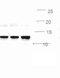
Application: Western BlotSample Tested: Hela whole cell lysateSpecies: HumanVerified Customer | Posted 11/03/2016Western blot for INSL5, Hela cell lysates 10 ug, dillution 1:2000, predicted band size : 13 kDa.

There are no reviews that match your criteria.
Protocols
Find general support by application which include: protocols, troubleshooting, illustrated assays, videos and webinars.
- Antigen Retrieval Protocol (PIER)
- Antigen Retrieval for Frozen Sections Protocol
- Appropriate Fixation of IHC/ICC Samples
- Cellular Response to Hypoxia Protocols
- Chromogenic IHC Staining of Formalin-Fixed Paraffin-Embedded (FFPE) Tissue Protocol
- Chromogenic Immunohistochemistry Staining of Frozen Tissue
- ClariTSA™ Fluorophore Kits
- Detection & Visualization of Antibody Binding
- Fluorescent IHC Staining of Frozen Tissue Protocol
- Graphic Protocol for Heat-induced Epitope Retrieval
- Graphic Protocol for the Preparation and Fluorescent IHC Staining of Frozen Tissue Sections
- Graphic Protocol for the Preparation and Fluorescent IHC Staining of Paraffin-embedded Tissue Sections
- Graphic Protocol for the Preparation of Gelatin-coated Slides for Histological Tissue Sections
- IHC Sample Preparation (Frozen sections vs Paraffin)
- Immunofluorescent IHC Staining of Formalin-Fixed Paraffin-Embedded (FFPE) Tissue Protocol
- Immunohistochemistry (IHC) and Immunocytochemistry (ICC) Protocols
- Immunohistochemistry Frozen Troubleshooting
- Immunohistochemistry Paraffin Troubleshooting
- Preparing Samples for IHC/ICC Experiments
- Preventing Non-Specific Staining (Non-Specific Binding)
- Primary Antibody Selection & Optimization
- Protocol for Heat-Induced Epitope Retrieval (HIER)
- Protocol for Making a 4% Formaldehyde Solution in PBS
- Protocol for VisUCyte™ HRP Polymer Detection Reagent
- Protocol for the Preparation & Fixation of Cells on Coverslips
- Protocol for the Preparation and Chromogenic IHC Staining of Frozen Tissue Sections
- Protocol for the Preparation and Chromogenic IHC Staining of Frozen Tissue Sections - Graphic
- Protocol for the Preparation and Chromogenic IHC Staining of Paraffin-embedded Tissue Sections
- Protocol for the Preparation and Chromogenic IHC Staining of Paraffin-embedded Tissue Sections - Graphic
- Protocol for the Preparation and Fluorescent IHC Staining of Frozen Tissue Sections
- Protocol for the Preparation and Fluorescent IHC Staining of Paraffin-embedded Tissue Sections
- Protocol for the Preparation of Gelatin-coated Slides for Histological Tissue Sections
- R&D Systems Quality Control Western Blot Protocol
- TUNEL and Active Caspase-3 Detection by IHC/ICC Protocol
- The Importance of IHC/ICC Controls
- Troubleshooting Guide: Immunohistochemistry
- Troubleshooting Guide: Western Blot Figures
- Western Blot Conditions
- Western Blot Protocol
- Western Blot Protocol for Cell Lysates
- Western Blot Troubleshooting
- Western Blot Troubleshooting Guide
- View all Protocols, Troubleshooting, Illustrated assays and Webinars
Loading...