mCherry Antibody - BSA Free
Novus Biologicals | Catalog # NBP2-25157
![Immunohistochemistry: mCherry Antibody [NBP2-25157]](https://resources.rndsystems.com/images/products/mCherry-Antibody-Immunocytochemistry-Immunofluorescence-NBP2-25157-img0011.jpg "Immunohistochemistry: mCherry Antibody [NBP2-25157]")
Key Product Details
Validated by
Biological Validation
Species Reactivity
Validated:
Non-species specific
Cited:
Human, Mouse, Rat, Bacteria, Insect - Drosophila, Invertebrate, Non-species specific
Applications
Validated:
Immunohistochemistry, Immunohistochemistry-Paraffin, Immunohistochemistry Whole-Mount, Western Blot, Immunocytochemistry/ Immunofluorescence, Live Imaging Microscopy, Fluorescence Imaging, Knockdown Validated
Cited:
Immunohistochemistry, Immunohistochemistry-Paraffin, Immunohistochemistry-Frozen, Immunohistochemistry Whole-Mount, Western Blot, Immunocytochemistry/ Immunofluorescence, Immunoprecipitation, IF/IHC, Knockdown Validated
Label
Unconjugated
Antibody Source
Polyclonal Rabbit
Format
BSA Free
Loading...
Product Specifications
Immunogen
This mCherry Antibody was developed against full length recombinant mCherry protein
Specificity
This antibody has cross-reactivity to TDtomato but not GFP.
Clonality
Polyclonal
Host
Rabbit
Theoretical MW
27 kDa.
Disclaimer note: The observed molecular weight of the protein may vary from the listed predicted molecular weight due to post translational modifications, post translation cleavages, relative charges, and other experimental factors.
Disclaimer note: The observed molecular weight of the protein may vary from the listed predicted molecular weight due to post translational modifications, post translation cleavages, relative charges, and other experimental factors.
Scientific Data Images for mCherry Antibody - BSA Free
Immunohistochemistry: mCherry Antibody [NBP2-25157]
mCherry-Antibody-Immunocytochemistry-Immunofluorescence-NBP2-25157-img0011.jpg![Immunohistochemistry: mCherry Antibody [NBP2-25157]](https://resources.rndsystems.com/images/products/mCherry-Antibody-Immunohistochemistry-NBP2-25157-img0010.jpg "Immunohistochemistry: mCherry Antibody [NBP2-25157]")
![Immunohistochemistry-Paraffin: mCherry Antibody [NBP2-25157]](https://resources.rndsystems.com/images/products/mCherry-Antibody-Immunohistochemistry-Paraffin-NBP2-25157-img0013.jpg "Immunohistochemistry-Paraffin: mCherry Antibody [NBP2-25157]")
Immunohistochemistry-Paraffin: mCherry Antibody [NBP2-25157]
mCherry-Antibody-Immunohistochemistry-Paraffin-NBP2-25157-img0013.jpg
Western Blot: Rabbit Polyclonal mCherry Antibody [NBP2-25157] -
Western Blot: Rabbit Polyclonal mCherry Antibody [NBP2-25157] - Analysis of HEK293 cell lysates using rabbit polyclonal mCherry antibody, dilution 1:3000, in green, and mouse mAb to GAPDH, dilution 1:2000, in red: [1] protein standard, [2] HEK293 control cells, [3] HEK293 cells transfected with pCI-Neo-mod vector expressing two tdTomato protein domains, [4] HEK293 cells transfected with pCI-mod vector expressing one mCherry-HA protein domain, and [5] HEK293 cells transfected with pCI-Neo-mod vector expressing one GFP domain. The rabbit polyclonal mCherry antibody recognizes tdTomato and mCherry proteins revealing major bands at about 60kDa and 30kDa, in green, respectively, but does not recognize GFP. The red band at 37kDa corresponds to GAPDH protein here used as a loading control.![Immunocytochemistry/ Immunofluorescence: mCherry Antibody [NBP2-25157]](https://resources.rndsystems.com/images/products/mCherry-Antibody-Immunocytochemistry-Immunofluorescence-NBP2-25157-img0012.jpg "Immunocytochemistry/ Immunofluorescence: mCherry Antibody [NBP2-25157]")
Immunocytochemistry/ Immunofluorescence: mCherry Antibody [NBP2-25157]
mCherry-Antibody-Immunocytochemistry-Immunofluorescence-NBP2-25157-img0012.jpg![Western Blot: mCherry Antibody [NBP2-25157]](https://resources.rndsystems.com/images/products/mCherry-Antibody-Western-Blot-NBP2-25157-img0014.jpg "Western Blot: mCherry Antibody [NBP2-25157]")
![Immunohistochemistry-Paraffin: mCherry Antibody [NBP2-25157]](https://resources.rndsystems.com/images/products/mCherry-Antibody-Immunohistochemistry-Paraffin-NBP2-25157-img0006.jpg "Immunohistochemistry-Paraffin: mCherry Antibody [NBP2-25157]")
Immunohistochemistry-Paraffin: mCherry Antibody [NBP2-25157]
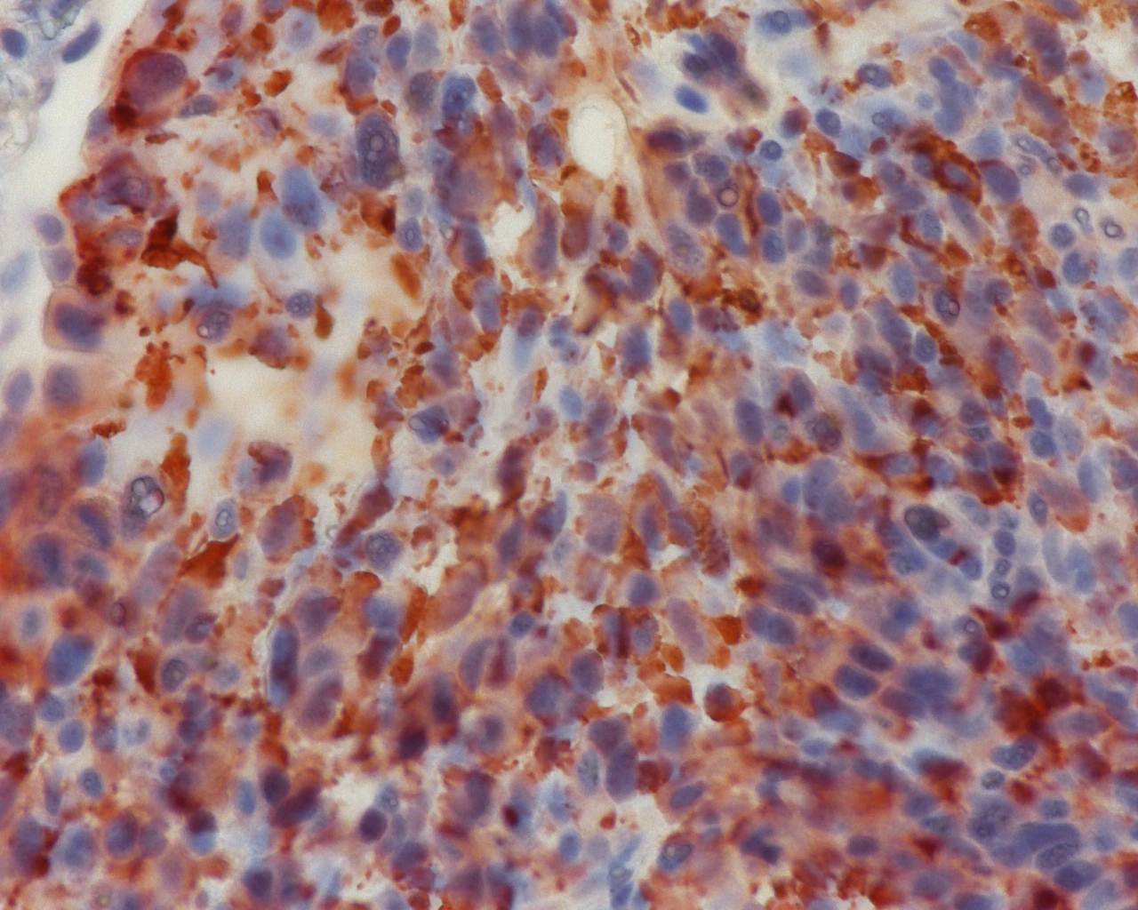
Immunohistochemistry-Paraffin: mCherry Antibody [NBP2-25157] - Staining of a mouse melanoma with over expression of mCherry in lung tissue. IHC-P image submitted by a verified customer review.![Western Blot: mCherry Antibody [NBP2-25157]](https://resources.rndsystems.com/images/products/mCherry-Antibody-Western-Blot-NBP2-25157-img0003.jpg "Western Blot: mCherry Antibody [NBP2-25157]")
Western Blot: mCherry Antibody [NBP2-25157]
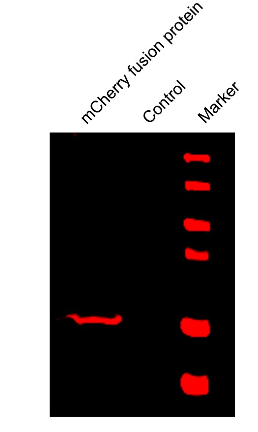
Western Blot: mCherry Antibody [NBP2-25157] - 3 cells transfected with pFin-EF1-mCherry vector, in the lane marked '+'. HEK293 cells which were not transfected with this vector show no protein band in lane marked '-'.![Western Blot: mCherry Antibody [NBP2-25157]](https://resources.rndsystems.com/images/products/mCherry-Antibody-Western-Blot-NBP2-25157-img0008.jpg "Western Blot: mCherry Antibody [NBP2-25157]")
Western Blot: mCherry Antibody [NBP2-25157]
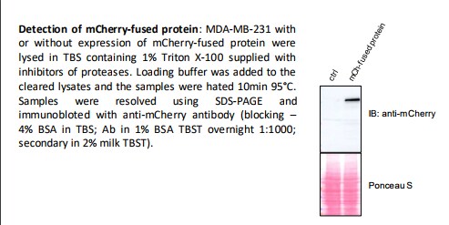
Western Blot: mCherry Antibody [NBP2-25157] - Detection of mCherry-fused protein: MDA-MB-231 cell lysates with and without expression of mCherry-fused protein. WB image submitted by a verified customer review.![Immunocytochemistry/ Immunofluorescence: mCherry Antibody [NBP2-25157]](https://resources.rndsystems.com/images/products/mCherry-Antibody-Immunocytochemistry-Immunofluorescence-NBP2-25157-img0004.jpg "Immunocytochemistry/ Immunofluorescence: mCherry Antibody [NBP2-25157]")
Immunocytochemistry/ Immunofluorescence: mCherry Antibody [NBP2-25157]
Immunocytochemistry/Immunofluorescence: mCherry Antibody [NBP2-25157] - HEK293 cells transfected in the same way and viewed in the confocal microscope. Most HEK293 cells are not transfected so only the nucleus of these cells can be visualized with a blue DNA stain. Cells which are transfected with mCherry are red. Staining with NBP2-25157 is shown in Green. Green antibody staining is only seen in cells which express mCherry, as expected, and the superimposition of the green and red signals results in an orange signal. Interestingly, stronger mCherry staining is seen in the nucleus, possibly due to degradation of some mCherry molecules to release the low molecular weight mCherry fluorochrome. Blot and transfected cells courtesy of the Semple-Rowland lab at the University of Florida.![Fluorescence Imaging: mCherry Antibody [NBP2-25157]](https://resources.rndsystems.com/images/products/mCherry-Antibody-Fluorescence-Imaging-NBP2-25157-img0007.jpg "Fluorescence Imaging: mCherry Antibody [NBP2-25157]")
Fluorescence Imaging: mCherry Antibody [NBP2-25157]
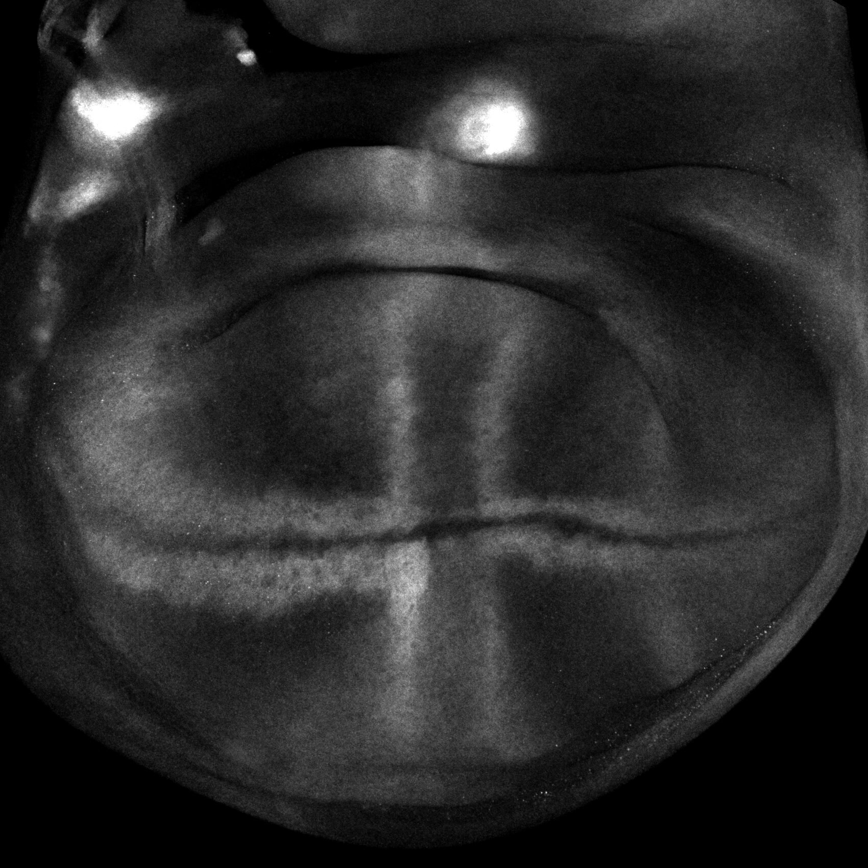
Fluorescence Imaging: mCherry Antibody [NBP2-25157] - Analysis of a Drosophila wing imaginal disc. Image submitted by a verified customer review.
Immunocytochemistry/ Immunofluorescence: mCherry Antibody [NBP2-25157] -
PKN3 activity is important for stress fibers formation & is stimulated by the expression of p130Cas. (A) p130Cas−/−MEFs growing on FN‐coated cover slips were co‐transfected by GFP‐p130Cas & mCherry‐PKN3 fusion variant (WT, mPR, KD) or mCherry. After 48 h, cells were fixed & imaged by Leica TCS SP2 microscope (63×/1.45 oil objective). Stress fibers were visualized by Phalloidin (405) & focal adhesions by anti‐Paxillin staining (2nd 633). Representative images are shown. Scale bars represent 20 μm. (B) p130Cas−/−MEFs or p130Cas−/−MEFs re‐expressing p130Cas or transfected by GFP‐fused p130Cas variants (WT, YE, dCCH) were lysed in RIPA buffer, blotted to nitrocellulose membrane, & analyzed for endogenous PKN3 activity by antibody anti‐phosphoThr849 of PKN3 (pT849 PKN3). Expression of p130Cas mutants was verified by anti‐p130Cas antibody & loading by anti‐PKN3 & anti‐actin antibody. (C) Densitometric quantification of PKN3 activity (pT849 PKN3 phosphorylation). The effect of p130Cas re‐expression on PKN3 T849 phosphorylation was analyzed separately from the effect of transfected p130Cas mutants (indicated by a dotted line). Error bars indicate means ± SD from three independent experiments (four experiments for the left part). Statistical significance was evaluated by one‐way repeated ANOVA followed by Turkey's post hoc test (*P < 0.05; **P < 0.01). (D) Lysates or (E) immunoprecipitates (by Flag sepharose) from p130Cas−/−MEFs re‐expressing p130Cas & overexpressing PKN3 variants (WT, mPR, KD) were immunoblotted by anti‐PKN3, anti‐pT849 PKN3, & anti‐Akt antibodies (loading control). Image collected & cropped by CiteAb from the following publication (https://pubmed.ncbi.nlm.nih.gov/30422386), licensed under a CC-BY license. Not internally tested by Novus Biologicals.
Immunocytochemistry/ Immunofluorescence: mCherry Antibody [NBP2-25157] -
Immunocytochemistry/ Immunofluorescence: mCherry Antibody [NBP2-25157] - Brn3b-mCherry expression in other regions of the CNS. (A) Optic tracts at P0 visualized by mCherry staining (white arrows). (B,B’) Co-localization experiments with mCherry (red) & pan-Brn3 (green) antibodies at P0. (C,C’) Co-localization with mCherry (red) & Tuj1 (green) in the trigeminal ganglia at E13.5. Arrows indicate mCherry+ Tuj+ neurons. (D) Low magnification image of a horizontal E13.5 whole-head section. E–F’) White arrows indicate mCherry+ Tuj1- cells present in the optic stalk (E) & optic recess (F). Aq: Aqueduct. soSC: stratum opticum of the Superior Colliculus. SGS: Stratum Griseum Superficiale. Tg: Trigeminal ganglion. L: Lens. ONH: Optic Nerve Head. OR: optic recess. III: 3rd ventricle. Scale bar: 100 microns in C,C’, E,E’ & F,F’, 200 microns in B,B’, 300 microns in D, & 500 microns in A. Image collected & cropped by CiteAb from the following publication (https://pubmed.ncbi.nlm.nih.gov/31197108), licensed under a CC-BY license. Not internally tested by Novus Biologicals.
Immunocytochemistry/ Immunofluorescence: mCherry Antibody [NBP2-25157] -
Immunocytochemistry/ Immunofluorescence: mCherry Antibody [NBP2-25157] - Brn3b-mCherry expression in the adult retina. (A) Flat-mounted retina labeled with anti-mCherry antibody. (B,B’) mCherry (red) & Brn3 (teal) colocalization. Yellow arrow indicates a Brn3+ mCherry- cell. (C,C’) mCherry (red) & RBPMS (teal) colocalization. Yellow arrows indicate RBPMS+ mCherry- cell bodies. (D,D’) mCherry (red) & Tuj1 (teal) colocalization. White arrows indicate Tuj1+ mCherry+ axons. (E–E’’’) Cross-section of an adult retina labeled with mCherry (red), RBPMS (gray), DAPI (blue), & Pax6 (green). All mCherry+ cells (white arrows) are RBPMS+. Amacrine cells are labeled with yellow stars & are mCherry- (Pax6+ RBPMS− mCherry− cells). Yellow arrowhead corresponds to an mCherry− RGC (RBPMS+ Pax6− mCherry− cell). ONH: Optic Nerve Head. ONL: Outer Nuclear Layer. INL: Inner Nuclear Layer. GCL: Ganglion Cell Layer. Scale bars: 300 microns in A, 50 microns in B–E’’’. Image collected & cropped by CiteAb from the following publication (https://pubmed.ncbi.nlm.nih.gov/31197108), licensed under a CC-BY license. Not internally tested by Novus Biologicals.
Immunocytochemistry/ Immunofluorescence: mCherry Antibody [NBP2-25157] -
Immunocytochemistry/ Immunofluorescence: mCherry Antibody [NBP2-25157] - Brn3b-mCherry expression in other regions of the CNS. (A) Optic tracts at P0 visualized by mCherry staining (white arrows). (B,B’) Co-localization experiments with mCherry (red) & pan-Brn3 (green) antibodies at P0. (C,C’) Co-localization with mCherry (red) & Tuj1 (green) in the trigeminal ganglia at E13.5. Arrows indicate mCherry+ Tuj+ neurons. (D) Low magnification image of a horizontal E13.5 whole-head section. E–F’) White arrows indicate mCherry+ Tuj1- cells present in the optic stalk (E) & optic recess (F). Aq: Aqueduct. soSC: stratum opticum of the Superior Colliculus. SGS: Stratum Griseum Superficiale. Tg: Trigeminal ganglion. L: Lens. ONH: Optic Nerve Head. OR: optic recess. III: 3rd ventricle. Scale bar: 100 microns in C,C’, E,E’ & F,F’, 200 microns in B,B’, 300 microns in D, & 500 microns in A. Image collected & cropped by CiteAb from the following publication (https://pubmed.ncbi.nlm.nih.gov/31197108), licensed under a CC-BY license. Not internally tested by Novus Biologicals.
Western Blot: mCherry Antibody [NBP2-25157] -
Western Blot: mCherry Antibody [NBP2-25157] - PKN3 overexpression regulates growth of MEFs, & this effect requires PKN3–p130Cas interaction. (A) Immunoblotted lysates from MEFs p130Cas−/−/MEFs p130Cas−/− re‐expressing p130Cas (p130Cas+) treated by Doxycycline (Dox) to induce expression of mCherry‐PKN3/mCherry alone. p130Cas presence detected by ani‐p130Cas antibody & mCherry epitope by anti‐mCherry antibody. (B) Dynamics of mCherry‐PKN3 expression after supplementation w/ Dox shown by immunoblot w/ anti‐mCherry antibody. (C–E) Effect of induced mCherry‐PKN3 expression on cell growth. Representative graphs showing growth of MEFs p130Cas−/− re‐expressing p130Cas (p130Cas+) (C)/MEFs p130Cas−/− (D) measured in real‐time using xCELLigence RTCA (real‐time cell analysis) system instrument. (E) Quantification of cell growth change induced by mCherry‐PKN3 expression (‘−’ indicates inducible mCherry expression used as negative control). Slope ratios reflecting cell growth calculated from log growth phase of cell growth (indicated by dotted lines; see C & D). (F) Immunoblotted lysates from MEFs p130Cas−/− re‐expressing p130Cas (p130Cas+) treated/not treated by Dox which induced expression of Flag‐fused PKN3 variants (WT, mPR, KD, empty vector). Stimulated overexpression of PKN3 detected by anti‐PKN3 antibody & its activity by antibody anti‐pT849 PKN3. (G) Quantification of cell growth change stimulated by Dox‐inducible expression of Flag‐fused PKN3 variants (WT, mPR, KD) in MEFs p130Cas−/− re‐expressing p130Cas (p130Cas+). All error bars indicate means ± SD calculated from 3 to 5 independent experiments (each in triplicates). Statistical significance always calculated between induced & noninduced cells & evaluated by one‐way repeated ANOVA followed by Turkey post hoc test (***P < 0.001). Image collected & cropped by CiteAb from following publication (https://pubmed.ncbi.nlm.nih.gov/30422386), licensed under a CC-BY license. Not internally tested by Novus Biologicals.
Immunocytochemistry/ Immunofluorescence: mCherry Antibody [NBP2-25157] -
Immunocytochemistry/ Immunofluorescence: mCherry Antibody [NBP2-25157] - PKN3 colocalizes with p130Cas in lamellipodia & podosome rosettes. Representative images are shown. (A) p130Cas−/−MEFs plated on fibronectin (FN) were transfected by GFP‐p130Cas, CFP‐LifeAct, & mCherry‐PKN3WT or mCherry‐PKN3 mPR & imaged live 24 h after transfection. White arrow indicates lamellipodia. Histogram of dotted straight line is shown. (B) Quantification of mCherry‐PKN3 WT, mCherry‐PKN3 mPR, & mCherry localization to lamellipodia (LifeAct as marker) was calculated as described in methods (values are mean ± SD from three independent experiments, n > 50 measurements – 3 per cell; ***P < 0.001, one‐way ANOVA on ranks followed by Dunn's post hoc test). (C) Src‐transformed p130Cas−/−MEFs co‐expressing p130Cas (SC) & mouse Flag tagged PKN3 WT or Flag‐PKN3 mPR are shown. Cells were grown on FN‐coated coverslips for 48 h, fixed, & stained for p130Cas by anti‐pTyr165 p130Cas antibody (pY165 p130Cas; 2nd 405), for actin by Phalloidin 488 & for Flag‐PKN3 by anti‐Flag antibody (2nd 633). Reflection (670 nm) indicates fibronectin degradation. All scale bars represent 20 μm. Cell were imaged by Leica TCS SP8 microscope system equipped with Leica 63×/1.45 oil objective. Image collected & cropped by CiteAb from the following publication (https://pubmed.ncbi.nlm.nih.gov/30422386), licensed under a CC-BY license. Not internally tested by Novus Biologicals.
Immunocytochemistry/ Immunofluorescence: mCherry Antibody [NBP2-25157] -
Immunocytochemistry/ Immunofluorescence: mCherry Antibody [NBP2-25157] - Brn3b-mCherry expression in the adult retina. (A) Flat-mounted retina labeled with anti-mCherry antibody. (B,B’) mCherry (red) & Brn3 (teal) colocalization. Yellow arrow indicates a Brn3+ mCherry- cell. (C,C’) mCherry (red) & RBPMS (teal) colocalization. Yellow arrows indicate RBPMS+ mCherry- cell bodies. (D,D’) mCherry (red) & Tuj1 (teal) colocalization. White arrows indicate Tuj1+ mCherry+ axons. (E–E’’’) Cross-section of an adult retina labeled with mCherry (red), RBPMS (gray), DAPI (blue), & Pax6 (green). All mCherry+ cells (white arrows) are RBPMS+. Amacrine cells are labeled with yellow stars & are mCherry- (Pax6+ RBPMS− mCherry− cells). Yellow arrowhead corresponds to an mCherry− RGC (RBPMS+ Pax6− mCherry− cell). ONH: Optic Nerve Head. ONL: Outer Nuclear Layer. INL: Inner Nuclear Layer. GCL: Ganglion Cell Layer. Scale bars: 300 microns in A, 50 microns in B–E’’’. Image collected & cropped by CiteAb from the following publication (https://pubmed.ncbi.nlm.nih.gov/31197108), licensed under a CC-BY license. Not internally tested by Novus Biologicals.Applications for mCherry Antibody - BSA Free
Application
Recommended Usage
Immunocytochemistry/ Immunofluorescence
1:500
Immunohistochemistry
1:500
Western Blot
1:1000
Application Notes
Use in Immunohistochemistry Whole-Mount reported in scientific literature (PMID:35013168) This mCherry antibody is useful for Immunocytochemistry/Immunofluorescence and Western Blot, where a band can be seen at ~28 kDa.
Use in IHC and IHC-P reported in scientific literature (PMID: 27396338 and 28891816 respectively).
Use in Live Imaging Microscopy was reported from a verified customer review.
Knockdown validation (PMID: 32494070).
Use in IHC and IHC-P reported in scientific literature (PMID: 27396338 and 28891816 respectively).
Use in Live Imaging Microscopy was reported from a verified customer review.
Knockdown validation (PMID: 32494070).
Reviewed Applications
Read 4 reviews rated 4.8 using NBP2-25157 in the following applications:
Formulation, Preparation, and Storage
Purification
Immunogen affinity purified
Formulation
50% PBS, 50% glycerol
Format
BSA Free
Preservative
0.035% Sodium Azide
Concentration
1 mg/ml
Shipping
The product is shipped with polar packs. Upon receipt, store it immediately at the temperature recommended below.
Stability & Storage
Store at 4C short term. Aliquot and store at -20C long term. Avoid freeze-thaw cycles.
Background: mCherry
mCherry can be used as a long-wavelength hetero-FRET (fluorescence resonance energy transfer) acceptor and probe for homoFRET experiments given its high peak molar absorptivity, folding efficiency, and superior spectral properties (4). Additionally, because mCherry does not interfere with other plasmids or alter the growth of Legionella species during intracellular growth, it can be used for constitutive gene expression in a variety of gram-negative bacterial species (5). For example, a plasmid developed to constitutively express mCherry under the Ptac promoter has been used in several Legionella species including L. pneumophila, the causative agent of Legionnaires' disease (5).
References
1. Shaner, N. C., Steinbach, P. A., & Tsien, R. Y. (2005). A guide to choosing fluorescent proteins. Nature Methods, 2(12), 905-909. doi:10.1038/nmeth819
2. Bevis, B. J., & Glick, B. S. (2002). Rapidly maturing variants of the Discosoma red fluorescent protein (DsRed). Nature Biotechnology, 20(1), 83-87. https://doi.org/10.1038/nbt0102-83
3. Wall, M. A., Socolich, M., & Ranganathan, R. (2000). The structural basis for red fluorescence in the tetrameric GFP homolog DsRed. Nature Structural Biology, 7(12), 1133-1138. https://doi.org/10.1038/81992
4. Akrap, N., Seidel, T., & Barisas, B. G. (2010). Forster distances for fluorescence resonant energy transfer between mCherry and other visible fluorescent proteins. Analytical Biochemistry, 402(1), 105-106. https://doi.org/10.1016/j.ab.2010.03.026
5. Gebhardt, M. J., Jacobson, R. K., & Shuman, H. A. (2017). Seeing red; the development of pON.mCherry, a broad-host range constitutive expression plasmid for Gram-negative bacteria. Plos One, 12(3), e0173116. https://doi.org/10.1371/journal.pone.0173116
Long Name
mCherry
Alternate Names
DSRED, red fluorescent protein mCherry, Red Fluoroscent Protein
Additional mCherry Products
Product Documents for mCherry Antibody - BSA Free
Certificate of Analysis
To download a Certificate of Analysis, please enter a lot or batch number in the search box below.
Product Specific Notices for mCherry Antibody - BSA Free
This product is for research use only and is not approved for use in humans or in clinical diagnosis. Primary Antibodies are guaranteed for 1 year from date of receipt.
Citations for mCherry Antibody - BSA Free
 Powered by Bioz
Customer Reviews for mCherry Antibody - BSA Free (4)
4.8 out of 5
4 Customer Ratings
Have you used mCherry Antibody - BSA Free?
Submit a review and receive an Amazon gift card!
$25/€18/£15/$25CAN/¥2500 Yen for a review with an image
$10/€7/£6/$10CAN/¥1110 Yen for a review without an image
Submit a review
Customer Images
Showing
1
-
4 的
4 reviews
Showing All
Filter By:
-
Application: Western BlotSample Tested: 293T whole cell lysate, Sample Amount: 20ngSpecies: HumanVerified Customer | Posted 11/18/2019Specificity is good. But it needs low dilution, 1:250.

-
Application: Western BlotSample Tested: MDA-MB-231 Cell LysateSpecies: HumanVerified Customer | Posted 08/22/2017
-
Application: Immunohistochemistry (whole mount)Sample Tested: Wing imaginal discSpecies: DrosophilaVerified Customer | Posted 12/08/2016The expression of mCherry under the control of the endogenous Sulf1 in the wing disc.1:1000 dilution
-
Application: ImmunofluorescenceSample Tested: Mouse melanoma with mCherry overexpressionSpecies: MouseVerified Customer | Posted 07/23/2015Mouse melanoma in lung tissue
There are no reviews that match your criteria.
Protocols
Find general support by application which include: protocols, troubleshooting, illustrated assays, videos and webinars.
- Antigen Retrieval Protocol (PIER)
- Antigen Retrieval for Frozen Sections Protocol
- Appropriate Fixation of IHC/ICC Samples
- Cellular Response to Hypoxia Protocols
- Chromogenic IHC Staining of Formalin-Fixed Paraffin-Embedded (FFPE) Tissue Protocol
- Chromogenic Immunohistochemistry Staining of Frozen Tissue
- ClariTSA™ Fluorophore Kits
- Detection & Visualization of Antibody Binding
- Fluorescent IHC Staining of Frozen Tissue Protocol
- Graphic Protocol for Heat-induced Epitope Retrieval
- Graphic Protocol for the Preparation and Fluorescent IHC Staining of Frozen Tissue Sections
- Graphic Protocol for the Preparation and Fluorescent IHC Staining of Paraffin-embedded Tissue Sections
- Graphic Protocol for the Preparation of Gelatin-coated Slides for Histological Tissue Sections
- ICC Cell Smear Protocol for Suspension Cells
- ICC Immunocytochemistry Protocol Videos
- ICC for Adherent Cells
- IHC Sample Preparation (Frozen sections vs Paraffin)
- Immunocytochemistry (ICC) Protocol
- Immunocytochemistry Troubleshooting
- Immunofluorescence of Organoids Embedded in Cultrex Basement Membrane Extract
- Immunofluorescent IHC Staining of Formalin-Fixed Paraffin-Embedded (FFPE) Tissue Protocol
- Immunohistochemistry (IHC) and Immunocytochemistry (ICC) Protocols
- Immunohistochemistry Frozen Troubleshooting
- Immunohistochemistry Paraffin Troubleshooting
- Preparing Samples for IHC/ICC Experiments
- Preventing Non-Specific Staining (Non-Specific Binding)
- Primary Antibody Selection & Optimization
- Protocol for Heat-Induced Epitope Retrieval (HIER)
- Protocol for Making a 4% Formaldehyde Solution in PBS
- Protocol for VisUCyte™ HRP Polymer Detection Reagent
- Protocol for the Fluorescent ICC Staining of Cell Smears - Graphic
- Protocol for the Fluorescent ICC Staining of Cultured Cells on Coverslips - Graphic
- Protocol for the Preparation & Fixation of Cells on Coverslips
- Protocol for the Preparation and Chromogenic IHC Staining of Frozen Tissue Sections
- Protocol for the Preparation and Chromogenic IHC Staining of Frozen Tissue Sections - Graphic
- Protocol for the Preparation and Chromogenic IHC Staining of Paraffin-embedded Tissue Sections
- Protocol for the Preparation and Chromogenic IHC Staining of Paraffin-embedded Tissue Sections - Graphic
- Protocol for the Preparation and Fluorescent ICC Staining of Cells on Coverslips
- Protocol for the Preparation and Fluorescent ICC Staining of Non-adherent Cells
- Protocol for the Preparation and Fluorescent ICC Staining of Stem Cells on Coverslips
- Protocol for the Preparation and Fluorescent IHC Staining of Frozen Tissue Sections
- Protocol for the Preparation and Fluorescent IHC Staining of Paraffin-embedded Tissue Sections
- Protocol for the Preparation of Gelatin-coated Slides for Histological Tissue Sections
- Protocol for the Preparation of a Cell Smear for Non-adherent Cell ICC - Graphic
- R&D Systems Quality Control Western Blot Protocol
- TUNEL and Active Caspase-3 Detection by IHC/ICC Protocol
- The Importance of IHC/ICC Controls
- Troubleshooting Guide: Immunohistochemistry
- Troubleshooting Guide: Western Blot Figures
- Western Blot Conditions
- Western Blot Protocol
- Western Blot Protocol for Cell Lysates
- Western Blot Troubleshooting
- Western Blot Troubleshooting Guide
- View all Protocols, Troubleshooting, Illustrated assays and Webinars
FAQs for mCherry Antibody - BSA Free
Showing
1
-
3 的
3 FAQs
Showing All
-
Q: Does this antibody cross-react with GFP epitopes? As I would like to use both GFP and mCherry antibodies during histochemistry I would not like them to cross-react.
A: mCherry and GFP share just 29% sequence similarity, so this antibody is not predicted to cross-react to GFP and has never shown any ability to detect GFP in testing.
-
Q: I was wondering if you had any literature on whether the polyclonal mCherry antibody NBP2-25157 shows cross-reaction with td-tomato?
A: We do see 88% homology between the mCherry and the dTomato sequences and therefore, the antibody will likely see both of these RFP proteins. The antibody is a polyclonal, which then increases the likelihood that it will have multiple binding sites along the homologous sequences. There really are no regions of sequence where one could make an antibody that did not see both proteins.
-
Q: I would like to know the isotype of our NBP2-25157 and the secondary antibody that would be suggested for it.
A: This antibody is a rabbit polyclonal of the isotype IgG. Of our rabbit IgG isotype control products, NB810-56910 should meet your requirements.
-
Q: Does this antibody cross-react with GFP epitopes? As I would like to use both GFP and mCherry antibodies during histochemistry I would not like them to cross-react.
A: mCherry and GFP share just 29% sequence similarity, so this antibody is not predicted to cross-react to GFP and has never shown any ability to detect GFP in testing.
-
Q: I was wondering if you had any literature on whether the polyclonal mCherry antibody NBP2-25157 shows cross-reaction with td-tomato?
A: We do see 88% homology between the mCherry and the dTomato sequences and therefore, the antibody will likely see both of these RFP proteins. The antibody is a polyclonal, which then increases the likelihood that it will have multiple binding sites along the homologous sequences. There really are no regions of sequence where one could make an antibody that did not see both proteins.
-
Q: I would like to know the isotype of our NBP2-25157 and the secondary antibody that would be suggested for it.
A: This antibody is a rabbit polyclonal of the isotype IgG. Of our rabbit IgG isotype control products, NB810-56910 should meet your requirements.
Loading...