
Agouti-Related Protein (AgRP), the protein product of the Agouti-Related Transcript (ART), is a neuroprotein that regulates energy metabolism and the development of obesity by antagonizing alpha -melanocyte stimulating hormone ( alpha -MSH) action on MC-3 and MC-4 receptors (1-4). AgRP is predominantly expressed in the hypothalamus and adrenal medulla (5). Mature mouse AgRP is a 111 amino acid (aa) polypeptide; its C-terminal portion contains ten conserved cysteines that form five disulfide bonds (5, 6). Within the C-terminal region, mouse AgRP shares 80% and 90% aa sequence identity with human and rat AgRP, respectively. It also shares 44% aa sequence identity with Agouti. As with Agouti, the C-terminal cysteine-rich region is sufficient for biological activity (7). AgRP, however, is 100 times more potent than Agouti in antagonizing MC-3 and MC-4 receptors (8). AgRP also induces the beta -arrestin dependent endocytosis of MC-3 and MC-4 (9). Hypothalamic expression of AgRP is upregulated in obesity and diabetes (5, 10), and chronic AgRP administration increases food intake and weight gain in rats (11). Genetically-linked polymorphisms of AgRP in humans are associated with susceptibility to anorexia nervosa (12, 13). In addition, AgRP inhibits the ACTH-induced synthesis of steroid hormones via a mechanism that does not involve melanocortin receptors (14).
Mouse AgRP/ART Antibody
R&D Systems | Catalog # AF634

Scientific images may be resized, cropped, or adjusted for brightness or contrast for presentation purposes. These updates are limited to presentation and do not affect the underlying scientific interpretation of the data. Where available, additional source data may be provided upon request.
Key Product Details
Species Reactivity
Validated:
Cited:
Applications
Validated:
Cited:
Label
Antibody Source
Product Specifications
Immunogen
Val25-Thr131
Accession # P56473
Specificity
Clonality
Host
Isotype
Scientific Data Images for Mouse AgRP/ART Antibody
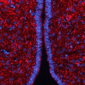
AgRP/ART in Mouse Brain.
AgRP/ART was detected in perfusion fixed frozen sections of mouse brain (hypothalamus) using Goat Anti-Mouse AgRP/ART Antigen Affinity-purified Polyclonal Antibody (Catalog # AF634) at 1.7 µg/mL overnight at 4 °C. Tissue was stained using the NorthernLights™ 557-conjugated Anti-Goat IgG Secondary Antibody (red; NL001) and counterstained with DAPI (blue). View our protocol for Fluorescent IHC Staining of Frozen Tissue Sections.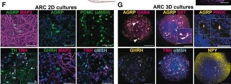
Detection of AgRP by Immunohistochemistry
Signaling factors required for generation of human ARC in vitro(A) Hypothalamic development in vivo and in vitro differentiation of hPSC into hypothalamic cells.(B) Principal-component analysis (PCA) of day 16 RT-qPCR data (RC17), clustering according to ARC- and PVN-associated genes. Each dot represents one differentiation batch (n = 113).(C) Estimated marginal means assessing the effect of factors on PC1, adjusted for covariates.(D) ICC/ISH at days 16 and 50 for ARC protocol with or without BMP4. Scale bars: 100 μm.(E) ARC differentiation protocol and maturation options.(F–H) ICC of day 50 in 2D (F) and 3D (G and H), scale bars: 50 and 100 μm.(I) Quantification of day 50 3D spheroids (AGRP: n = 14, 4.53% ± 0.69%, mean ± SEM; alpha MSH: n = 6, 5.90% ± 1.19%).(J) Xenotransplantation of day 16 ARC progenitors into nude rats.(J′) Top: ICC for human neural cell adhesion molecule (NCAM) highlights graft site, scale bar: 2 mm. Bottom: IF for human nuclear antigen (HuNu), scale bar: 200 μm.(K–Q) IF for ARC neuronal markers in grafts. Scale bars: 100 μm (K and O), 50 μm (N), 25 μm (L, M, P, and Q). Image collected and cropped by CiteAb from the following open publication (https://linkinghub.elsevier.com/retrieve/pii/S1934590926001979), licensed under a CC-BY license. Not internally tested by R&D Systems.Applications for Mouse AgRP/ART Antibody
Immunohistochemistry
Sample: Perfusion fixed frozen sections of mouse brain (hypothalamus)
Formulation, Preparation, and Storage
Purification
Reconstitution
Reconstitute at 0.2 mg/mL in sterile PBS. For liquid material, refer to CoA for concentration.
Formulation
Shipping
Stability & Storage
- 12 months from date of receipt, -20 to -70 °C as supplied.
- 1 month, 2 to 8 °C under sterile conditions after reconstitution.
- 6 months, -20 to -70 °C under sterile conditions after reconstitution.
Calculators
Background: AgRP/ART
References
- Martin, N.M. et al. (2006) Peptides 27:333.
- Fan, W. et al. (1997) Nature 385:165.
- Ollmann, M.M. et al. (1997) Science 278:135.
- Arora, S. and Anubhuti (2006) Neuropeptides 40:375.
- Shutter, J.R. et al. (1997) Genes Dev. 11:593.
- Kiefer, L.L. et al. (1998) Biochemistry 37:991.
- Jackson, P.J. et al. (2002) Biochemistry 41:7565.
- Fong, T.M. et al. (1997) Biochem. Biophys. Res. Commun. 237:629.
- Breit, A. et al. (2006) J. Biol. Chem. 281:37447.
- Katsuki, A. et al. (2001) J. Clin. Endocrinol. Metab. 86:1921.
- Small, C.K. et al. (2001) Diabetes 50:248.
- Vink, T. et al. (2001) Mol. Psychiatry 6:325.
- Dardennes, R.M. et al. (2007) Psychoneuroendocrinology 32:106.
- Doghman, M. et al. (2007) Mol. Cell. Endocrinol. 265-266:108.
Long Name
Alternate Names
Gene Symbol
UniProt
Additional AgRP/ART Products
Product Documents for Mouse AgRP/ART Antibody
Certificate of Analysis
To download a Certificate of Analysis, please enter a lot or batch number in the search box below.
Note: Certificate of Analysis not available for kit components.
Product Specific Notices for Mouse AgRP/ART Antibody
For research use only
Related Research Areas
Citations for Mouse AgRP/ART Antibody
 Powered by Bioz
Customer Reviews for Mouse AgRP/ART Antibody
There are currently no reviews for this product. Be the first to review Mouse AgRP/ART Antibody and earn rewards!
Have you used Mouse AgRP/ART Antibody?
Submit a review and receive an Amazon gift card!
$25/€18/£15/$25CAN/¥2500 for a review with an image
$10/€7/£6/$10CAN/¥1110 for a review without an image
Submit a review
Protocols
Find general support by application which include: protocols, troubleshooting, illustrated assays, videos and webinars.
- Antigen Retrieval Protocol (PIER)
- Antigen Retrieval for Frozen Sections Protocol
- Appropriate Fixation of IHC/ICC Samples
- Cellular Response to Hypoxia Protocols
- Chromogenic IHC Staining of Formalin-Fixed Paraffin-Embedded (FFPE) Tissue Protocol
- Chromogenic Immunohistochemistry Staining of Frozen Tissue
- ClariTSA™ Fluorophore Kits
- Detection & Visualization of Antibody Binding
- Fluorescent IHC Staining of Frozen Tissue Protocol
- Graphic Protocol for Heat-induced Epitope Retrieval
- Graphic Protocol for the Preparation and Fluorescent IHC Staining of Frozen Tissue Sections
- Graphic Protocol for the Preparation and Fluorescent IHC Staining of Paraffin-embedded Tissue Sections
- Graphic Protocol for the Preparation of Gelatin-coated Slides for Histological Tissue Sections
- IHC Sample Preparation (Frozen sections vs Paraffin)
- Immunofluorescent IHC Staining of Formalin-Fixed Paraffin-Embedded (FFPE) Tissue Protocol
- Immunohistochemistry (IHC) and Immunocytochemistry (ICC) Protocols
- Immunohistochemistry Frozen Troubleshooting
- Immunohistochemistry Paraffin Troubleshooting
- Preparing Samples for IHC/ICC Experiments
- Preventing Non-Specific Staining (Non-Specific Binding)
- Primary Antibody Selection & Optimization
- Protocol for Heat-Induced Epitope Retrieval (HIER)
- Protocol for Making a 4% Formaldehyde Solution in PBS
- Protocol for VisUCyte™ HRP Polymer Detection Reagent
- Protocol for the Preparation & Fixation of Cells on Coverslips
- Protocol for the Preparation and Chromogenic IHC Staining of Frozen Tissue Sections
- Protocol for the Preparation and Chromogenic IHC Staining of Frozen Tissue Sections - Graphic
- Protocol for the Preparation and Chromogenic IHC Staining of Paraffin-embedded Tissue Sections
- Protocol for the Preparation and Chromogenic IHC Staining of Paraffin-embedded Tissue Sections - Graphic
- Protocol for the Preparation and Fluorescent IHC Staining of Frozen Tissue Sections
- Protocol for the Preparation and Fluorescent IHC Staining of Paraffin-embedded Tissue Sections
- Protocol for the Preparation of Gelatin-coated Slides for Histological Tissue Sections
- TUNEL and Active Caspase-3 Detection by IHC/ICC Protocol
- The Importance of IHC/ICC Controls
- Troubleshooting Guide: Immunohistochemistry
- View all Protocols, Troubleshooting, Illustrated assays and Webinars