
CD38, also known as ADP-ribosyl cyclase and cyclic ADP-ribose hydrolase, is a Type II integral membrane protein. The enzyme is able to transform NAD(P)+ into three different products with calcium mobilizing ability; cyclic ADP-ribose, NAADP+, and ADP-ribose (1). CD38 is expressed in B and T lymphocytes, osteoclasts, and in cardiac, pancreatic, liver and kidney cells (2, 3). Through its production of cyclic ADP-ribose, CD38 modulates calcium-mediated signal transduction in many types of cells, including neutrophils and pancreatic beta cells (4, 5). CD38 has been shown to regulate oxytocin secretion, and may be involved in the development of complex social behaviors in mammals (6).

Scientific images may be resized, cropped, or adjusted for brightness or contrast for presentation purposes. These updates are limited to presentation and do not affect the underlying scientific interpretation of the data. Where available, additional source data may be provided upon request.
Key Product Details
Species Reactivity
Validated:
Mouse
Cited:
Mouse, Transgenic Mouse
Applications
Validated:
Western Blot, Flow Cytometry, Simple Western, CyTOF-ready
Cited:
Immunohistochemistry, Western Blot
Label
Unconjugated
Antibody Source
Polyclonal Sheep IgG
Loading...
Product Specifications
Immunogen
Mouse myeloma cell line NS0-derived recombinant mouse CD38
Leu45-Thr304
Accession # EDL37596
Leu45-Thr304
Accession # EDL37596
Specificity
Detects mouse CD38 in direct ELISAs and Western blots. In direct ELISAs and Western blots, approximately 20% cross‑reactivity with recombinant human CD38 is observed.
Clonality
Polyclonal
Host
Sheep
Isotype
IgG
Scientific Data Images for Mouse CD38 Antibody
Detection of Mouse CD38 by Western Blot.
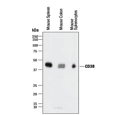
Western blot shows lysates of mouse spleen tissue, mouse colon tissue, and mouse splenocytes. PVDF membrane was probed with 0.5 µg/mL of Sheep Anti-Mouse CD38 Antigen Affinity-purified Polyclonal Antibody (Catalog # AF4947) followed by HRP-conjugated Anti-Sheep IgG Secondary Antibody (Catalog # HAF016). A specific band was detected for CD38 at approximately 45 kDa (as indicated). This experiment was conducted under reducing conditions and using Immunoblot Buffer Group 1.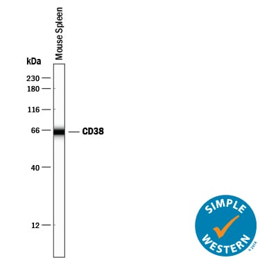
Detection of Mouse CD38 by Simple WesternTM.
Simple Western lane view shows lysates of mouse spleen tissue, loaded at 0.2 mg/mL. A specific band was detected for CD38 at approximately 64 kDa (as indicated) using 10 µg/mL of Sheep Anti-Mouse CD38 Antigen Affinity-purified Polyclonal Antibody (Catalog # AF4947) followed by 1:50 dilution of HRP-conjugated Anti-Goat IgG Secondary Antibody (Catalog # HAF109). This experiment was conducted under reducing conditions and using the 12-230 kDa separation system.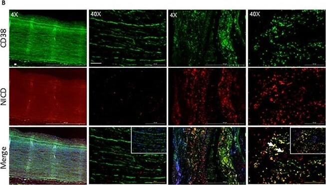
Detection of CD38 by Immunohistochemistry
NICD and CD38 expression is upregulated in the aorta of AAA human subjects. (A) Representative H&E images of abdominal aortic sections from non-AAA and AAA human subjects (4X and 40X). (B) Representative double immunofluorescence images showing NICD and CD38 immunostaining in the aorta of non-AAA and AAA human subjects (4X and 40X). (C) Pearson’s correlation graphs showing the coefficient values of NICD/CD38 expression. Scale bar = 50 µm. Image collected and cropped by CiteAb from the following open publication (https://pubmed.ncbi.nlm.nih.gov/31530833), licensed under a CC-BY license. Not internally tested by R&D Systems.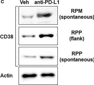
Detection of CD38 by Western Blot
Expression of CD38 by treatment with anti-PD-L1 antibody and anti-tumor effect of anti-CD38 antibody with anti-PD-L1 antibody in vivo. (A) Boxplots from bulk RNAseq analysis corresponding to the CD38 expression (B) Quantitative mRNA expression of CD38 by real-time RT-PCR analysis (C) Western blot showing expression of CD38 and actin (loading control)- in RPP (flank and orthotopic tumors) and RPM flank tumors, which were treated with vehicle or anti-PD-L1 antibody (300µg/body, once weekly). (D) Tumor growth curves (mean ± standard error [SE]) of IgG control, anti-PD-L1 antibody alone (200 µg/body, once weekly), anti-CD38 antibody (300 µg/body, once weekly), and anti-CD38 antibody plus anti-PD-L1 antibody groups in B6129F1 mice injected with RPP cells (SCLC cells with conditional loss of Trp53, p130, and Rb1; n = 6 in each group). P values were calculated by student-T test. Image collected and cropped by CiteAb from the following open publication (https://www.frontiersin.org/articles/10.3389/fimmu.2024.1348982/full), licensed under a CC-BY license. Not internally tested by R&D Systems.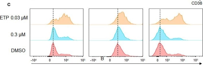
Detection of CD38 by Flow Cytometry
Change of expression of CD38 by chemotherapy and anti-tumor effect of anti-CD38 antibody with anti-PD-L1 antibody and chemotherapy. (A) CD38 expression on CD45+ total immune cell, CD4+ T cell, and CD8+ T cell in clinical specimen from the patients with SCLC. (B, C) Flow cytometry analysis was performed on H82, DMS114, and H187 cell lines treated with 0.3 or 3 µM cisplatin (upper) and 0.03 or 0.3 µM etoposide for 72 hours. (D) Tumor growth curves (mean ± standard error [SE]) of IgG control and vehicle, anti-PD-L1 antibody alone (200 µg/body, once weekly) and anti-CD38 antibody (300 µg/body, once weekly), cisplatin (CDDP) (2mg/kg) and etoposide (ETP) (3mg/kg), and anti-CD38 antibody, anti-PD-L1 antibody, CDDP and ETP groups in B6129F1 mice injected with RPP cells (SCLC cells with conditional loss of Trp53, p130, and Rb1; n = = 6 in each group). P values were calculated by Mann-Whiteney for (A), * p<0.0332 and student-T test for (D). Image collected and cropped by CiteAb from the following open publication (https://www.frontiersin.org/articles/10.3389/fimmu.2024.1348982/full), licensed under a CC-BY license. Not internally tested by R&D Systems.Applications for Mouse CD38 Antibody
Application
Recommended Usage
CyTOF-ready
Ready to be labeled using established conjugation methods. No BSA or other carrier proteins that could interfere with conjugation.
Flow Cytometry
0.25 µg/106 cells
Sample: Mouse splenocytes
Sample: Mouse splenocytes
Simple Western
10 µg/mL
Sample: Mouse spleen tissue
Sample: Mouse spleen tissue
Western Blot
0.5 µg/mL
Sample: Mouse spleen tissue, mouse colon tissue, and mouse splenocytes
Sample: Mouse spleen tissue, mouse colon tissue, and mouse splenocytes
Flow Cytometry Panel Builder
Bio-Techne Knows Flow Cytometry
Save time and reduce costly mistakes by quickly finding compatible reagents using the Panel Builder Tool.
Advanced Features
- Spectra Viewer - Custom analysis of spectra from multiple fluorochromes
- Spillover Popups - Visualize the spectra of individual fluorochromes
- Antigen Density Selector - Match fluorochrome brightness with antigen density

Formulation, Preparation, and Storage
Purification
Antigen Affinity-purified
Reconstitution
Reconstitute at 0.2 mg/mL in sterile PBS. For liquid material, refer to CoA for concentration.
Loading...
Formulation
Lyophilized from a 0.2 μm filtered solution in PBS with Trehalose. *Small pack size (SP) is supplied either lyophilized or as a 0.2 µm filtered solution in PBS.
Shipping
Lyophilized product is shipped at ambient temperature. Liquid small pack size (-SP) is shipped with polar packs. Upon receipt, store immediately at the temperature recommended below.
Stability & Storage
Use a manual defrost freezer and avoid repeated freeze-thaw cycles.
- 12 months from date of receipt, -20 to -70 °C as supplied.
- 1 month, 2 to 8 °C under sterile conditions after reconstitution.
- 6 months, -20 to -70 °C under sterile conditions after reconstitution.
Calculators
Background: CD38
References
- Schuber, F. and F.E. Lund (2004) Curr. Mol. Med. 4:249.
- Jackson, D.G. and J.I. Bell (1990) J. Immunol. 144:2811.
- Sun, L. et al. (1999) J. Cell Biol. 146:1161.
- Partida-Sanchez, S. et al. (2001) Nature Med. 7:1209.
- Kato, I. et al. (1995) J. Biol. Chem. 270:30045.
- Jin, D. et al. (2007) Nature 446:41.
Long Name
Cluster of Differentiation 38
Alternate Names
ADP-ribosyl Cyclase, CD38, Cyclic ADP-ribose Hydrolase
Gene Symbol
CD38
UniProt
Additional CD38 Products
Product Documents for Mouse CD38 Antibody
Certificate of Analysis
To download a Certificate of Analysis, please enter a lot or batch number in the search box below.
Note: Certificate of Analysis not available for kit components.
Product Specific Notices for Mouse CD38 Antibody
For research use only
Citations for Mouse CD38 Antibody
 Powered by Bioz
Customer Reviews for Mouse CD38 Antibody
There are currently no reviews for this product. Be the first to review Mouse CD38 Antibody and earn rewards!
Have you used Mouse CD38 Antibody?
Submit a review and receive an Amazon gift card!
$25/€18/£15/$25CAN/¥2500 for a review with an image
$10/€7/£6/$10CAN/¥1110 for a review without an image
Submit a review
Protocols
Find general support by application which include: protocols, troubleshooting, illustrated assays, videos and webinars.
- 7-Amino Actinomycin D (7-AAD) Cell Viability Flow Cytometry Protocol
- Cellular Response to Hypoxia Protocols
- Extracellular Membrane Flow Cytometry Protocol
- Flow Cytometry Protocol for Cell Surface Markers
- Flow Cytometry Protocol for Staining Membrane Associated Proteins
- Flow Cytometry Staining Protocols
- Flow Cytometry Troubleshooting Guide
- Intracellular Flow Cytometry Protocol Using Alcohol (Methanol)
- Intracellular Flow Cytometry Protocol Using Detergents
- Intracellular Nuclear Staining Flow Cytometry Protocol Using Detergents
- Intracellular Staining Flow Cytometry Protocol Using Alcohol Permeabilization
- Intracellular Staining Flow Cytometry Protocol Using Detergents to Permeabilize Cells
- Propidium Iodide Cell Viability Flow Cytometry Protocol
- Protocol for Liperfluo
- Protocol for the Characterization of Human Th22 Cells
- Protocol for the Characterization of Human Th9 Cells
- Protocol: Annexin V and PI Staining by Flow Cytometry
- Protocol: Annexin V and PI Staining for Apoptosis by Flow Cytometry
- R&D Systems Quality Control Western Blot Protocol
- Troubleshooting Guide: Fluorokine Flow Cytometry Kits
- Troubleshooting Guide: Western Blot Figures
- Western Blot Conditions
- Western Blot Protocol
- Western Blot Protocol for Cell Lysates
- Western Blot Troubleshooting
- Western Blot Troubleshooting Guide
- View all Protocols, Troubleshooting, Illustrated assays and Webinars
Loading...
Associated Pathways

