Scientific images may be resized, cropped, or adjusted for brightness or contrast for presentation purposes. These updates are limited to presentation and do not affect the underlying scientific interpretation of the data. Where available, additional source data may be provided upon request.
Key Product Details
Species Reactivity
Validated:
Cited:
Applications
Validated:
Cited:
Label
Antibody Source
Product Specifications
Immunogen
Specificity
Clonality
Host
Isotype
Endotoxin Level
Scientific Data Images for Mouse IL-6 Antibody (MP5-20F3)
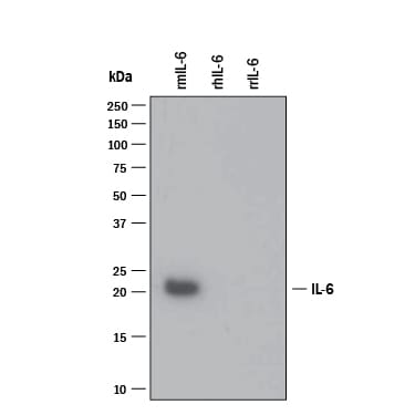
Detection of Recombinant Mouse IL‑6 by Western Blot.
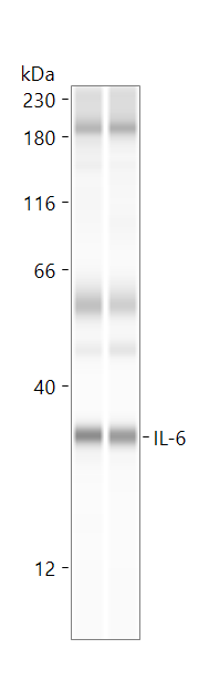
Western blot shows 25 ng of Recombinant Mouse IL-6 (406-ML), Recombinant Human IL-6 (206-IL) and Recombinant Rat IL-6 (506-RL). PVDF Membrane was probed with 1 µg/mL of Rat Anti-Mouse IL-6 Monoclonal Antibody (Catalog # MAB406) followed by HRP-conjugated Anti-Rat IgG Secondary Antibody (HAF005). A specific band was detected for IL-6 at approximately 18 kDa (as indicated). This experiment was conducted under reducing conditions and using Immunoblot Buffer Group 3.
Cell Proliferation Induced by IL‑6 and Neutralization by Mouse IL‑6 Antibody.
Recombinant Mouse IL-6 (406-ML) stimulates proliferation in the T1165.85.2.1 mouse plasmacytoma cell line in a dose-dependent manner (orange line). Proliferation elicited by Recombinant Mouse IL-6 (0.25 ng/mL) is neutralized (green line) by increasing concentrations of Mouse IL-6 Monoclonal Antibody (Catalog # MAB406). The ND50 is typically 0.005-0.025 µg/mL.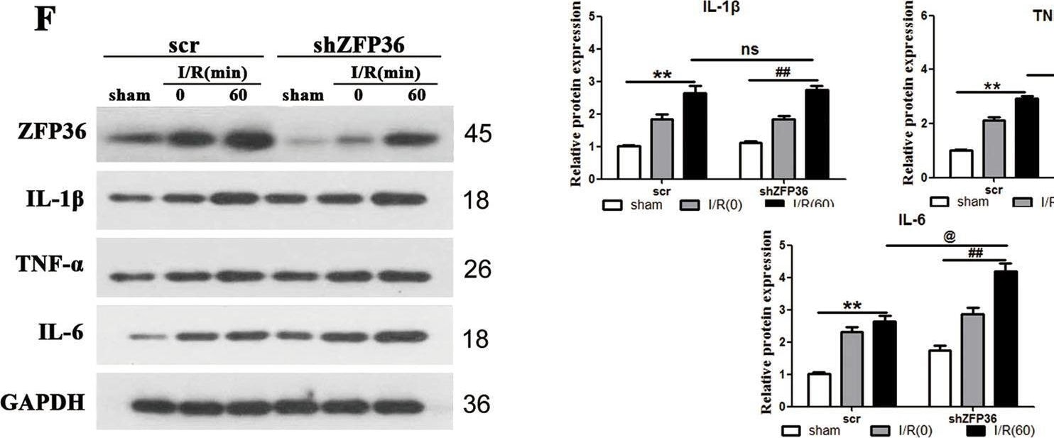
Detection of IL-6 by Western Blot
Role of ZFP36 in intestinal ischemia–reperfusion (I/R)-induced acute lung injury.C57BL/6 mice were subjected to 60 min of intestinal ischemia followed by 0, 30, 60, and 90 min of reperfusion as indicated. Sham mice were included as the control. A ZFP36 mRNA and protein expression in lung tissues were analyzed by RT-qPCR and Western blot (n = 6 per group, **P < 0.01). B Immunohistochemical staining of lung tissues for ZFP36 of sham and I/R 60 min. Scale bars: 50 μm. C–F Levels of IL-1 beta (C), TNF-alpha (D), and IL-6 (E) were measured by ELISA and Western blotting (F). G H&E staining of lung tissues. Red arrows outline collapsed alveoli, blue arrows outline multiple inflammatory cells infilitration, black arrows outline bronchial hemorrhage. Scale bars: 50 μm. H–J Arterial blood PaO2 (H), lung water content (I), and BALF protein content (J) were measured. Image collected and cropped by CiteAb from the following open publication (https://pubmed.ncbi.nlm.nih.gov/34238924), licensed under a CC-BY license. Not internally tested by R&D Systems.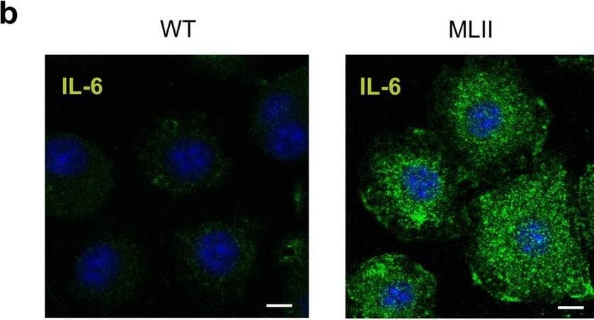
Detection of IL-6 by Immunocytochemistry/ Immunofluorescence
Increased expression of IL-6 in primary cultured osteoblasts and chondrocytes from MLII mice. (a) Signal log ratio (SLR) of differentially expressed genes (SLR ≥ 2 or ≤ -2) in terminally differentiated osteoblasts from wild-type (WT) and MLII mice related to the gene ontology (GO) "Bone biological processes": "Bone mineralization" (GO 0030282) and "Osteoclast differentiation" (GO 0030316). (b) IL-6 immunostaining (green) of WT and MLII osteoblasts. Nuclei were visualized by 4′,6-diamidino-2-phenylindole (DAPI) staining (blue). Scale bar: 10 μm. (c) Concentration of IL-6 in conditioned media from WT and MLII osteoblasts (n = 5, mean ± SD, ***p ≤ 0.001). (d) Expression levels of Il6 mRNA related to Gapdh in primary osteoblasts, chondrocytes and osteoclasts from WT and MLII mice (n = 3, mean ± SD, ***p ≤ 0.001). Image collected and cropped by CiteAb from the following open publication (https://pubmed.ncbi.nlm.nih.gov/33574442), licensed under a CC-BY license. Not internally tested by R&D Systems.
Mouse IL-6 ELISA Standard Curve
Recombinant Mouse IL‑6 (Catalog # 406-ML) was serially diluted and captured by Rat Anti-Mouse IL‑6 Monoclonal Antibody (Catalog # MAB406) coated on a Clear Polystyrene Microplate (Catalog # DY990). Goat Anti-Mouse IL‑6 Antigen Affinity-purified Polyclonal Antibody (Catalog # AF-406-NA) was biotinylated and incubated with the protein captured on the plate. Detection of the standard curve was achieved by incubating Streptavidin-HRP (Catalog # DY998)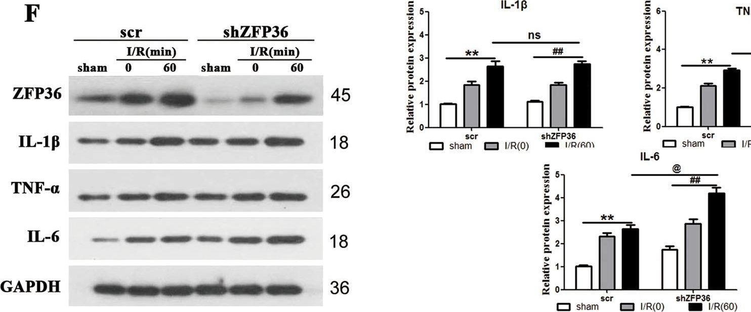
Detection of IL-6 by Western Blot
Role of ZFP36 in intestinal ischemia–reperfusion (I/R)-induced acute lung injury.C57BL/6 mice were subjected to 60 min of intestinal ischemia followed by 0, 30, 60, and 90 min of reperfusion as indicated. Sham mice were included as the control. A ZFP36 mRNA and protein expression in lung tissues were analyzed by RT-qPCR and Western blot (n = 6 per group, **P < 0.01). B Immunohistochemical staining of lung tissues for ZFP36 of sham and I/R 60 min. Scale bars: 50 μm. C–F Levels of IL-1 beta (C), TNF-alpha (D), and IL-6 (E) were measured by ELISA and Western blotting (F). G H&E staining of lung tissues. Red arrows outline collapsed alveoli, blue arrows outline multiple inflammatory cells infilitration, black arrows outline bronchial hemorrhage. Scale bars: 50 μm. H–J Arterial blood PaO2 (H), lung water content (I), and BALF protein content (J) were measured. Image collected and cropped by CiteAb from the following open publication (https://pubmed.ncbi.nlm.nih.gov/34238924), licensed under a CC-BY license. Not internally tested by R&D Systems.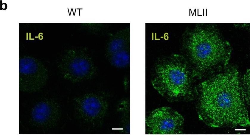
Detection of IL-6 by Immunocytochemistry/ Immunofluorescence
Increased expression of IL-6 in primary cultured osteoblasts and chondrocytes from MLII mice. (a) Signal log ratio (SLR) of differentially expressed genes (SLR ≥ 2 or ≤ -2) in terminally differentiated osteoblasts from wild-type (WT) and MLII mice related to the gene ontology (GO) "Bone biological processes": "Bone mineralization" (GO 0030282) and "Osteoclast differentiation" (GO 0030316). (b) IL-6 immunostaining (green) of WT and MLII osteoblasts. Nuclei were visualized by 4′,6-diamidino-2-phenylindole (DAPI) staining (blue). Scale bar: 10 μm. (c) Concentration of IL-6 in conditioned media from WT and MLII osteoblasts (n = 5, mean ± SD, ***p ≤ 0.001). (d) Expression levels of Il6 mRNA related to Gapdh in primary osteoblasts, chondrocytes and osteoclasts from WT and MLII mice (n = 3, mean ± SD, ***p ≤ 0.001). Image collected and cropped by CiteAb from the following open publication (https://pubmed.ncbi.nlm.nih.gov/33574442), licensed under a CC-BY license. Not internally tested by R&D Systems.Applications for Mouse IL-6 Antibody (MP5-20F3)
Western Blot
Sample: Recombinant Mouse IL‑6 (Catalog # 406-ML)
Neutralization
Mouse IL-6 Sandwich Immunoassay
Reviewed Applications
Read 6 reviews rated 4.7 using MAB406 in the following applications:
Formulation, Preparation, and Storage
Purification
Reconstitution
Reconstitute at 0.5 mg/mL in sterile PBS. For liquid material, refer to CoA for concentration.
Formulation
Shipping
Stability & Storage
- 12 months from date of receipt, -20 to -70 °C as supplied.
- 1 month, 2 to 8 °C under sterile conditions after reconstitution.
- 6 months, -20 to -70 °C under sterile conditions after reconstitution.
Calculators
Background: IL-6
Long Name
Alternate Names
Entrez Gene IDs
Gene Symbol
Additional IL-6 Products
Product Documents for Mouse IL-6 Antibody (MP5-20F3)
Certificate of Analysis
To download a Certificate of Analysis, please enter a lot or batch number in the search box below.
Note: Certificate of Analysis not available for kit components.
Product Specific Notices for Mouse IL-6 Antibody (MP5-20F3)
For research use only
Related Research Areas
Citations for Mouse IL-6 Antibody (MP5-20F3)
 Powered by Bioz
Customer Reviews for Mouse IL-6 Antibody (MP5-20F3) (6)
Have you used Mouse IL-6 Antibody (MP5-20F3)?
Submit a review and receive an Amazon gift card!
$25/€18/£15/$25CAN/¥2500 for a review with an image
$10/€7/£6/$10CAN/¥1110 for a review without an image
Submit a review
Customer Images
-
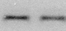
Application: Simple WesternSample Tested: Lung tissueVerified Customer | Posted 06/03/2025IL-6 expression in WT animals1:20 in antibody diluent 2. Incubation time - 60min.
 Bio-Techne ResponseThis review reflects a new species or application tested on a primary antibody.
 -
Application: Western BlotSample Tested: Heart tissueSpecies: MouseVerified Customer | Posted 08/12/2021
-
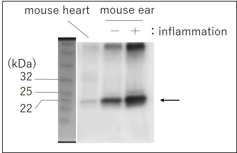
Application: Western BlotSample Tested: Heart tissue and Ear tissueSpecies: MouseVerified Customer | Posted 02/10/2021
-
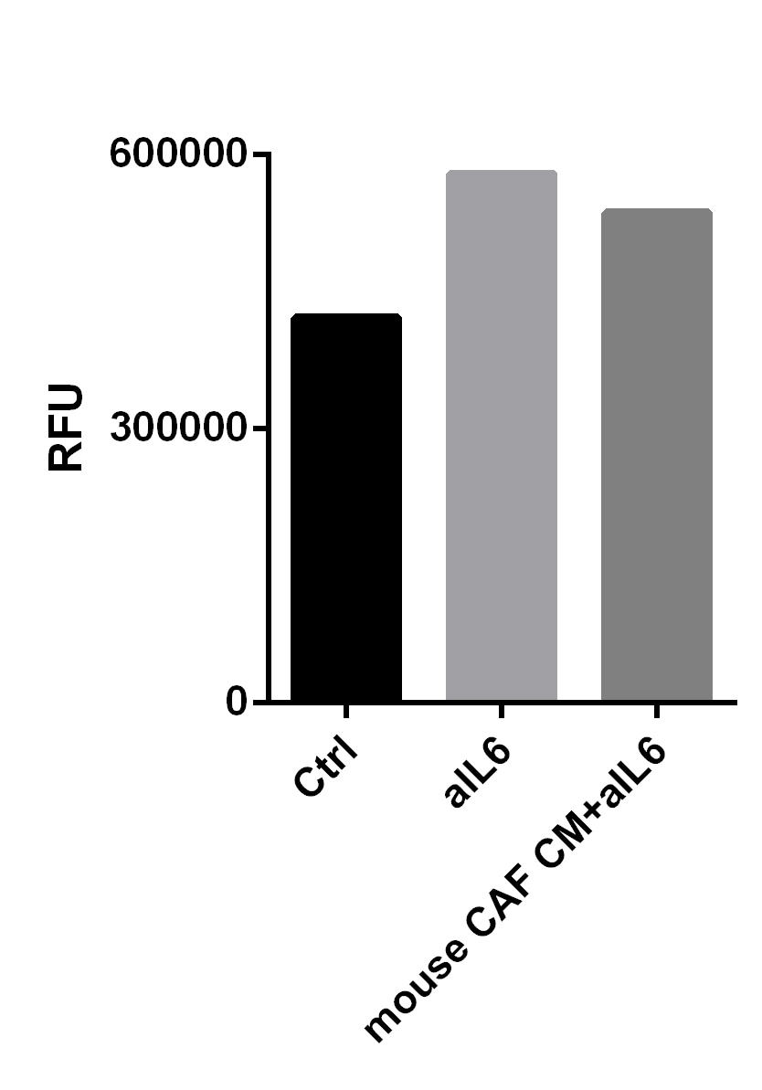
Application: Cell InvasionSample Tested: Cancer-associated Fibroblasts and tumor cellsSpecies: MouseVerified Customer | Posted 04/11/2019
-
Application: Cell depletionSample Tested: Pancreatic cancer cellsSpecies: MouseVerified Customer | Posted 05/11/2018
-
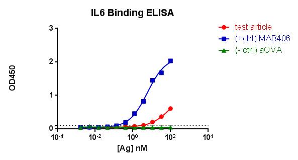
Application: ELISASample Tested: Human recombinant antibodySpecies: MouseVerified Customer | Posted 12/08/2017
There are no reviews that match your criteria.
Protocols
Find general support by application which include: protocols, troubleshooting, illustrated assays, videos and webinars.
- Cellular Response to Hypoxia Protocols
- R&D Systems Quality Control Western Blot Protocol
- Troubleshooting Guide: Western Blot Figures
- Western Blot Conditions
- Western Blot Protocol
- Western Blot Protocol for Cell Lysates
- Western Blot Troubleshooting
- Western Blot Troubleshooting Guide
- View all Protocols, Troubleshooting, Illustrated assays and Webinars
Associated Pathways