
Matrix metalloproteinases (MMPs) are a family of zinc and calcium dependent endopeptidases with the combined ability to degrade all the components of the extracellular matrix. MMP-7 (matrilysin) is expressed in epithelial cells of normal and diseased tissues, and is capable of digesting a large series of proteins of the extracellular matrix including collagen IV and X, gelatin, casein, laminin, aggrecan, entactin, elastin and versican (1). MMP-7 is implicated in the activation of other proteinases such as plasminogen, MMP-1, MMP-2, and MMP-9. In addition to its roles in connective tissue remodeling and cancer, MMP-7 also regulates intestinal alpha ‑defensin activation in innate host defense, releases tumor necrosis factor-alpha in a model of herniated disc resorption, and cleaves FasL to generate a soluble form in a model of prostate involution. Structurally, MMP-7 is the smallest of the MMPs and consists of two domains: a pro-domain that is cleaved upon activation and a catalytic domain containing the zinc-binding site.


Scientific images may be resized, cropped, or adjusted for brightness or contrast for presentation purposes. These updates are limited to presentation and do not affect the underlying scientific interpretation of the data. Where available, additional source data may be provided upon request.
Key Product Details
Species Reactivity
Validated:
Cited:
Applications
Validated:
Cited:
Label
Antibody Source
Product Specifications
Immunogen
Leu18-Leu264
Accession # AAA99983
Specificity
Clonality
Host
Isotype
Scientific Data Images for Mouse MMP-7 Antibody
MMP‑7 in Mouse Intestine.
MMP‑7 was detected in perfusion fixed frozen sections of mouse small intestine using Goat Anti-Mouse MMP‑7 Antigen Affinity-purified Polyclonal Antibody (Catalog # AF2967) at 15 µg/mL overnight at 4 °C. Tissue was stained using the Anti-Goat HRP-DAB Cell & Tissue Staining Kit (brown; Catalog # CTS008) and counterstained with hematoxylin (blue). Specific labeling was localized to the cytoplasm of cells in intestinal glands. View our protocol for Chromogenic IHC Staining of Frozen Tissue Sections.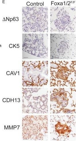
Detection of MMP-7 by Immunohistochemistry
Deletion of Nkx2-1, Foxa1 and Foxa2 at initiation blocks gastric differentiation and induces expression of squamocolumnar junctional markers in lung neoplasia.(A) Correlation between differentially expressed genes identified in single-cell analysis (C1 vs. C2, X axis) and population analysis (KrasLSL-G12D/+(K) vs. KrasLSL-G12D/+; Nkx2-1F/F (KN), Y axis). (B) tSNE plot of single high-quality tumor cells (this study) and E18.5 type two pneumocytes (AT2) from murine lung (Treutlein et al.). Color indicates cluster. Shape indicates source of cell. (C) Principal component analysis (PCA) of cancer cell clusters (C1–C3) based on genes comprising invasive mucinous adenocarcinoma signature (Guo et al). (D) Hierarchical clustering on principal components (HCPC) of the three cancer cell clusters (C1–C3) and normal murine tissue. The first two dimensions of a principal component analysis (PCA) is overlayed with the hierarchical clustering of the first five PCA dimensions. Dot color indicates cluster to which sample was assigned in K-means clustering. Gray ovals highlight multiple samples from the same tissue. ‘Glandular’ indicates glandular stomach. (E) IHC for indicated proteins in lung neoplasia 5 weeks after initiation with PGK-Cre lentivirus in KrasLSL-G12D/+; Nkx2-1F/F; Foxa1F/F; Foxa2F/F mice and controls. Scale bar: 100 microns. (F) IHC for FoxA1 and FoxA2 at the squamocolumnar junction (SCJ) of glandular stomach and forestomach (left panels) and in proximal forestomach (right panels). Arrows point to SCJ. Scale bars: 100 microns. Image collected and cropped by CiteAb from the following open publication (https://pubmed.ncbi.nlm.nih.gov/30475207), licensed under a CC-BY license. Not internally tested by R&D Systems.Applications for Mouse MMP-7 Antibody
Immunohistochemistry
Sample: Perfusion fixed frozen sections of mouse small intestine
Reviewed Applications
Read 1 review rated 5 using AF2967 in the following applications:
Formulation, Preparation, and Storage
Purification
Reconstitution
Reconstitute at 0.2 mg/mL in sterile PBS. For liquid material, refer to CoA for concentration.
Formulation
Shipping
Stability & Storage
- 12 months from date of receipt, -20 to -70 °C as supplied.
- 1 month, 2 to 8 °C under sterile conditions after reconstitution.
- 6 months, -20 to -70 °C under sterile conditions after reconstitution.
Calculators
Background: MMP-7
References
- Woessner, J.F. (2004) in Handbook of Proteolytic Enzymes, Barrett, A.J. et al. eds. p. 532.
Long Name
Alternate Names
Gene Symbol
UniProt
Additional MMP-7 Products
Product Documents for Mouse MMP-7 Antibody
Certificate of Analysis
To download a Certificate of Analysis, please enter a lot or batch number in the search box below.
Note: Certificate of Analysis not available for kit components.
Product Specific Notices for Mouse MMP-7 Antibody
For research use only
Related Research Areas
Citations for Mouse MMP-7 Antibody
 Powered by Bioz
Customer Reviews for Mouse MMP-7 Antibody (1)
Have you used Mouse MMP-7 Antibody?
Submit a review and receive an Amazon gift card!
$25/€18/£15/$25CAN/¥2500 for a review with an image
$10/€7/£6/$10CAN/¥1110 for a review without an image
Submit a review
Customer Images
-
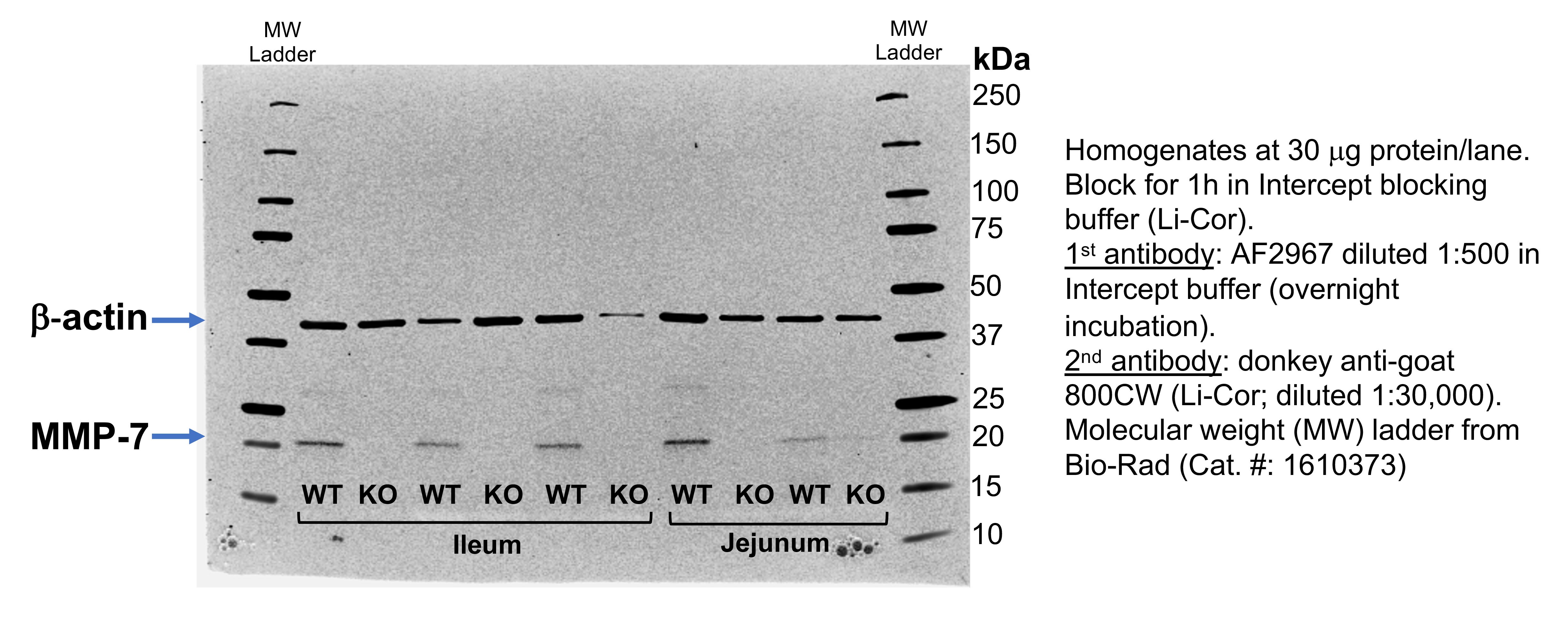
Application: Western BlotSample Tested: Ileum and jejunum homogenatesSpecies: MouseVerified Customer | Posted 09/03/2020

There are no reviews that match your criteria.
Protocols
Find general support by application which include: protocols, troubleshooting, illustrated assays, videos and webinars.
- Antigen Retrieval Protocol (PIER)
- Antigen Retrieval for Frozen Sections Protocol
- Appropriate Fixation of IHC/ICC Samples
- Cellular Response to Hypoxia Protocols
- Chromogenic IHC Staining of Formalin-Fixed Paraffin-Embedded (FFPE) Tissue Protocol
- Chromogenic Immunohistochemistry Staining of Frozen Tissue
- ClariTSA™ Fluorophore Kits
- Detection & Visualization of Antibody Binding
- Fluorescent IHC Staining of Frozen Tissue Protocol
- Graphic Protocol for Heat-induced Epitope Retrieval
- Graphic Protocol for the Preparation and Fluorescent IHC Staining of Frozen Tissue Sections
- Graphic Protocol for the Preparation and Fluorescent IHC Staining of Paraffin-embedded Tissue Sections
- Graphic Protocol for the Preparation of Gelatin-coated Slides for Histological Tissue Sections
- IHC Sample Preparation (Frozen sections vs Paraffin)
- Immunofluorescent IHC Staining of Formalin-Fixed Paraffin-Embedded (FFPE) Tissue Protocol
- Immunohistochemistry (IHC) and Immunocytochemistry (ICC) Protocols
- Immunohistochemistry Frozen Troubleshooting
- Immunohistochemistry Paraffin Troubleshooting
- Preparing Samples for IHC/ICC Experiments
- Preventing Non-Specific Staining (Non-Specific Binding)
- Primary Antibody Selection & Optimization
- Protocol for Heat-Induced Epitope Retrieval (HIER)
- Protocol for Making a 4% Formaldehyde Solution in PBS
- Protocol for VisUCyte™ HRP Polymer Detection Reagent
- Protocol for the Preparation & Fixation of Cells on Coverslips
- Protocol for the Preparation and Chromogenic IHC Staining of Frozen Tissue Sections
- Protocol for the Preparation and Chromogenic IHC Staining of Frozen Tissue Sections - Graphic
- Protocol for the Preparation and Chromogenic IHC Staining of Paraffin-embedded Tissue Sections
- Protocol for the Preparation and Chromogenic IHC Staining of Paraffin-embedded Tissue Sections - Graphic
- Protocol for the Preparation and Fluorescent IHC Staining of Frozen Tissue Sections
- Protocol for the Preparation and Fluorescent IHC Staining of Paraffin-embedded Tissue Sections
- Protocol for the Preparation of Gelatin-coated Slides for Histological Tissue Sections
- TUNEL and Active Caspase-3 Detection by IHC/ICC Protocol
- The Importance of IHC/ICC Controls
- Troubleshooting Guide: Immunohistochemistry
- View all Protocols, Troubleshooting, Illustrated assays and Webinars