
Reelin is a 400 kDa, extracellular matrix glycoprotein secreted by several neurons. It is a serine protease that degrades fibronectin and laminin. Reelin also binds lipoprotein receptor superfamily members APOER2 and VLDLR which transduce signals important for neuronal positioning during brain development and synaptic plasticity in the adult brain. In vivo, Reelin undergoes proteolytic processing at two sites, generating three Reelin fragments. The N-terminal domain binds integrin alpha 3/ beta 1, leading to a disruption of neuronal glial cell interactions and inhibition of neuronal migration. Mouse Reelin shares 94% and 96% amino acid identity with human and rat Reelin, respectively in the region immunized.
Mouse Reelin Antibody
R&D Systems | Catalog # AF3820


Scientific images may be resized, cropped, or adjusted for brightness or contrast for presentation purposes. These updates are limited to presentation and do not affect the underlying scientific interpretation of the data. Where available, additional source data may be provided upon request.
Key Product Details
Species Reactivity
Validated:
Mouse
Cited:
Human, Mouse, Rat, Chicken, Transgenic Mouse
Applications
Validated:
Immunohistochemistry, Western Blot
Cited:
Immunohistochemistry, Immunohistochemistry-Paraffin, Western Blot, Immunocytochemistry
Label
Unconjugated
Antibody Source
Polyclonal Goat IgG
Loading...
Product Specifications
Immunogen
Mouse myeloma cell line NS0-derived recombinant mouse Reelin
Ala27-Met862
Accession # Q60841
Ala27-Met862
Accession # Q60841
Specificity
Detects mouse Reelin in direct ELISAs and Western blots.
Clonality
Polyclonal
Host
Goat
Isotype
IgG
Scientific Data Images for Mouse Reelin Antibody
Reelin in Mouse Embryo.
Reelin was detected in immersion fixed frozen sections of mouse embryo (13 d.p.c.) using Goat Anti-Mouse Reelin Antigen Affinity-purified Polyclonal Antibody (Catalog # AF3820) at 1.7 µg/mL overnight at 4 °C. Tissue was stained using the Anti-Goat HRP-DAB Cell & Tissue Staining Kit (brown; Catalog # CTS008) and counterstained with hematoxylin (blue). Specific staining was localized to developing brain. View our protocol for Chromogenic IHC Staining of Frozen Tissue Sections.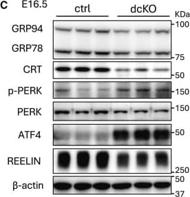
Detection of Reelin by Western Blot
Expression of UPR genes in control and dcKO brains(A) Total RNA isolated from E16.5 control or dcKO cerebral cortex was subjected to RNA-sequencing (n = 3 brains per group). Heatmaps show gene expressions in each GO term or in ATF6 branch including molecular chaperones in the ER. Color scale bars indicate Z score by row.(B) RT-qPCR. Brains samples from E16.5 control or dcKO cerebral cortex were subjected to RT-qPCR with specific primers for the indicated genes (n = 6–7 brains per group). Expressions levels of molecular chaperones in the ER are downregulated, while those of IRE1-and PERK-downstream genes are upregulated in dcKO brains.(C) Western blotting. Protein was extracted from cerebral cortices of E16.5, and subjected to western blotting with indicated antibodies (n = 3–7 brains per group). Expression of REELIN was reduced in dcKO brain.(D and E) Immunohistochemistry of brain section from E16.5 control and dcKO embryos for p-PERK and SOX2. p-PERK(+) cells are distributed around ventricular zone (D) and majority of them are co-localized with SOX2(+) cells (E) (n = 4 brains per group). Cx, cortex; CPu, caudate putamen. Data are represented as mean ± SEM. ∗p < 0.05, ∗∗p < 0.01, and ∗∗∗p < 0.001 by Mann-Whitney U test. Scale bars: 500 μm (D), 50 μm (E). Image collected and cropped by CiteAb from the following open publication (https://linkinghub.elsevier.com/retrieve/pii/S2589004225008302), licensed under a CC-BY license. Not internally tested by R&D Systems.Applications for Mouse Reelin Antibody
Application
Recommended Usage
Immunohistochemistry
5-15 µg/mL
Sample: Immersion fixed frozen sections of mouse embryo (13 d.p.c.)
Sample: Immersion fixed frozen sections of mouse embryo (13 d.p.c.)
Western Blot
0.1 µg/mL
Sample: Recombinant Mouse Reelin
Sample: Recombinant Mouse Reelin
Formulation, Preparation, and Storage
Purification
Antigen Affinity-purified
Reconstitution
Reconstitute at 0.2 mg/mL in sterile PBS. For liquid material, refer to CoA for concentration.
Loading...
Formulation
Lyophilized from a 0.2 μm filtered solution in PBS with Trehalose. *Small pack size (SP) is supplied either lyophilized or as a 0.2 µm filtered solution in PBS.
Shipping
Lyophilized product is shipped at ambient temperature. Liquid small pack size (-SP) is shipped with polar packs. Upon receipt, store immediately at the temperature recommended below.
Stability & Storage
Use a manual defrost freezer and avoid repeated freeze-thaw cycles.
- 12 months from date of receipt, -20 to -70 °C as supplied.
- 1 month, 2 to 8 °C under sterile conditions after reconstitution.
- 6 months, -20 to -70 °C under sterile conditions after reconstitution.
Calculators
Background: Reelin
Alternate Names
Reeler, RELN, RL
Gene Symbol
RELN
UniProt
Additional Reelin Products
Product Documents for Mouse Reelin Antibody
Certificate of Analysis
To download a Certificate of Analysis, please enter a lot or batch number in the search box below.
Note: Certificate of Analysis not available for kit components.
Product Specific Notices for Mouse Reelin Antibody
For research use only
Related Research Areas
Citations for Mouse Reelin Antibody
 Powered by Bioz
Customer Reviews for Mouse Reelin Antibody
There are currently no reviews for this product. Be the first to review Mouse Reelin Antibody and earn rewards!
Have you used Mouse Reelin Antibody?
Submit a review and receive an Amazon gift card!
$25/€18/£15/$25CAN/¥2500 for a review with an image
$10/€7/£6/$10CAN/¥1110 for a review without an image
Submit a review
Protocols
Find general support by application which include: protocols, troubleshooting, illustrated assays, videos and webinars.
- Antigen Retrieval Protocol (PIER)
- Antigen Retrieval for Frozen Sections Protocol
- Appropriate Fixation of IHC/ICC Samples
- Cellular Response to Hypoxia Protocols
- Chromogenic IHC Staining of Formalin-Fixed Paraffin-Embedded (FFPE) Tissue Protocol
- Chromogenic Immunohistochemistry Staining of Frozen Tissue
- ClariTSA™ Fluorophore Kits
- Detection & Visualization of Antibody Binding
- Fluorescent IHC Staining of Frozen Tissue Protocol
- Graphic Protocol for Heat-induced Epitope Retrieval
- Graphic Protocol for the Preparation and Fluorescent IHC Staining of Frozen Tissue Sections
- Graphic Protocol for the Preparation and Fluorescent IHC Staining of Paraffin-embedded Tissue Sections
- Graphic Protocol for the Preparation of Gelatin-coated Slides for Histological Tissue Sections
- IHC Sample Preparation (Frozen sections vs Paraffin)
- Immunofluorescent IHC Staining of Formalin-Fixed Paraffin-Embedded (FFPE) Tissue Protocol
- Immunohistochemistry (IHC) and Immunocytochemistry (ICC) Protocols
- Immunohistochemistry Frozen Troubleshooting
- Immunohistochemistry Paraffin Troubleshooting
- Preparing Samples for IHC/ICC Experiments
- Preventing Non-Specific Staining (Non-Specific Binding)
- Primary Antibody Selection & Optimization
- Protocol for Heat-Induced Epitope Retrieval (HIER)
- Protocol for Making a 4% Formaldehyde Solution in PBS
- Protocol for VisUCyte™ HRP Polymer Detection Reagent
- Protocol for the Preparation & Fixation of Cells on Coverslips
- Protocol for the Preparation and Chromogenic IHC Staining of Frozen Tissue Sections
- Protocol for the Preparation and Chromogenic IHC Staining of Frozen Tissue Sections - Graphic
- Protocol for the Preparation and Chromogenic IHC Staining of Paraffin-embedded Tissue Sections
- Protocol for the Preparation and Chromogenic IHC Staining of Paraffin-embedded Tissue Sections - Graphic
- Protocol for the Preparation and Fluorescent IHC Staining of Frozen Tissue Sections
- Protocol for the Preparation and Fluorescent IHC Staining of Paraffin-embedded Tissue Sections
- Protocol for the Preparation of Gelatin-coated Slides for Histological Tissue Sections
- R&D Systems Quality Control Western Blot Protocol
- TUNEL and Active Caspase-3 Detection by IHC/ICC Protocol
- The Importance of IHC/ICC Controls
- Troubleshooting Guide: Immunohistochemistry
- Troubleshooting Guide: Western Blot Figures
- Western Blot Conditions
- Western Blot Protocol
- Western Blot Protocol for Cell Lysates
- Western Blot Troubleshooting
- Western Blot Troubleshooting Guide
- View all Protocols, Troubleshooting, Illustrated assays and Webinars
Loading...