![Western Blot: NOXO1 Antibody [NBP1-77899]](https://resources.rndsystems.com/images/products/NOXO1-Antibody-Western-Blot-NBP1-77899-img0001.jpg "Western Blot: NOXO1 Antibody [NBP1-77899]")
Loading...
Key Product Details
Species Reactivity
Human, Mouse
Applications
Immunohistochemistry, Immunohistochemistry-Paraffin, Western Blot, ELISA, Simple Western
Label
Unconjugated
Antibody Source
Polyclonal Rabbit IgG
Format
BSA Free
Loading...
Product Specifications
Immunogen
NOXO1 Antibody was prepared from whole rabbit serum produced by repeated immunizations with a synthetic peptide corresponding to amino acids 238-252 of human NOXO1 protein. (Uniprot: Q8NFA2)
Reactivity Notes
A BLAST analysis was used to suggest cross reactivity with NOXO1 protein from human and chimpanzee (100% homology). Expect reactivity with alpha, delta and gamma isoforms of NOXO1. Also expect partial reactivity against NOXO1 homologues from dog and guinea pig (87%), as well as rat (75%) and mouse (68%). Reactivity against homologues from other sources is not known. Mouse reactivity reported in verified customer review.
Specificity
Anti-NOXO-1 antibody is directed against human NOXO1 protein. A BLAST analysis was used to suggest cross reactivity with NOXO1 protein from human and chimpanzee (100% homology). Expect reactivity with alpha, delta and gamma isoforms of NOXO1. Also expect partial reactivity against NOXO1 homologues from dog and guinea pig (87%), as well as rat (75%) and mouse (68%). Reactivity against homologues from other sources is not known.
Clonality
Polyclonal
Host
Rabbit
Isotype
IgG
Description
The product was affinity purified from monospecific antiserum by immunoaffinity purification
Store vial at -20C prior to opening. Aliquot contents and freeze at -20C or below for extended storage. Avoid cycles of freezing and thawing. Centrifuge product if not completely clear after standing at room temperature. This product is stable for several weeks at 4C as an undiluted liquid. Dilute only prior to immediate use.
Store vial at -20C prior to opening. Aliquot contents and freeze at -20C or below for extended storage. Avoid cycles of freezing and thawing. Centrifuge product if not completely clear after standing at room temperature. This product is stable for several weeks at 4C as an undiluted liquid. Dilute only prior to immediate use.
Scientific Data Images for NOXO1 Antibody - BSA Free
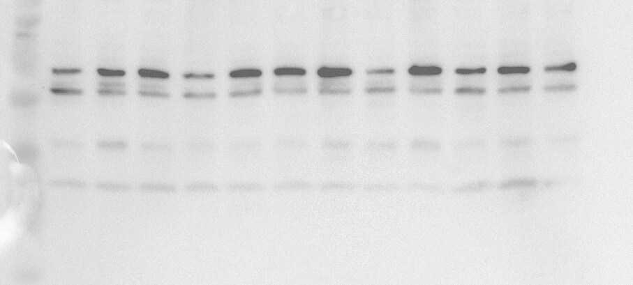
Western Blot: NOXO1 Antibody [NBP1-77899]
Western Blot: NOXO1 Antibody [NBP1-77899] - NOXO1 antibody shows detection of a band ~50 kDa corresponding to human NOXO1 (arrowhead). Reactivity was observed in transfected human 293 cells and human HT-29 colon carcinoma cells (endogenous). Under these conditions endogenous NOXO1 detection was not observed in HeLa, HL-60, untransfected 293 or WT MEF cells. A 1:1,000 dilution of the primary antibody was used for detection followed by secondary antibody reactivity. Specific band reactivity was competed away when the antibody was pre-incubated with the peptide immunogen (data not shown).![Immunohistochemistry: NOXO1 Antibody [NBP1-77899]](https://resources.rndsystems.com/images/products/NOXO1-Antibody-Immunohistochemistry-NBP1-77899-img0003.jpg "Immunohistochemistry: NOXO1 Antibody [NBP1-77899]")
Immunohistochemistry: NOXO1 Antibody [NBP1-77899]
Immunohistochemistry: NOXO1 Antibody [NBP1-77899] - Used at 5 ug/ml to detect signal in a variety of tissues including multi-human, multi-brain and multi-cancer slides. This image shows moderate positive staining of the lamina propia in human colon epithelium and macrophages at 40X. Tissue was formalin-fixed and paraffin embedded. The image shows localization of the antibody as the precipitated red signal, with a hematoxylin purple nuclear counterstain. Personal Communi-cation, Tina Roush, LifeSpanBiosciences, Seattle, WA.
NOXO1 Antibody
Western blot using Affinity Purified anti-NOXO1 antibody shows detection of a band ~50 kDa corresponding to human NOXO1. Reactivity was observed in transfected human 293 cells and human HT-29 colon carcinoma cells (endogenous). Under these conditions endogenous NOXO1 detection was not observed in HeLa, HL-60, 3T3, untransfected 293 or WT MEF cells. A 1:1,000 dilution of the primary antibody was used for detection followed by secondary antibody reactivity. Specific band reactivity was competed away when the antibody was pre-incubated with the peptide immunogen (data not shown). Personal Communication, Zhenggang Liu, NIH, CCR, Bethesda, MD.
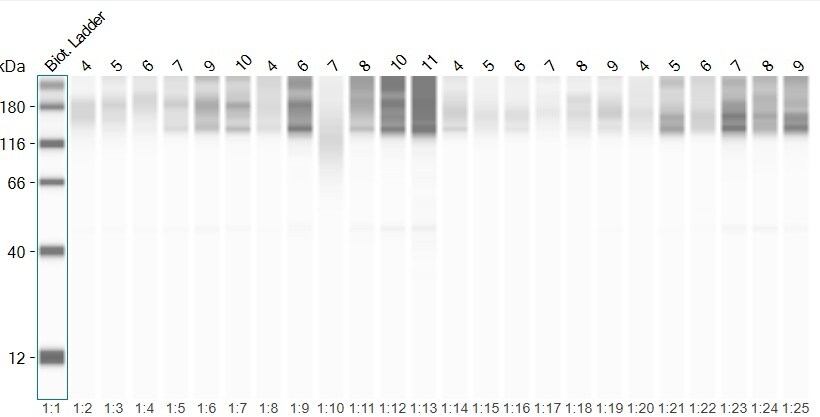
Simple Western: Rabbit Polyclonal NOXO1 Antibody [NBP1-77899]
NOXO1 in mouse lungs. The first lane is a control; the second lane is treated. Image from a verified customer review.Applications for NOXO1 Antibody - BSA Free
Application
Recommended Usage
ELISA
1:20000-1:100000
Immunohistochemistry
5-10 ug/ml
Immunohistochemistry-Paraffin
1:10-1:500
Simple Western
Validated for Simple Western from a verified customer review.
Western Blot
1:1000-1:10000
Application Notes
This product has been tested for use in ELISA, immunohistochemistry and western blot. Specific conditions for reactivity should be optimized by the end user. Expect a band approximately 50 kDa in size corresponding to NOXO1 protein by western blotting in the appropriate cell lysate or extract.
Reviewed Applications
Read 3 reviews rated 3.3 using NBP1-77899 in the following applications:
Formulation, Preparation, and Storage
Purification
Immunogen affinity purified
Formulation
0.02 M Potassium Phosphate, 0.15 M Sodium Chloride, pH 7.2
Format
BSA Free
Preservative
0.01% Sodium Azide
Concentration
Please see the vial label for concentration. If unlisted please contact technical services.
Shipping
The product is shipped with polar packs. Upon receipt, store it immediately at the temperature recommended below.
Stability & Storage
Store at -20C short term. Aliquot and store at -80C long term. Avoid freeze-thaw cycles.
Background: NOXO1
Alternate Names
MGC20258, NADPH oxidase organizer 1, Nox organizer 1, Nox-organizing protein 1, P41NOX, P41NOXA, P41NOXB, P41NOXC, regulatory protein P41NOX, SH3 and PX domain-containing protein 5, SH3PXD5NADPH oxidase regulatory protein, SNX28
Gene Symbol
NOXO1
UniProt
Additional NOXO1 Products
Product Documents for NOXO1 Antibody - BSA Free
Certificate of Analysis
To download a Certificate of Analysis, please enter a lot or batch number in the search box below.
Product Specific Notices for NOXO1 Antibody - BSA Free
This product is for research use only and is not approved for use in humans or in clinical diagnosis. Primary Antibodies are guaranteed for 1 year from date of receipt.
Customer Reviews for NOXO1 Antibody - BSA Free (3)
3.3 out of 5
3 Customer Ratings
Have you used NOXO1 Antibody - BSA Free?
Submit a review and receive an Amazon gift card!
$25/€18/£15/$25CAN/¥2500 Yen for a review with an image
$10/€7/£6/$10CAN/¥1110 Yen for a review without an image
Submit a review
Customer Images
_2770f6ed-280b-4647-ad5e-3e80fbed019c.png)
Showing
1
-
3 的
3 reviews
Showing All
Filter By:
-
Application: Western BlotSample Tested: Lung tissueSpecies: MouseVerified Customer | Posted 09/08/2025Noxo1 expression in mouse lung homogenates using Western blotI tested the protein expression of noxo1 in mouse lung homogenates after deletion of a NADPH oxidase upon smoke exposure

-
Application: Simple WesternSample Tested: Lung tissueSpecies: Mouse, Mouse and MouseVerified Customer | Posted 09/08/2025Noxo1 protein expression in mouse lung homogenatesNoxo1 protein expression in mouse lung homogenates using JESS
Bio-Techne ResponseThank you for reviewing our product. We are sorry to hear that this product did not perform as expected. We have been in touch with the customer to resolve this issue according to our Product Guarantee and to the customer’s satisfaction.
-
Application: Simple WesternSample Tested: Lung tissueSpecies: MouseVerified Customer | Posted 06/03/2025NOXO1 in mouse lungs. The first lane is a control, the second lane is treated.Antibodies were diluted in antibody diluent 2 in 1:20 concentration. Incubation 60min.
Bio-Techne ResponseThis review reflects a new species or application tested on a primary antibody.
There are no reviews that match your criteria.
Protocols
Find general support by application which include: protocols, troubleshooting, illustrated assays, videos and webinars.
- Antigen Retrieval Protocol (PIER)
- Antigen Retrieval for Frozen Sections Protocol
- Appropriate Fixation of IHC/ICC Samples
- Cellular Response to Hypoxia Protocols
- Chromogenic IHC Staining of Formalin-Fixed Paraffin-Embedded (FFPE) Tissue Protocol
- Chromogenic Immunohistochemistry Staining of Frozen Tissue
- ClariTSA™ Fluorophore Kits
- Detection & Visualization of Antibody Binding
- ELISA Sample Preparation & Collection Guide
- ELISA Troubleshooting Guide
- Fluorescent IHC Staining of Frozen Tissue Protocol
- Graphic Protocol for Heat-induced Epitope Retrieval
- Graphic Protocol for the Preparation and Fluorescent IHC Staining of Frozen Tissue Sections
- Graphic Protocol for the Preparation and Fluorescent IHC Staining of Paraffin-embedded Tissue Sections
- Graphic Protocol for the Preparation of Gelatin-coated Slides for Histological Tissue Sections
- How to Run an R&D Systems DuoSet ELISA
- How to Run an R&D Systems Quantikine ELISA
- How to Run an R&D Systems Quantikine™ QuicKit™ ELISA
- IHC Sample Preparation (Frozen sections vs Paraffin)
- Immunofluorescent IHC Staining of Formalin-Fixed Paraffin-Embedded (FFPE) Tissue Protocol
- Immunohistochemistry (IHC) and Immunocytochemistry (ICC) Protocols
- Immunohistochemistry Frozen Troubleshooting
- Immunohistochemistry Paraffin Troubleshooting
- Preparing Samples for IHC/ICC Experiments
- Preventing Non-Specific Staining (Non-Specific Binding)
- Primary Antibody Selection & Optimization
- Protocol for Heat-Induced Epitope Retrieval (HIER)
- Protocol for Making a 4% Formaldehyde Solution in PBS
- Protocol for VisUCyte™ HRP Polymer Detection Reagent
- Protocol for the Preparation & Fixation of Cells on Coverslips
- Protocol for the Preparation and Chromogenic IHC Staining of Frozen Tissue Sections
- Protocol for the Preparation and Chromogenic IHC Staining of Frozen Tissue Sections - Graphic
- Protocol for the Preparation and Chromogenic IHC Staining of Paraffin-embedded Tissue Sections
- Protocol for the Preparation and Chromogenic IHC Staining of Paraffin-embedded Tissue Sections - Graphic
- Protocol for the Preparation and Fluorescent IHC Staining of Frozen Tissue Sections
- Protocol for the Preparation and Fluorescent IHC Staining of Paraffin-embedded Tissue Sections
- Protocol for the Preparation of Gelatin-coated Slides for Histological Tissue Sections
- Quantikine HS ELISA Kit Assay Principle, Alkaline Phosphatase
- Quantikine HS ELISA Kit Principle, Streptavidin-HRP Polymer
- R&D Systems Quality Control Western Blot Protocol
- Sandwich ELISA (Colorimetric) – Biotin/Streptavidin Detection Protocol
- Sandwich ELISA (Colorimetric) – Direct Detection Protocol
- TUNEL and Active Caspase-3 Detection by IHC/ICC Protocol
- The Importance of IHC/ICC Controls
- Troubleshooting Guide: ELISA
- Troubleshooting Guide: Immunohistochemistry
- Troubleshooting Guide: Western Blot Figures
- Western Blot Conditions
- Western Blot Protocol
- Western Blot Protocol for Cell Lysates
- Western Blot Troubleshooting
- Western Blot Troubleshooting Guide
- View all Protocols, Troubleshooting, Illustrated assays and Webinars
Loading...