PGAM5 Antibody - BSA Free
Novus Biologicals | Catalog # NBP1-92257
![Western Blot: PGAM5 Antibody [NBP1-92257]](https://resources.rndsystems.com/images/products/PGAM5-Antibody-Western-Blot-NBP1-92257-img0018.jpg "Western Blot: PGAM5 Antibody [NBP1-92257]")
Key Product Details
Validated by
Species Reactivity
Validated:
Cited:
Predicted:
Applications
Validated:
Cited:
Label
Antibody Source
Format
Product Specifications
Immunogen
Clonality
Host
Isotype
Theoretical MW
Disclaimer note: The observed molecular weight of the protein may vary from the listed predicted molecular weight due to post translational modifications, post translation cleavages, relative charges, and other experimental factors.
Scientific Data Images for PGAM5 Antibody - BSA Free
![Immunohistochemistry-Paraffin: PGAM5 Antibody [NBP1-92257]](https://resources.rndsystems.com/images/products/PGAM5-Antibody-Immunohistochemistry-Paraffin-NBP1-92257-img0020.jpg "Immunohistochemistry-Paraffin: PGAM5 Antibody [NBP1-92257]")
Immunohistochemistry-Paraffin: PGAM5 Antibody [NBP1-92257]
Immunohistochemistry-Paraffin: PGAM5 Antibody [NBP1-92257] - Staining of human colon, liver, testis and tonsil using Anti-PGAM5 antibody NBP1-92257 (A) shows similar protein distribution across tissues to independent antibody NBP1-92258 (B).![Immunocytochemistry/ Immunofluorescence: PGAM5 Antibody [NBP1-92257]](https://resources.rndsystems.com/images/products/PGAM5-Antibody-Immunocytochemistry-Immunofluorescence-NBP1-92257-img0016.jpg "Immunocytochemistry/ Immunofluorescence: PGAM5 Antibody [NBP1-92257]")
Immunocytochemistry/ Immunofluorescence: PGAM5 Antibody [NBP1-92257]
Immunocytochemistry/Immunofluorescence: PGAM5 Antibody [NBP1-92257] - Staining of human cell line MCF7 shows localization to mitochondria. Antibody staining is shown in green.![Immunohistochemistry-Paraffin: PGAM5 Antibody [NBP1-92257]](https://resources.rndsystems.com/images/products/PGAM5-Antibody-Immunohistochemistry-Paraffin-NBP1-92257-img0021.jpg "Immunohistochemistry-Paraffin: PGAM5 Antibody [NBP1-92257]")
Immunohistochemistry-Paraffin: PGAM5 Antibody [NBP1-92257]
Immunohistochemistry-Paraffin: PGAM5 Antibody [NBP1-92257] - Staining of human liver shows moderate granular cytoplasmic positivity in hepatocytes.![Immunohistochemistry-Paraffin: PGAM5 Antibody [NBP1-92257]](https://resources.rndsystems.com/images/products/PGAM5-Antibody-Immunohistochemistry-Paraffin-NBP1-92257-img0022.jpg "Immunohistochemistry-Paraffin: PGAM5 Antibody [NBP1-92257]")
Immunohistochemistry-Paraffin: PGAM5 Antibody [NBP1-92257]
Immunohistochemistry-Paraffin: PGAM5 Antibody [NBP1-92257] - Staining of human testis shows moderate granular cytoplasmic positivity in cells in seminiferous ducts.![Immunohistochemistry-Paraffin: PGAM5 Antibody [NBP1-92257]](https://resources.rndsystems.com/images/products/PGAM5-Antibody-Immunohistochemistry-Paraffin-NBP1-92257-img0023.jpg "Immunohistochemistry-Paraffin: PGAM5 Antibody [NBP1-92257]")
Immunohistochemistry-Paraffin: PGAM5 Antibody [NBP1-92257]
Immunohistochemistry-Paraffin: PGAM5 Antibody [NBP1-92257] - Staining of human tonsil shows moderate granular cytoplasmic positivity in germinal center cells![Simple Western: PGAM5 Antibody [NBP1-92257]](https://resources.rndsystems.com/images/products/PGAM5-Antibody-Simple-Western-NBP1-92257-img0007.jpg "Simple Western: PGAM5 Antibody [NBP1-92257]")
Simple Western: PGAM5 Antibody [NBP1-92257]
Simple Western: PGAM5 Antibody [NBP1-92257] - Simple Western lane view shows a specific band for PGAM5 in 0.2 mg/ml of RT-4 lysate. This experiment was performed under reducing conditions using the 12-230 kDa separation system.![Simple Western: PGAM5 Antibody [NBP1-92257]](https://resources.rndsystems.com/images/products/PGAM5-Antibody-Simple-Western-NBP1-92257-img0009.jpg "Simple Western: PGAM5 Antibody [NBP1-92257]")
Simple Western: PGAM5 Antibody [NBP1-92257]
Simple Western: PGAM5 Antibody [NBP1-92257] - Electropherogram image(s) of corresponding Simple Western lane view. PGAM5 antibody was used at 1:60 dilution on RT-4 lysate(s).
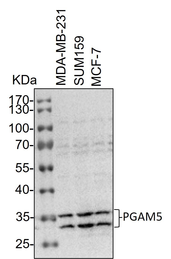
Western Blot: Rabbit Polyclonal PGAM5 Antibody [NBP1-92257] -
Western Blot: Rabbit Polyclonal PGAM5 Antibody [NBP1-92257] - Whole cell lysates from MDA-MB-231, SUM159 and MCF-7 cells were loaded with 50 ug/lane. 10% SDS-PAGE. PGAM5 Antibody (NBP1-92257) was used for primary antibody: 1:1000, 4C, overnight. Image from a verified customer review.![PGAM5 Antibody - BSA Free Immunohistochemistry: PGAM5 Antibody - BSA Free [NBP1-92257]](https://resources.rndsystems.com/images/products/nbp1-92257_rabbit-polyclonal-pgam5-antibody-29520251622212.jpg "Immunohistochemistry: PGAM5 Antibody - BSA Free [NBP1-92257]")
Immunohistochemistry: PGAM5 Antibody - BSA Free [NBP1-92257]
Staining of human liver shows moderate granular cytoplasmic positivity in hepatocytes.![PGAM5 Antibody - BSA Free Immunohistochemistry: PGAM5 Antibody - BSA Free [NBP1-92257]](https://resources.rndsystems.com/images/products/nbp1-92257_rabbit-polyclonal-pgam5-antibody-29520251621575.jpg "Immunohistochemistry: PGAM5 Antibody - BSA Free [NBP1-92257]")
Immunohistochemistry: PGAM5 Antibody - BSA Free [NBP1-92257]
Staining of human colon shows moderate granular cytoplasmic positivity in glandular cells.![PGAM5 Antibody - BSA Free Western Blot: PGAM5 Antibody - BSA Free [NBP1-92257]](https://resources.rndsystems.com/images/products/nbp1-92257_rabbit-polyclonal-pgam5-antibody-295202516222118.jpg "Western Blot: PGAM5 Antibody - BSA Free [NBP1-92257]")
![PGAM5 Antibody - BSA Free Immunohistochemistry: PGAM5 Antibody - BSA Free [NBP1-92257]](https://resources.rndsystems.com/images/products/nbp1-92257_rabbit-polyclonal-pgam5-antibody-3052025104383.jpg "Immunohistochemistry: PGAM5 Antibody - BSA Free [NBP1-92257]")
Immunohistochemistry: PGAM5 Antibody - BSA Free [NBP1-92257]
Staining of human tonsil shows moderate granular cytoplasmic positivity in germinal center cells.Applications for PGAM5 Antibody - BSA Free
Immunocytochemistry/ Immunofluorescence
Immunohistochemistry
Immunohistochemistry-Paraffin
Simple Western
Western Blot
In Simple Western only 10 - 15 uL of the recommended dilution is used per data point.
See Simple Western Antibody Database for Simple Western validation: Tested in RT-4, separated by Size, antibody dilution of 1:60, apparent MW was 38 kDa. Separated by Size-Wes, Sally Sue/Peggy Sue.
Reviewed Applications
Read 1 review rated 5 using NBP1-92257 in the following applications:
Formulation, Preparation, and Storage
Purification
Formulation
Format
Preservative
Concentration
Shipping
Stability & Storage
Background: PGAM5
Alternate Names
Entrez Gene IDs
Gene Symbol
UniProt
Additional PGAM5 Products
Product Documents for PGAM5 Antibody - BSA Free
Certificate of Analysis
To download a Certificate of Analysis, please enter a lot or batch number in the search box below.
Product Specific Notices for PGAM5 Antibody - BSA Free
This product is for research use only and is not approved for use in humans or in clinical diagnosis. Primary Antibodies are guaranteed for 1 year from date of receipt.
Citations for PGAM5 Antibody - BSA Free
 Powered by Bioz
Customer Reviews for PGAM5 Antibody - BSA Free (1)
Have you used PGAM5 Antibody - BSA Free?
Submit a review and receive an Amazon gift card!
$25/€18/£15/$25CAN/¥2500 Yen for a review with an image
$10/€7/£6/$10CAN/¥1110 Yen for a review without an image
Submit a review
Customer Images
-
Application: Western BlotVerified Customer | Posted 12/20/2023Western Blot: whole cell lysates from MDA-MB-231, SUM159 and MCF-7 cells were loaded with 50 ug/lane. 10% SDS-PAGE. PGAM5 Antibody (NBP1-92257) was used for primary antibody: 1:1000, 4℃, overnight.

There are no reviews that match your criteria.
Protocols
Find general support by application which include: protocols, troubleshooting, illustrated assays, videos and webinars.
- Antigen Retrieval Protocol (PIER)
- Antigen Retrieval for Frozen Sections Protocol
- Appropriate Fixation of IHC/ICC Samples
- Cellular Response to Hypoxia Protocols
- Chromogenic IHC Staining of Formalin-Fixed Paraffin-Embedded (FFPE) Tissue Protocol
- Chromogenic Immunohistochemistry Staining of Frozen Tissue
- ClariTSA™ Fluorophore Kits
- Detection & Visualization of Antibody Binding
- Fluorescent IHC Staining of Frozen Tissue Protocol
- Graphic Protocol for Heat-induced Epitope Retrieval
- Graphic Protocol for the Preparation and Fluorescent IHC Staining of Frozen Tissue Sections
- Graphic Protocol for the Preparation and Fluorescent IHC Staining of Paraffin-embedded Tissue Sections
- Graphic Protocol for the Preparation of Gelatin-coated Slides for Histological Tissue Sections
- ICC Cell Smear Protocol for Suspension Cells
- ICC Immunocytochemistry Protocol Videos
- ICC for Adherent Cells
- IHC Sample Preparation (Frozen sections vs Paraffin)
- Immunocytochemistry (ICC) Protocol
- Immunocytochemistry Troubleshooting
- Immunofluorescence of Organoids Embedded in Cultrex Basement Membrane Extract
- Immunofluorescent IHC Staining of Formalin-Fixed Paraffin-Embedded (FFPE) Tissue Protocol
- Immunohistochemistry (IHC) and Immunocytochemistry (ICC) Protocols
- Immunohistochemistry Frozen Troubleshooting
- Immunohistochemistry Paraffin Troubleshooting
- Preparing Samples for IHC/ICC Experiments
- Preventing Non-Specific Staining (Non-Specific Binding)
- Primary Antibody Selection & Optimization
- Protocol for Heat-Induced Epitope Retrieval (HIER)
- Protocol for Making a 4% Formaldehyde Solution in PBS
- Protocol for VisUCyte™ HRP Polymer Detection Reagent
- Protocol for the Fluorescent ICC Staining of Cell Smears - Graphic
- Protocol for the Fluorescent ICC Staining of Cultured Cells on Coverslips - Graphic
- Protocol for the Preparation & Fixation of Cells on Coverslips
- Protocol for the Preparation and Chromogenic IHC Staining of Frozen Tissue Sections
- Protocol for the Preparation and Chromogenic IHC Staining of Frozen Tissue Sections - Graphic
- Protocol for the Preparation and Chromogenic IHC Staining of Paraffin-embedded Tissue Sections
- Protocol for the Preparation and Chromogenic IHC Staining of Paraffin-embedded Tissue Sections - Graphic
- Protocol for the Preparation and Fluorescent ICC Staining of Cells on Coverslips
- Protocol for the Preparation and Fluorescent ICC Staining of Non-adherent Cells
- Protocol for the Preparation and Fluorescent ICC Staining of Stem Cells on Coverslips
- Protocol for the Preparation and Fluorescent IHC Staining of Frozen Tissue Sections
- Protocol for the Preparation and Fluorescent IHC Staining of Paraffin-embedded Tissue Sections
- Protocol for the Preparation of Gelatin-coated Slides for Histological Tissue Sections
- Protocol for the Preparation of a Cell Smear for Non-adherent Cell ICC - Graphic
- R&D Systems Quality Control Western Blot Protocol
- TUNEL and Active Caspase-3 Detection by IHC/ICC Protocol
- The Importance of IHC/ICC Controls
- Troubleshooting Guide: Immunohistochemistry
- Troubleshooting Guide: Western Blot Figures
- Western Blot Conditions
- Western Blot Protocol
- Western Blot Protocol for Cell Lysates
- Western Blot Troubleshooting
- Western Blot Troubleshooting Guide
- View all Protocols, Troubleshooting, Illustrated assays and Webinars
FAQs for PGAM5 Antibody - BSA Free
-
Q: Could you please tell me the concentration of your anti-PGAM5 antibody NBP1-92257? It was purchased on 5-14-13. Our batch has been aliquoted, so we no longer have the original tube with the concentration and lot#.
A: Our current lot of this antibody is at 0.3 mg/mL. It looks like you may have received a previous lot and I have emailed the lab for the concentration of that lot. It is most likely very close to the concentration of the current lot though. If you need to proceed with your studies immediately, I would recommend assuming that your lot is close to 0.3 mg/mL. I have confirmed that the concentration of the lot I suspect you received is also 0.3 mg/mL.
-
Q: I am interested in PGAM5 antibody (NBP1-92257) for use in mouse cells. However, as described in the information on the website and in the data sheet, the reactivity is only for human cells. Furthermore, the images of the western-blot show many nonspecific bands. I wonder if you may send me a small sample of this antibody to test the reactivity of mouse cell lines? If possible, I could test reactivity of its antibody and send images and optimized protocol. Can you please let the customer know the cross reactivity % for this?
A: The cross reactivity with mouse is 94% and rat is 92% however because we have not tested it we can't guarantee it. As per company policy, we do not provide free samples; however, we do have a program that may be of interest to you called Innovators Reward Program.
-
Q: Could you please tell me the concentration of your anti-PGAM5 antibody NBP1-92257? It was purchased on 5-14-13. Our batch has been aliquoted, so we no longer have the original tube with the concentration and lot#.
A: Our current lot of this antibody is at 0.3 mg/mL. It looks like you may have received a previous lot and I have emailed the lab for the concentration of that lot. It is most likely very close to the concentration of the current lot though. If you need to proceed with your studies immediately, I would recommend assuming that your lot is close to 0.3 mg/mL. I have confirmed that the concentration of the lot I suspect you received is also 0.3 mg/mL.
-
Q: I am interested in PGAM5 antibody (NBP1-92257) for use in mouse cells. However, as described in the information on the website and in the data sheet, the reactivity is only for human cells. Furthermore, the images of the western-blot show many nonspecific bands. I wonder if you may send me a small sample of this antibody to test the reactivity of mouse cell lines? If possible, I could test reactivity of its antibody and send images and optimized protocol. Can you please let the customer know the cross reactivity % for this?
A: The cross reactivity with mouse is 94% and rat is 92% however because we have not tested it we can't guarantee it. As per company policy, we do not provide free samples; however, we do have a program that may be of interest to you called Innovators Reward Program.