
Rat Jagged 1 was the first ligand for Notch identified in mammals. Since both the ligands and receptors are transmembrane proteins, direct cell-cell interactions are thought to be required for activating Notch signaling. Jagged 1 is synthesized as a precursor protein that contains a 21 aa signal sequence, a 1048 aa extracellular region, a 25 aa transmembrane (TM) segment and a short, 226 aa cytoplasmic domain. The large extracellular region has a DSL (Delta, Serrate, Lag-2 consensus sequence) domain followed by 16 EGF-like repeats, and a cysteine-rich (CR) region (1). The extracellular region of rJagged 1 binds to multiple Notch receptors on the cell surface as well as in solid phase binding studies. The DSL motif is necessary for binding to Notch receptors and the EGF repeats modulate the affinity of the interaction with Notch receptors (2). Notch signaling is implicated in many developmental processes in a variety of cell types. Jagged-Notch signaling specifies cell fate, regulates pattern formation, defines boundaries between different cell types, and modulates cell proliferation and differentiation. Some specific areas where Jagged is involved include hematopoiesis, myogenesis, neurogenesis and development of the vasculature (3). For instance soluble, non-transmembrane forms of Jagged 1 influence behavior in fibroblast cells leading to characteristics exhibited by endothelial cells during angiogenesis (4). Soluble Jagged 1 is also capable of maintaining the survival and enhancing the expansion of human stem cells that are capable of reconstituting hematopoietic lineages in vivo (5). Furthermore, Jagged 1 is implicated in human disease: Alagille syndrome, an autosomal dominant disorder characterized by defects in liver, heart, eye, skeletal, craniofacial tissues, and kidney, is caused by mutations in Jagged 1 (6). Depending on cell types and how soluble forms of the ligand are presented, ligand binding can result in activation or inhibition of Notch signaling (7). Rat Jagged 1 shows 98% and 99% aa identity to human and mouse Jagged 1 extracellular domains respectively. Relative to the extracellular region of rat Jagged 2, the aa identity is 58%.

Scientific images may be resized, cropped, or adjusted for brightness or contrast for presentation purposes. These updates are limited to presentation and do not affect the underlying scientific interpretation of the data. Where available, additional source data may be provided upon request.
Key Product Details
Species Reactivity
Validated:
Cited:
Applications
Validated:
Cited:
Label
Antibody Source
Product Specifications
Immunogen
Ser32-Asp1068 (Gly57-Arg59 del, Asp63Thr, Arg64Leu, and Val-Arg-Pro-Tyr ins before Lys69)
Accession # Q63722
Specificity
Clonality
Host
Isotype
Endotoxin Level
Scientific Data Images for Rat Jagged 1 Antibody
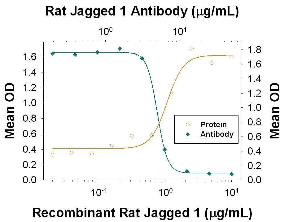
Differentiation Induced by Jagged 1 and Neutralization by Rat Jagged 1 Antibody.
Recombinant Rat Jagged 1 Fc Chimera (Catalog # 599-JG) induces differentiation in the C3H10T1/2 mouse embryonic fibroblast cell line in the presence of Recombinant Human BMP-2 (Catalog # 355-BM) in a dose-dependent manner (orange line), as measured by alkaline phosphatase activity. Differentiation elicited by Recombinant Rat Jagged 1 Fc Chimera (3 µg/mL) in the presence of Recombinant Human BMP-2 (150 ng/mL) is neutralized (green line) by increasing concentrations of Goat Anti-Rat Jagged 1 Antigen Affinity-purified Polyclonal Antibody (Catalog # AF599). The ND50 is typically 3-15 µg/mL.
Rat Jagged 1 ELISA Standard Curve
Recombinant Rat Jagged 1 Fc Chimera (Catalog # 599-JG) was serially diluted and captured by Goat Anti-Rat Jagged 1 Antigen Affinity-purified Polyclonal Antibody (Catalog # AF599) coated on a Clear Polystyrene Microplate (Catalog # DY990). Goat Anti-Rat Jagged 1 Antigen Affinity-purified Polyclonal Antibody (Catalog # AF599) was biotinylated and incubated with the protein captured on the plate. Detection of the standard curve was achieved by incubating Streptavidin-HRP (Catalog # DY998)
Rat Jagged 1 ELISA Standard Curve
Recombinant Rat Jagged 1 Fc Chimera (Catalog # 599-JG) was serially diluted and captured by Goat Anti-Rat Jagged 1 Antigen Affinity-purified Polyclonal Antibody (Catalog # AF599) coated on a Clear Polystyrene Microplate (Catalog # DY990). Goat Anti-Rat Jagged 1 Antigen Affinity-purified Polyclonal Antibody (Catalog # AF599) was biotinylated and incubated with the protein captured on the plate. Detection of the standard curve was achieved by incubating Streptavidin-HRP (Catalog # DY998)Applications for Rat Jagged 1 Antibody
CyTOF-ready
Flow Cytometry
Sample: Rat cortical stem cells
Western Blot
Sample: Recombinant Rat Jagged 1 Fc Chimera (Catalog # 599-JG)
under non-reducing conditions only
Neutralization
Rat Jagged 1 Sandwich Immunoassay
Reviewed Applications
Read 1 review rated 5 using AF599 in the following applications:
Flow Cytometry Panel Builder
Bio-Techne Knows Flow Cytometry
Save time and reduce costly mistakes by quickly finding compatible reagents using the Panel Builder Tool.
Advanced Features
- Spectra Viewer - Custom analysis of spectra from multiple fluorochromes
- Spillover Popups - Visualize the spectra of individual fluorochromes
- Antigen Density Selector - Match fluorochrome brightness with antigen density

Formulation, Preparation, and Storage
Purification
Reconstitution
Reconstitute at 0.2 mg/mL in sterile PBS. For liquid material, refer to CoA for concentration.
Formulation
Shipping
Stability & Storage
- 12 months from date of receipt, -20 to -70 °C as supplied.
- 1 month, 2 to 8 °C under sterile conditions after reconstitution.
- 6 months, -20 to -70 °C under sterile conditions after reconstitution.
Calculators
Background: Jagged 1
References
- Lindsell, C.E. et al. (1995) Cell 80:909.
- Shimizu, K. et al. (1999) J. Biol. Chem. 274:32961.
- Lewis, J. (1998) Stem Cell & Dev. Biol. 9:583.
- Small, D. et al. (2001) J. Biol. Chem. 276:32022.
- Karanu, F. et al. (2000) J. Exp. Med. 192:1365.
- Joutel, A. and E. Tounier-Lasserve (1998) Stem Cell & Dev. Biol. 9:619.
- Hicks, C. et al. (2002) J Neurosci. Res. 68:655.
Alternate Names
Gene Symbol
UniProt
Additional Jagged 1 Products
Product Documents for Rat Jagged 1 Antibody
Certificate of Analysis
To download a Certificate of Analysis, please enter a lot or batch number in the search box below.
Note: Certificate of Analysis not available for kit components.
Product Specific Notices for Rat Jagged 1 Antibody
For research use only
Related Research Areas
Citations for Rat Jagged 1 Antibody
 Powered by Bioz
Customer Reviews for Rat Jagged 1 Antibody (1)
Have you used Rat Jagged 1 Antibody?
Submit a review and receive an Amazon gift card!
$25/€18/£15/$25CAN/¥2500 for a review with an image
$10/€7/£6/$10CAN/¥1110 for a review without an image
Submit a review
Customer Images
-
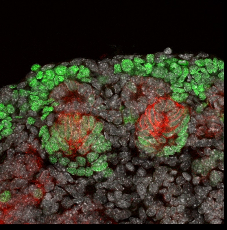
Application: Immunocytochemistry/ImmunofluorescenceSample Tested: Embryonic kidneySpecies: MouseVerified Customer | Posted 04/22/2016Jag1 (Red) Six2 (Green)

There are no reviews that match your criteria.
Protocols
Find general support by application which include: protocols, troubleshooting, illustrated assays, videos and webinars.
- 7-Amino Actinomycin D (7-AAD) Cell Viability Flow Cytometry Protocol
- Cellular Response to Hypoxia Protocols
- Extracellular Membrane Flow Cytometry Protocol
- Flow Cytometry Protocol for Cell Surface Markers
- Flow Cytometry Protocol for Staining Membrane Associated Proteins
- Flow Cytometry Staining Protocols
- Flow Cytometry Troubleshooting Guide
- Intracellular Flow Cytometry Protocol Using Alcohol (Methanol)
- Intracellular Flow Cytometry Protocol Using Detergents
- Intracellular Nuclear Staining Flow Cytometry Protocol Using Detergents
- Intracellular Staining Flow Cytometry Protocol Using Alcohol Permeabilization
- Intracellular Staining Flow Cytometry Protocol Using Detergents to Permeabilize Cells
- Propidium Iodide Cell Viability Flow Cytometry Protocol
- Protocol for Liperfluo
- Protocol for the Characterization of Human Th22 Cells
- Protocol for the Characterization of Human Th9 Cells
- Protocol: Annexin V and PI Staining by Flow Cytometry
- Protocol: Annexin V and PI Staining for Apoptosis by Flow Cytometry
- R&D Systems Quality Control Western Blot Protocol
- Troubleshooting Guide: Fluorokine Flow Cytometry Kits
- Troubleshooting Guide: Western Blot Figures
- Western Blot Conditions
- Western Blot Protocol
- Western Blot Protocol for Cell Lysates
- Western Blot Troubleshooting
- Western Blot Troubleshooting Guide
- View all Protocols, Troubleshooting, Illustrated assays and Webinars
Associated Pathways

