RbBP5 Antibody - BSA Free
Novus Biologicals | Catalog # NB600-252
![Western Blot: RbBP5 Antibody [NB600-252]](https://resources.rndsystems.com/images/products/RbBP5-Antibody-Western-Blot-NB600-252-img0012.jpg "Western Blot: RbBP5 Antibody [NB600-252]")
Key Product Details
Species Reactivity
Validated:
Human, Mouse
Cited:
Human
Applications
Validated:
Immunohistochemistry, Immunohistochemistry-Paraffin, Western Blot, Immunocytochemistry/ Immunofluorescence, Immunoprecipitation, Chromatin Immunoprecipitation (ChIP), Chromatin Immunoprecipitation Sequencing
Cited:
Western Blot
Label
Unconjugated
Antibody Source
Polyclonal Rabbit IgG
Format
BSA Free
Loading...
Product Specifications
Immunogen
The immunogen recognized by this antibody maps to a region between residue 500 and the C-terminus (residue 538) of human retinoblastoma binding protein 5 using the numbering given in entry NP_005048.2 (GeneID 5929).
Clonality
Polyclonal
Host
Rabbit
Isotype
IgG
Scientific Data Images for RbBP5 Antibody - BSA Free
Western Blot: RbBP5 Antibody [NB600-252]
Western Blot: RbBP5 Antibody [NB600-252] - Whole cell lysate (50 ug) from TCMK-1 and NIH 3T3 cells prepared using NETN lysis buffer. Antibody: Affinity purified rabbit anti-RbBP5 antibody used for WB at 0.04 ug/ml. Detection: Chemiluminescence with an exposure time of 10 seconds.![Immunohistochemistry-Paraffin: RbBP5 Antibody [NB600-252]](https://resources.rndsystems.com/images/products/RbBP5-Antibody-Immunohistochemistry-NB600-252-img0008.jpg "Immunohistochemistry-Paraffin: RbBP5 Antibody [NB600-252]")
Immunohistochemistry-Paraffin: RbBP5 Antibody [NB600-252]
Immunohistochemistry-Paraffin: RbBP5 Antibody [NB600-252] - Sample: FFPE section of human colon carcinoma. Antibody: Affinity purified rabbit anti- RbBP5 used at a dilution of 1:1,000 (1ug/ml). Detection: DAB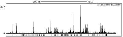
Chromatin Immunoprecipitation Sequencing: RbBP5 Antibody [NB600-252] - Localization of RbBP5 Binding Sites by ChIP-sequencing. Chromatin from K562 cells was immunoprecipitated with anti-RbBP5 antibody NB600-252 and analyzed by DNA sequencing. The figure illustrates the peak distribution of RbBP5 binding within a 500 Kb region of chromosome 1 as detected using anti-RbBP5 antibody NB600-252. ChIP-seq validation performed by Diogenode, Denville, NJ.
![Western Blot: RbBP5 Antibody [NB600-252]](https://resources.rndsystems.com/images/products/RbBP5-Antibody-Western-Blot-NB600-252-img0011.jpg "Western Blot: RbBP5 Antibody [NB600-252]")
Western Blot: RbBP5 Antibody [NB600-252]
Western Blot: RbBP5 Antibody [NB600-252] - Whole cell lysate (15 ug) from HeLa, HEK293T, and Jurkat cells prepared using NETN lysis buffer. Antibody: Affinity purified rabbit anti-RbBP5 antibody used for WB at 0.04 ug/ml. Detection: Chemiluminescence with an exposure time of 3 seconds.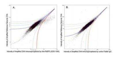
Chromatin Immunoprecipitation: RbBP5 Antibody [NB600-252] - Chromatin Immunoprecipitation: RbBP5 Antibody [NB600-252] - ChIP-chip scatter plot of anti-RbBP5 enriched DNA binding sites versus input reference DNA. A. 10 ug of NB600-252 was used to immunoprecipitate chromatin from K562 cells per Ren et al (Genes Dev. 2002 16: 245-256). Immunoprecipitated DNA and reference DNA were amplified via ligation-mediated PCR and the products labeled with fluorescent dNTPs. The labeled ChIP and reference DNA were pooled, hybridized to a DNA microarray, and analyzed. Data points below the +3 SD curve (red line) represent significantly enriched binding sites. B. As a control, a similar experiment was performed using normal rabbit IgG. Compared to the anti-RbBP5 ChIP, normal rabbit IgG showed little enrichment.
Applications for RbBP5 Antibody - BSA Free
Application
Recommended Usage
Chromatin Immunoprecipitation (ChIP)
10 ug
Chromatin Immunoprecipitation Sequencing
2 ug
Immunocytochemistry/ Immunofluorescence
1-4 ug/ml
Immunohistochemistry
1:500- 1:2000
Immunohistochemistry-Paraffin
1:500- 1:2000
Immunoprecipitation
2-10 ug/mg lysate
Western Blot
1:10000 - 1:25000
Application Notes
Reported use in ChIP assays (see Dou et al). Epitope retrieval with citrate buffer pH6.0 is recommended for FFPE tissue sections.
Reviewed Applications
Read 1 review rated 5 using NB600-252 in the following applications:
Formulation, Preparation, and Storage
Purification
Immunogen affinity purified
Formulation
Tris-Citrate/Phosphate (pH 7.0 - 8.0)
Format
BSA Free
Preservative
0.09% Sodium Azide
Concentration
1.0 mg/ml
Shipping
The product is shipped with polar packs. Upon receipt, store it immediately at the temperature recommended below.
Stability & Storage
Store at 4C. Do not freeze.
Background: RbBP5
Alternate Names
RBQ3RBBP-5, retinoblastoma binding protein 5, Retinoblastoma-binding protein RBQ-3, Set1c WD40 repeat protein, homolog, SWD1
Gene Symbol
RBBP5
UniProt
Additional RbBP5 Products
Product Documents for RbBP5 Antibody - BSA Free
Certificate of Analysis
To download a Certificate of Analysis, please enter a lot or batch number in the search box below.
Product Specific Notices for RbBP5 Antibody - BSA Free
This product is for research use only and is not approved for use in humans or in clinical diagnosis. Primary Antibodies are guaranteed for 1 year from date of receipt.
Citations for RbBP5 Antibody - BSA Free
 Powered by Bioz
Customer Reviews for RbBP5 Antibody - BSA Free (1)
5 out of 5
1 Customer Rating
Have you used RbBP5 Antibody - BSA Free?
Submit a review and receive an Amazon gift card!
$25/€18/£15/$25CAN/¥2500 Yen for a review with an image
$10/€7/£6/$10CAN/¥1110 Yen for a review without an image
Submit a review
Showing
1
-
1 的
1 review
Showing All
Filter By:
-
Application: Western BlotSample Tested:Species: HumanVerified Customer | Posted 03/26/2015
There are no reviews that match your criteria.
Protocols
Find general support by application which include: protocols, troubleshooting, illustrated assays, videos and webinars.
- Antigen Retrieval Protocol (PIER)
- Antigen Retrieval for Frozen Sections Protocol
- Appropriate Fixation of IHC/ICC Samples
- Cellular Response to Hypoxia Protocols
- ChIP Protocol Video
- Chromatin Immunoprecipitation (ChIP) Protocol
- Chromatin Immunoprecipitation Protocol
- Chromogenic IHC Staining of Formalin-Fixed Paraffin-Embedded (FFPE) Tissue Protocol
- Chromogenic Immunohistochemistry Staining of Frozen Tissue
- ClariTSA™ Fluorophore Kits
- Detection & Visualization of Antibody Binding
- Fluorescent IHC Staining of Frozen Tissue Protocol
- Graphic Protocol for Heat-induced Epitope Retrieval
- Graphic Protocol for the Preparation and Fluorescent IHC Staining of Frozen Tissue Sections
- Graphic Protocol for the Preparation and Fluorescent IHC Staining of Paraffin-embedded Tissue Sections
- Graphic Protocol for the Preparation of Gelatin-coated Slides for Histological Tissue Sections
- ICC Cell Smear Protocol for Suspension Cells
- ICC Immunocytochemistry Protocol Videos
- ICC for Adherent Cells
- IHC Sample Preparation (Frozen sections vs Paraffin)
- Immunocytochemistry (ICC) Protocol
- Immunocytochemistry Troubleshooting
- Immunofluorescence of Organoids Embedded in Cultrex Basement Membrane Extract
- Immunofluorescent IHC Staining of Formalin-Fixed Paraffin-Embedded (FFPE) Tissue Protocol
- Immunohistochemistry (IHC) and Immunocytochemistry (ICC) Protocols
- Immunohistochemistry Frozen Troubleshooting
- Immunohistochemistry Paraffin Troubleshooting
- Immunoprecipitation Protocol
- Preparing Samples for IHC/ICC Experiments
- Preventing Non-Specific Staining (Non-Specific Binding)
- Primary Antibody Selection & Optimization
- Protocol for Heat-Induced Epitope Retrieval (HIER)
- Protocol for Making a 4% Formaldehyde Solution in PBS
- Protocol for VisUCyte™ HRP Polymer Detection Reagent
- Protocol for the Fluorescent ICC Staining of Cell Smears - Graphic
- Protocol for the Fluorescent ICC Staining of Cultured Cells on Coverslips - Graphic
- Protocol for the Preparation & Fixation of Cells on Coverslips
- Protocol for the Preparation and Chromogenic IHC Staining of Frozen Tissue Sections
- Protocol for the Preparation and Chromogenic IHC Staining of Frozen Tissue Sections - Graphic
- Protocol for the Preparation and Chromogenic IHC Staining of Paraffin-embedded Tissue Sections
- Protocol for the Preparation and Chromogenic IHC Staining of Paraffin-embedded Tissue Sections - Graphic
- Protocol for the Preparation and Fluorescent ICC Staining of Cells on Coverslips
- Protocol for the Preparation and Fluorescent ICC Staining of Non-adherent Cells
- Protocol for the Preparation and Fluorescent ICC Staining of Stem Cells on Coverslips
- Protocol for the Preparation and Fluorescent IHC Staining of Frozen Tissue Sections
- Protocol for the Preparation and Fluorescent IHC Staining of Paraffin-embedded Tissue Sections
- Protocol for the Preparation of Gelatin-coated Slides for Histological Tissue Sections
- Protocol for the Preparation of a Cell Smear for Non-adherent Cell ICC - Graphic
- R&D Systems Quality Control Western Blot Protocol
- TUNEL and Active Caspase-3 Detection by IHC/ICC Protocol
- The Importance of IHC/ICC Controls
- Troubleshooting Guide: Immunohistochemistry
- Troubleshooting Guide: Western Blot Figures
- Western Blot Conditions
- Western Blot Protocol
- Western Blot Protocol for Cell Lysates
- Western Blot Troubleshooting
- Western Blot Troubleshooting Guide
- View all Protocols, Troubleshooting, Illustrated assays and Webinars
Loading...