RRM2 Antibody - Azide Free
Novus Biologicals | Catalog # NBP1-31661
![Immunocytochemistry/ Immunofluorescence: RRM2 Antibody [NBP1-31661]](https://resources.rndsystems.com/images/products/RRM2-Antibody-Immunocytochemistry-Immunofluorescence-NBP1-31661-img0011.jpg "Immunocytochemistry/ Immunofluorescence: RRM2 Antibody [NBP1-31661]")
Key Product Details
Validated by
Species Reactivity
Validated:
Cited:
Predicted:
Applications
Validated:
Cited:
Label
Antibody Source
Format
Product Specifications
Immunogen
Localization
Clonality
Host
Isotype
Theoretical MW
Disclaimer note: The observed molecular weight of the protein may vary from the listed predicted molecular weight due to post translational modifications, post translation cleavages, relative charges, and other experimental factors.
Scientific Data Images for RRM2 Antibody - Azide Free
Immunocytochemistry/ Immunofluorescence: RRM2 Antibody [NBP1-31661]
Immunocytochemistry/Immunofluorescence: RRM2 Antibody [NBP1-31661] - HeLa cells were fixed in 4% paraformaldehyde at RT for 15 min. Green: RRM2 protein stained by RRM2 antibody [N1C1] diluted at 1:500. Blue: Hoechst 33342 staining. Scale bar = 10 um.![Immunohistochemistry-Paraffin: RRM2 Antibody [NBP1-31661]](https://resources.rndsystems.com/images/products/RRM2-Antibody-Immunohistochemistry-Paraffin-NBP1-31661-img0012.jpg "Immunohistochemistry-Paraffin: RRM2 Antibody [NBP1-31661]")
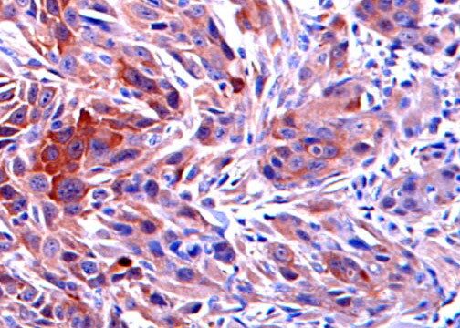
Immunohistochemistry-Paraffin: RRM2 Antibody [NBP1-31661]
Immunohistochemistry-Paraffin: RRM2 Antibody [NBP1-31661] - Mouse tumor tissues were stained with RRM2 antibody at 1:50 dilution. Image submitted by a verified customer review.![Immunocytochemistry/ Immunofluorescence: RRM2 Antibody [NBP1-31661]](https://resources.rndsystems.com/images/products/RRM2-Antibody-Immunocytochemistry-Immunofluorescence-NBP1-31661-img0009.jpg "Immunocytochemistry/ Immunofluorescence: RRM2 Antibody [NBP1-31661]")
Immunocytochemistry/ Immunofluorescence: RRM2 Antibody [NBP1-31661]
Immunocytochemistry/Immunofluorescence: RRM2 Antibody [NBP1-31661] - HeLa cells were fixed in ice-cold MeOH for 5 min. Green: RRM2 protein stained by RRM2 antibody [N1C1] diluted at 1:1000. Blue: Hoechst 33342 staining. Scale bar = 10 um.![Immunohistochemistry-Paraffin: RRM2 Antibody [NBP1-31661]](https://resources.rndsystems.com/images/products/RRM2-Antibody-Immunohistochemistry-Paraffin-NBP1-31661-img0006.jpg "Immunohistochemistry-Paraffin: RRM2 Antibody [NBP1-31661]")
Immunohistochemistry-Paraffin: RRM2 Antibody [NBP1-31661]
Immunohistochemistry-Paraffin: RRM2 Antibody [NBP1-31661] - Mouse muscle. RRM2 antibody [N1C1] diluted at 1:500. Antigen Retrieval: Trilogy™ (EDTA based, pH 8.0) buffer, 15min.![Immunohistochemistry-Paraffin: RRM2 Antibody [NBP1-31661]](https://resources.rndsystems.com/images/products/RRM2-Antibody-Immunohistochemistry-Paraffin-NBP1-31661-img0007.jpg "Immunohistochemistry-Paraffin: RRM2 Antibody [NBP1-31661]")
Immunohistochemistry-Paraffin: RRM2 Antibody [NBP1-31661]
Immunohistochemistry-Paraffin: RRM2 Antibody [NBP1-31661] - Rat intestine. RRM2 antibody [N1C1] iluted at 1:500. Antigen Retrieval: Citrate buffer, pH 6.0, 15 min.![Immunohistochemistry-Paraffin: RRM2 Antibody [NBP1-31661]](https://resources.rndsystems.com/images/products/RRM2-Antibody-Immunohistochemistry-Paraffin-NBP1-31661-img0008.jpg "Immunohistochemistry-Paraffin: RRM2 Antibody [NBP1-31661]")
Immunohistochemistry-Paraffin: RRM2 Antibody [NBP1-31661]
Immunohistochemistry-Paraffin: RRM2 Antibody [NBP1-31661] - Mouse muscle. RRM2 antibody [N1C1] diluted at 1:500. Antigen Retrieval: Citrate buffer, pH 6.0, 15 min.
Western Blot: RRM2 Antibody [NBP1-31661] -
Western Blot: RRM2 Antibody [NBP1-31661] - Various tissue extracts (30 ug) were separated by 10% SDS-PAGE, and the membrane was blotted with RRM2 antibody [N1C1] diluted at 1:1000.
Immunohistochemistry: RRM2 Antibody [NBP1-31661] -
Immunohistochemistry: RRM2 Antibody [NBP1-31661] - Immunohistochemical analysis of paraffin-embedded zebrafish tissue, using RRM2 antibody [N1C1] (NBP1-31661) at 1:300 dilution.
Immunohistochemistry-Paraffin: RRM2 Antibody [NBP1-31661] -
Immunohistochemistry-Paraffin: RRM2 Antibody [NBP1-31661] - RRM2 antibody [N1C1] detects RRM2 protein at cytoplasm in rat intestine by immunohistochemical analysis.Sample: Paraffin-embedded rat intestine.
RRM2 antibody [N1C1] (NBP1-31661) diluted at 1:500.
Antigen Retrieval: Citrate buffer, pH 6.0, 15 min

Western Blot: RRM2 Antibody [NBP1-31661] -
Various whole cell extracts (30 ug) were separated by 10% SDS-PAGE, and the membrane was blotted with RRM2 antibody [N1C1] (NBP1-31661) diluted at 1:1000. The HRP-conjugated anti-rabbit IgG antibody was used to detect the primary antibody. Corresponding RNA expression data for the same cell lines are based on Human Protein Atlas program.
Western Blot: RRM2 Antibody - Azide Free [NBP1-31661] -
SARS-CoV-2 causes CHK1 and RRM2 reduction leading to cell cycle progression impairment.A) IF images of infected HNEpC; nuclei were stained with Hoechst. Scale bar, 10 μm. B) Quantification of the percentage of RRM2-expressing cells in infected (SARS-CoV-2 N + ) or not (SARS-CoV-2 N-) HNEpC. C) Immunoblotting of whole cell lysates of Calu-3 infected, or not, with SARS-CoV-2 and analyzed at different time points post-infection. D) Quantification of protein levels shown in C; values are shown as relative to mock-infected samples. E,F) RT–qPCR of CHK1 and RRM2 mRNA expression in infected (V+ ) or mock-infected (V−) Huh7 and Calu-3 cells, respectively. G) Immunoblotting of CDT1 in Huh7 and Calu-3 cells treated as indicated. The experiment was repeated three times with similar results. Source numerical data and unprocessed blots are available in source data.Source data Image collected and cropped by CiteAb from the following open publication (https://pubmed.ncbi.nlm.nih.gov/36894671), licensed under a CC-BY license. Not internally tested by Novus Biologicals.
Western Blot: RRM2 Antibody - Azide Free [NBP1-31661] -
SARS-CoV-2 reduces CHK1 and RRM2 levels leading to dNTP shortage.a, Immunoblotting of whole cell lysates of Huh7 infected, or not, with SARS-CoV-2 and analysed at different timepoints post-infection. b, Quantification of protein levels shown in a; values are shown as relative to mock-infected samples. c, Immunofluorescence (IF) images of infected (V+) or mock-infected (V−) Huh7 cells fixed 48 h post-infection; nuclei were stained with DAPI. Scale bar, 10 μm. d, Quantification of CHK1- or RRM2-positive cells shown in c; n = 3 independent experiments. e, dNTP concentration was measured in V− or V+ Huh7 and Calu-3; values are shown as relative to V−. f, Histograms show the percentage of cells in each phase of the cell cycle in V− or V+ Huh7 fixed 48 h post-infection. g, Fraction of V− or V+ Huh7 cells that did not incorporate BrdU (BrdU−) measured by flow cytometry 48 h post-infection. Source numerical data and unprocessed blots are available in source data.Source data Image collected and cropped by CiteAb from the following open publication (https://pubmed.ncbi.nlm.nih.gov/36894671), licensed under a CC-BY license. Not internally tested by Novus Biologicals.
Western Blot: RRM2 Antibody - Azide Free [NBP1-31661] -
CHK1 depletion is sufficient to recapitulate the effects of SARS-CoV-2 infection.A) Huh7 transfected with siRNAs against CHK1 mRNA (siCHK1) or siCTRL were stained for CHK1 and propidium iodide (PI) prior to flow cytometry. B) DNA content analysis of V− or V+ Huh7 fixed 48 h post-infection and siCHK1- or siCTRL-transfected Huh7. C) Histograms show the percentage of cells in each phase of the cell cycle upon siCHK1 or siCTRL treatment. D) Bivariate plot showing DNA content (PI) and BrdU incorporation measured by flow cytometry of V− or V+ Huh7 fixed 48 h post-infection and siCHK1- or siCTRL-transfected Huh7. E) Immunoblots of siCHK1- or siCTRL-transfected Huh7. F) Quantification of protein levels shown in E; values are the means +/- s.e.m. of two independent experiments and shown as relative to the siCTRL-transfected sample. G) IF images of Calu-3 transfected with the indicated siRNAs; nuclei were stained with DAPI. Scale bar, 10 μm. H) Quantification of gamma H2AX foci per cell shown in G. I) IF images of cGAS staining in samples as in G; nuclei were stained with DAPI. Scale bar, 10 μm. J) Micronuclei and cGAS+ micronuclei quantifications on total cell number; at least 300 nuclei were scored for each sample. K,L) RT–qPCR for pro-inflammatory cytokines and CHK1 mRNA expression in siCHK1-treated Calu-3 and Huh7 cells, respectively. Values are shown as relative to siCTRL-transfected samples. M) Quantification of the amounts of secreted cytokines and chemokines from siCHK1- or siCTRL-transfected Calu-3 by Bio-Plex multiplex immunoassays. Source numerical data and unprocessed blots are available in source data.Source data Image collected and cropped by CiteAb from the following open publication (https://pubmed.ncbi.nlm.nih.gov/36894671), licensed under a CC-BY license. Not internally tested by Novus Biologicals.Applications for RRM2 Antibody - Azide Free
Immunocytochemistry/ Immunofluorescence
Immunohistochemistry
Immunohistochemistry-Paraffin
Simple Western
Western Blot
Reviewed Applications
Read 1 review rated 5 using NBP1-31661 in the following applications:
Formulation, Preparation, and Storage
Purification
Formulation
Format
Preservative
Concentration
Shipping
Stability & Storage
Background: RRM2
Long Name
Alternate Names
Entrez Gene IDs
Gene Symbol
UniProt
Additional RRM2 Products
Product Documents for RRM2 Antibody - Azide Free
Certificate of Analysis
To download a Certificate of Analysis, please enter a lot or batch number in the search box below.
Product Specific Notices for RRM2 Antibody - Azide Free
This product is for research use only and is not approved for use in humans or in clinical diagnosis. Primary Antibodies are guaranteed for 1 year from date of receipt.
Citations for RRM2 Antibody - Azide Free
 Powered by Bioz
Customer Reviews for RRM2 Antibody - Azide Free (1)
Have you used RRM2 Antibody - Azide Free?
Submit a review and receive an Amazon gift card!
$25/€18/£15/$25CAN/¥2500 Yen for a review with an image
$10/€7/£6/$10CAN/¥1110 Yen for a review without an image
Submit a review
Customer Images
-
Application: Immunohistochemistry-ParaffinSample Tested: formalin fixed paraffin embedded mouse tumor tissuesSpecies: MouseVerified Customer | Posted 08/11/2017Formalin fixed parafin embedded mouse tumor tissues were stained with RRM2 antibody at 1:50 dilution.

There are no reviews that match your criteria.
Protocols
Find general support by application which include: protocols, troubleshooting, illustrated assays, videos and webinars.
- Antigen Retrieval Protocol (PIER)
- Antigen Retrieval for Frozen Sections Protocol
- Appropriate Fixation of IHC/ICC Samples
- Cellular Response to Hypoxia Protocols
- Chromogenic IHC Staining of Formalin-Fixed Paraffin-Embedded (FFPE) Tissue Protocol
- Chromogenic Immunohistochemistry Staining of Frozen Tissue
- ClariTSA™ Fluorophore Kits
- Detection & Visualization of Antibody Binding
- Fluorescent IHC Staining of Frozen Tissue Protocol
- Graphic Protocol for Heat-induced Epitope Retrieval
- Graphic Protocol for the Preparation and Fluorescent IHC Staining of Frozen Tissue Sections
- Graphic Protocol for the Preparation and Fluorescent IHC Staining of Paraffin-embedded Tissue Sections
- Graphic Protocol for the Preparation of Gelatin-coated Slides for Histological Tissue Sections
- ICC Cell Smear Protocol for Suspension Cells
- ICC Immunocytochemistry Protocol Videos
- ICC for Adherent Cells
- IHC Sample Preparation (Frozen sections vs Paraffin)
- Immunocytochemistry (ICC) Protocol
- Immunocytochemistry Troubleshooting
- Immunofluorescence of Organoids Embedded in Cultrex Basement Membrane Extract
- Immunofluorescent IHC Staining of Formalin-Fixed Paraffin-Embedded (FFPE) Tissue Protocol
- Immunohistochemistry (IHC) and Immunocytochemistry (ICC) Protocols
- Immunohistochemistry Frozen Troubleshooting
- Immunohistochemistry Paraffin Troubleshooting
- Preparing Samples for IHC/ICC Experiments
- Preventing Non-Specific Staining (Non-Specific Binding)
- Primary Antibody Selection & Optimization
- Protocol for Heat-Induced Epitope Retrieval (HIER)
- Protocol for Making a 4% Formaldehyde Solution in PBS
- Protocol for VisUCyte™ HRP Polymer Detection Reagent
- Protocol for the Fluorescent ICC Staining of Cell Smears - Graphic
- Protocol for the Fluorescent ICC Staining of Cultured Cells on Coverslips - Graphic
- Protocol for the Preparation & Fixation of Cells on Coverslips
- Protocol for the Preparation and Chromogenic IHC Staining of Frozen Tissue Sections
- Protocol for the Preparation and Chromogenic IHC Staining of Frozen Tissue Sections - Graphic
- Protocol for the Preparation and Chromogenic IHC Staining of Paraffin-embedded Tissue Sections
- Protocol for the Preparation and Chromogenic IHC Staining of Paraffin-embedded Tissue Sections - Graphic
- Protocol for the Preparation and Fluorescent ICC Staining of Cells on Coverslips
- Protocol for the Preparation and Fluorescent ICC Staining of Non-adherent Cells
- Protocol for the Preparation and Fluorescent ICC Staining of Stem Cells on Coverslips
- Protocol for the Preparation and Fluorescent IHC Staining of Frozen Tissue Sections
- Protocol for the Preparation and Fluorescent IHC Staining of Paraffin-embedded Tissue Sections
- Protocol for the Preparation of Gelatin-coated Slides for Histological Tissue Sections
- Protocol for the Preparation of a Cell Smear for Non-adherent Cell ICC - Graphic
- R&D Systems Quality Control Western Blot Protocol
- TUNEL and Active Caspase-3 Detection by IHC/ICC Protocol
- The Importance of IHC/ICC Controls
- Troubleshooting Guide: Immunohistochemistry
- Troubleshooting Guide: Western Blot Figures
- Western Blot Conditions
- Western Blot Protocol
- Western Blot Protocol for Cell Lysates
- Western Blot Troubleshooting
- Western Blot Troubleshooting Guide
- View all Protocols, Troubleshooting, Illustrated assays and Webinars
FAQs for RRM2 Antibody - Azide Free
-
Q: What is the isotype of this antibody?
A: NBP1-31661 is a polyclonal antibody made in rabbit. It is expected that the majority of the antibodies present will be IgG.