

![Western Blot: SLC4A11 Antibody [NBP1-46156]](https://resources.rndsystems.com/images/products/SLC4A11-Antibody-Western-Blot-NBP1-46156-img0004.jpg "Western Blot: SLC4A11 Antibody [NBP1-46156]")
Loading...
Key Product Details
Species Reactivity
Validated:
Human
Predicted:
Bovine (100%), Canine (100%), Mouse (100%), Rat (100%). Backed by our 100% Guarantee.
Applications
Immunohistochemistry, Immunohistochemistry-Paraffin, Western Blot, Peptide ELISA
Label
Unconjugated
Antibody Source
Polyclonal Goat IgG
Loading...
Product Specifications
Immunogen
This SLC4A11 Antibody was developed against a peptide with sequence C-HTSRKYLKLKNFKEE corresponding to internal region according to NP_114423.1.
Epitope
internal region
Reactivity Notes
Porcine reactivity reported from a verified customer review.
Clonality
Polyclonal
Host
Goat
Isotype
IgG
Scientific Data Images for SLC4A11 Antibody
Western Blot: SLC4A11 Antibody [NBP1-46156]
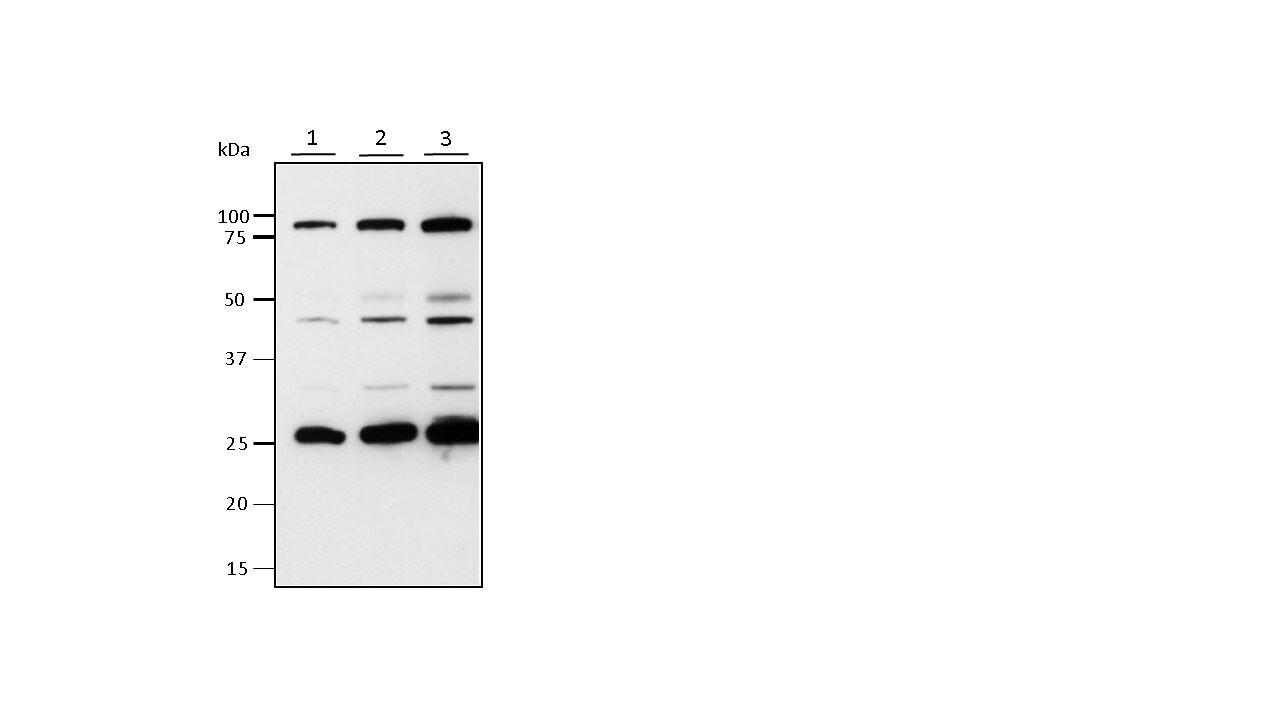
Western Blot: SLC4A11 Antibody [NBP1-46156] - Porcine kidney tissue lysates. Lane 1 - 15 ug. Lane 2 - 30 ug. Lane 3 - 60 ug. Antibody at 1:500. Secondary antibody at 1:1000. Western blot image submitted by a verified customer review.![Immunohistochemistry-Paraffin: SLC4A11 Antibody [NBP1-46156]](https://resources.rndsystems.com/images/products/SLC4A11-Antibody-Immunohistochemistry-Paraffin-NBP1-46156-img0003.jpg "Immunohistochemistry-Paraffin: SLC4A11 Antibody [NBP1-46156]")
Immunohistochemistry-Paraffin: SLC4A11 Antibody [NBP1-46156]
Immunohistochemistry-Paraffin: SLC4A11 Antibody [NBP1-46156] - Staining of paraffin embedded Human Kidney. Antibody at 3.75 ug/mL. Steamed antigen retrieval with citrate buffer pH 6, AP-staining.![Western Blot: SLC4A11 Antibody [NBP1-46156]](https://resources.rndsystems.com/images/products/SLC4A11-Antibody-Western-Blot-NBP1-46156-img0002.jpg "Western Blot: SLC4A11 Antibody [NBP1-46156]")
Western Blot: SLC4A11 Antibody [NBP1-46156]
Western Blot: SLC4A11 Antibody [NBP1-46156] - Staining of Human Kidney lysate (35 ug protein in RIPA buffer). Antibody at 0.3 ug/mL. Primary incubation was 1 hour. Detected by chemiluminescence. Approx 90 kDa band observed in Human Kidney lysates (calculated MW of 99.6 kDa according to NP_114423.1). An additional band of unknown identity was also consistently observed at 26 kDa. This band was successfully blocked by incubation with the immunizing peptide.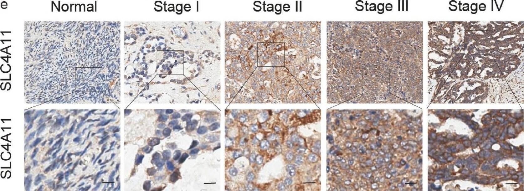
Immunohistochemistry: SLC4A11 Antibody [NBP1-46156] -
SLC4A11 is linked to ovarian cancer. (a) Fifty-two genes whose expression was elevated in ovarian cancer tissue compared with healthy tissue are displayed in a Venn diagram. Data were obtained from the GSE40505, GSE18520, GSE66057, TCGA and GTEx datasets. (b) SLC4A11 mRNA expression levels in 88 nontumor tissues and 426 OV tissues from the GEPIA database. (c) SLC4A11 expression is used to predict the overall prognosis of ovarian cancer patients. Data obtained from GEPIA. (d) The degree of SLC4A11 expression in the GEO database (GSE40595, 63 OV, and 14 nontumor tissue samples; GSE18520, 53 OV and 10 nontumor tissue samples; GSE66957, 57 OV and 13 nontumor tissue samples). Chi-square test: *p < 0.05, **p < 0.01, ***p < 0.001. (e) SLC4A11 expression in OV tissue microarrays at various disease stages and in normal tissues. Scale bar: 50 uM. (f) The number of samples expressing SLC4A11 in OV samples (n = 117) and normal tissue samples (n = 16) was determined by blinded IHC. Scoring is based on staining intensity: negative (no positive colouration): 0 points; weak positive (light yellow): 1 point; positive (brownish yellow): 2 points; and strong positive (tan): 3 points. A score greater than 1 is considered high. Image collected and cropped by CiteAb from the following open publication (https://pubmed.ncbi.nlm.nih.gov/40721447), licensed under a CC-BY license. Not internally tested by Novus Biologicals.Applications for SLC4A11 Antibody
Application
Recommended Usage
Immunohistochemistry-Paraffin
3.75 ug/mL
Peptide ELISA
Detection limit 1:32000
Western Blot
0.3 - 1 ug/mL
Reviewed Applications
Read 1 review rated 4 using NBP1-46156 in the following applications:
Formulation, Preparation, and Storage
Purification
Immunogen affinity purified
Formulation
Tris saline (20 mM Tris pH 7.3, 150 mM NaCl), 0.5% BSA
Preservative
0.02% Sodium Azide
Concentration
0.5 mg/ml
Shipping
The product is shipped with polar packs. Upon receipt, store it immediately at the temperature recommended below.
Stability & Storage
Store at -20C. Avoid freeze-thaw cycles.
Background: SLC4A11
Alternate Names
Bicarbonate transporter-related protein 1, BTR1bicarbonate transporter related protein 1, CDPD, CHED2, corneal endothelial dystrophy 2 (autosomal recessive), dJ794I6.2, FECD4, MGC126418, MGC126419, NABC1, sodium bicarbonate transporter-like protein 11, Sodium borate cotransporter 1, sodium-coupled borate cotransporter 1, Solute carrier family 4 member 11, solute carrier family 4, sodium bicarbonate transporter-like, member 11, solute carrier family 4, sodium borate transporter, member 11
Gene Symbol
SLC4A11
UniProt
Additional SLC4A11 Products
Product Documents for SLC4A11 Antibody
Certificate of Analysis
To download a Certificate of Analysis, please enter a lot or batch number in the search box below.
Product Specific Notices for SLC4A11 Antibody
This product is for research use only and is not approved for use in humans or in clinical diagnosis. Primary Antibodies are guaranteed for 1 year from date of receipt.
Citations for SLC4A11 Antibody
 Powered by Bioz
Customer Reviews for SLC4A11 Antibody (1)
4 out of 5
1 Customer Rating
Have you used SLC4A11 Antibody?
Submit a review and receive an Amazon gift card!
$25/€18/£15/$25CAN/¥2500 for a review with an image
$10/€7/£6/$10CAN/¥1110 for a review without an image
Submit a review
Customer Images
Showing
1
-
1 的
1 review
Showing All
Filter By:
-
Application: Western BlotSample Tested: Kidney tissueSpecies: PigVerified Customer | Posted 02/15/2021SLC4A11 antibody Validationlane 1 - 15ug lane 2 - 30ug lane 3 - 60ug Primary Antibody - 1:1500 Secondary Antibody - 1:1000 5% Blocking Solution
 Bio-Techne ResponseThis review was submitted through the legacy Novus Innovators Program, reflecting a new species or application tested on a primary antibody.
There are no reviews that match your criteria.
Protocols
Find general support by application which include: protocols, troubleshooting, illustrated assays, videos and webinars.
- Antigen Retrieval Protocol (PIER)
- Antigen Retrieval for Frozen Sections Protocol
- Appropriate Fixation of IHC/ICC Samples
- Cellular Response to Hypoxia Protocols
- Chromogenic IHC Staining of Formalin-Fixed Paraffin-Embedded (FFPE) Tissue Protocol
- Chromogenic Immunohistochemistry Staining of Frozen Tissue
- ClariTSA™ Fluorophore Kits
- Detection & Visualization of Antibody Binding
- ELISA Sample Preparation & Collection Guide
- ELISA Troubleshooting Guide
- Fluorescent IHC Staining of Frozen Tissue Protocol
- Graphic Protocol for Heat-induced Epitope Retrieval
- Graphic Protocol for the Preparation and Fluorescent IHC Staining of Frozen Tissue Sections
- Graphic Protocol for the Preparation and Fluorescent IHC Staining of Paraffin-embedded Tissue Sections
- Graphic Protocol for the Preparation of Gelatin-coated Slides for Histological Tissue Sections
- How to Run an R&D Systems DuoSet ELISA
- How to Run an R&D Systems Quantikine ELISA
- How to Run an R&D Systems Quantikine™ QuicKit™ ELISA
- IHC Sample Preparation (Frozen sections vs Paraffin)
- Immunofluorescent IHC Staining of Formalin-Fixed Paraffin-Embedded (FFPE) Tissue Protocol
- Immunohistochemistry (IHC) and Immunocytochemistry (ICC) Protocols
- Immunohistochemistry Frozen Troubleshooting
- Immunohistochemistry Paraffin Troubleshooting
- Preparing Samples for IHC/ICC Experiments
- Preventing Non-Specific Staining (Non-Specific Binding)
- Primary Antibody Selection & Optimization
- Protocol for Heat-Induced Epitope Retrieval (HIER)
- Protocol for Making a 4% Formaldehyde Solution in PBS
- Protocol for VisUCyte™ HRP Polymer Detection Reagent
- Protocol for the Preparation & Fixation of Cells on Coverslips
- Protocol for the Preparation and Chromogenic IHC Staining of Frozen Tissue Sections
- Protocol for the Preparation and Chromogenic IHC Staining of Frozen Tissue Sections - Graphic
- Protocol for the Preparation and Chromogenic IHC Staining of Paraffin-embedded Tissue Sections
- Protocol for the Preparation and Chromogenic IHC Staining of Paraffin-embedded Tissue Sections - Graphic
- Protocol for the Preparation and Fluorescent IHC Staining of Frozen Tissue Sections
- Protocol for the Preparation and Fluorescent IHC Staining of Paraffin-embedded Tissue Sections
- Protocol for the Preparation of Gelatin-coated Slides for Histological Tissue Sections
- Quantikine HS ELISA Kit Assay Principle, Alkaline Phosphatase
- Quantikine HS ELISA Kit Principle, Streptavidin-HRP Polymer
- R&D Systems Quality Control Western Blot Protocol
- Sandwich ELISA (Colorimetric) – Biotin/Streptavidin Detection Protocol
- Sandwich ELISA (Colorimetric) – Direct Detection Protocol
- TUNEL and Active Caspase-3 Detection by IHC/ICC Protocol
- The Importance of IHC/ICC Controls
- Troubleshooting Guide: ELISA
- Troubleshooting Guide: Immunohistochemistry
- Troubleshooting Guide: Western Blot Figures
- Western Blot Conditions
- Western Blot Protocol
- Western Blot Protocol for Cell Lysates
- Western Blot Troubleshooting
- Western Blot Troubleshooting Guide
- View all Protocols, Troubleshooting, Illustrated assays and Webinars
Loading...