SOD1/Cu-Zn SOD Antibody
Novus Biologicals | Catalog # NBP1-31204
![Western Blot: SOD1/Cu-Zn SOD Antibody [NBP1-31204]](https://resources.rndsystems.com/images/products/SOD1-Cu-Zn-SOD-Antibody-Western-Blot-NBP1-31204-img0027.jpg "Western Blot: SOD1/Cu-Zn SOD Antibody [NBP1-31204]")
Loading...
Key Product Details
Validated by
Knockout/Knockdown
Species Reactivity
Validated:
Human, Mouse, Rat, Porcine, Bovine
Cited:
Human, Bovine
Predicted:
Chimpanzee (100%). Backed by our 100% Guarantee.
Applications
Validated:
Knockout Validated, Immunohistochemistry-Frozen, Western Blot
Cited:
Western Blot
Label
Unconjugated
Antibody Source
Polyclonal Rabbit IgG
Loading...
Product Specifications
Immunogen
Carrier-protein conjugated synthetic peptide encompassing a sequence within the C-terminus region of human SOD1/Cu-Zn SOD. The exact sequence is proprietary.
Reactivity Notes
Bovine reactivity reported in scientific literature (PMID: 30738665). Porcine reactivity reported from a verified customer review.
Localization
Cytoplasm
Clonality
Polyclonal
Host
Rabbit
Isotype
IgG
Theoretical MW
16 kDa.
Disclaimer note: The observed molecular weight of the protein may vary from the listed predicted molecular weight due to post translational modifications, post translation cleavages, relative charges, and other experimental factors.
Disclaimer note: The observed molecular weight of the protein may vary from the listed predicted molecular weight due to post translational modifications, post translation cleavages, relative charges, and other experimental factors.
Scientific Data Images for SOD1/Cu-Zn SOD Antibody
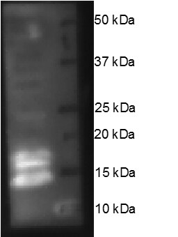
Western Blot: SOD1/Cu-Zn SOD Antibody [NBP1-31204]
Western Blot: SOD1/Cu-Zn SOD Antibody [NBP1-31204] - Various whole cell extracts (30 ug) were separated by 15% SDS-PAGE, and the membrane was blotted with SOD1 antibody diluted at 1:1000. The HRP-conjugated anti-rabbit IgG antibody (NBP2-19301) was used to detect the primary antibody.![Western Blot: SOD1/Cu-Zn SOD Antibody [NBP1-31204]](https://resources.rndsystems.com/images/products/SOD1-Cu-Zn-SOD-Antibody-Western-Blot-NBP1-31204-img0016.jpg "Western Blot: SOD1/Cu-Zn SOD Antibody [NBP1-31204]")
Western Blot: SOD1/Cu-Zn SOD Antibody [NBP1-31204]
Western Blot: SOD1/Cu-Zn SOD Antibody [NBP1-31204] - Sample (50 ug of whole cell lysate) A: Rat brain 15% SDS PAGE; antibody diluted at 1:1000.![Knockout Validated: SOD1/Cu-Zn SOD Antibody [NBP1-31204]](https://resources.rndsystems.com/images/products/SOD1-Cu-Zn-SOD-Antibody-Western-Blot-NBP1-31204-img0021.jpg "Western Blot: SOD1/Cu-Zn SOD Antibody [NBP1-31204]")

Western Blot: SOD1/Cu-Zn SOD Antibody [NBP1-31204] -
Western Blot: SOD1/Cu-Zn SOD Antibody [NBP1-31204] - Various whole cell extracts (30 ug) were separated by 15% SDS-PAGE, and the membrane was blotted with SOD1/Cu-Zn SOD antibody (NBP1-31204) diluted at 1:1000. The HRP-conjugated anti-rabbit IgG antibody was used to detect the primary antibody.Applications for SOD1/Cu-Zn SOD Antibody
Application
Recommended Usage
Immunohistochemistry-Frozen
Assay dependent
Western Blot
1:500-1:10000
Reviewed Applications
Read 1 review rated 5 using NBP1-31204 in the following applications:
Formulation, Preparation, and Storage
Purification
Antigen Affinity-purified
Formulation
PBS, 1% BSA, 20% Glycerol
Preservative
0.025% Proclin 300
Concentration
Concentrations vary lot to lot. See vial label for concentration. If unlisted please contact technical services.
Shipping
The product is shipped with polar packs. Upon receipt, store it immediately at the temperature recommended below.
Stability & Storage
Aliquot and store at -20C or -80C. Avoid freeze-thaw cycles.
Background: SOD1/Cu-Zn SOD
Long Name
Superoxide Dismutase-1
Alternate Names
Cu-Zn SOD, CuZn SOD, Ipo1, IPOA, SOD, cytosolic, SOD, Soluble
Gene Symbol
SOD1
Additional SOD1/Cu-Zn SOD Products
Product Documents for SOD1/Cu-Zn SOD Antibody
Certificate of Analysis
To download a Certificate of Analysis, please enter a lot or batch number in the search box below.
Product Specific Notices for SOD1/Cu-Zn SOD Antibody
This product is for research use only and is not approved for use in humans or in clinical diagnosis. Primary Antibodies are guaranteed for 1 year from date of receipt.
Citations for SOD1/Cu-Zn SOD Antibody
 Powered by Bioz
Customer Reviews for SOD1/Cu-Zn SOD Antibody (1)
5 out of 5
1 Customer Rating
Have you used SOD1/Cu-Zn SOD Antibody?
Submit a review and receive an Amazon gift card!
$25/€18/£15/$25CAN/¥2500 Yen for a review with an image
$10/€7/£6/$10CAN/¥1110 Yen for a review without an image
Submit a review
Customer Images
Showing
1
-
1 的
1 review
Showing All
Filter By:
-
Application: Western BlotSample Tested: Ovary tissueSpecies: PigVerified Customer | Posted 11/17/2016SOD1 on porcine whole ovarian homogenateSample Information: Treatment: None. Run on control tissue Controls: Positive Control: None Negative Control: Primary only, secondary only, and secondary with IgG all negative Loading Control: Ponceau S stain Total Protein Loaded: 40 ug/well Electrophoresis: Gel Percentage: Gradient, 4-20% Electrophoresis Conditions: Voltage: 50V then 90V Time: 5 min then 1 hr Membrane Transfer: Method (Submersion/Semi-dry): Semi-dry using iBlot2 system Membrane Type (PVDF/Nitrocellulose): Nitrocellulose Time: 7 min (iBlot2 Protocol 0) Blocking: Blocking Solution: 5% BSA in.2% PBST Time: 1.5 hours Primary Antibody: Dilution: 1:1000 Diluent Buffer: 5% BSA in.2% PBST Incubation Time: overnight Incubation Temperature: 4°C Washing Conditions: Wash Solution:.2% PBST Time and Repetitions: 3X, 10 minutes each Secondary Antibody Manufacturer and Catalog #: Cell Signaling, #7074 Secondary description: goat anti-rabbit Dilution: 1:1000 Diluent Buffer: 5% BSA in.2% PBST Incubation Time: 1 hour Incubation Temperature: room temperature Detection Method: Detection: SignalFire ECL reagent (Cell Signaling) Procedure: ECL on blot 3 min Development Time: 5 min Molecular weight of band(s): 13 kDa

There are no reviews that match your criteria.
Protocols
Find general support by application which include: protocols, troubleshooting, illustrated assays, videos and webinars.
- Antigen Retrieval Protocol (PIER)
- Antigen Retrieval for Frozen Sections Protocol
- Appropriate Fixation of IHC/ICC Samples
- Cellular Response to Hypoxia Protocols
- Chromogenic IHC Staining of Formalin-Fixed Paraffin-Embedded (FFPE) Tissue Protocol
- Chromogenic Immunohistochemistry Staining of Frozen Tissue
- ClariTSA™ Fluorophore Kits
- Detection & Visualization of Antibody Binding
- Fluorescent IHC Staining of Frozen Tissue Protocol
- Graphic Protocol for Heat-induced Epitope Retrieval
- Graphic Protocol for the Preparation and Fluorescent IHC Staining of Frozen Tissue Sections
- Graphic Protocol for the Preparation and Fluorescent IHC Staining of Paraffin-embedded Tissue Sections
- Graphic Protocol for the Preparation of Gelatin-coated Slides for Histological Tissue Sections
- IHC Sample Preparation (Frozen sections vs Paraffin)
- Immunofluorescent IHC Staining of Formalin-Fixed Paraffin-Embedded (FFPE) Tissue Protocol
- Immunohistochemistry (IHC) and Immunocytochemistry (ICC) Protocols
- Immunohistochemistry Frozen Troubleshooting
- Immunohistochemistry Paraffin Troubleshooting
- Preparing Samples for IHC/ICC Experiments
- Preventing Non-Specific Staining (Non-Specific Binding)
- Primary Antibody Selection & Optimization
- Protocol for Heat-Induced Epitope Retrieval (HIER)
- Protocol for Making a 4% Formaldehyde Solution in PBS
- Protocol for VisUCyte™ HRP Polymer Detection Reagent
- Protocol for the Preparation & Fixation of Cells on Coverslips
- Protocol for the Preparation and Chromogenic IHC Staining of Frozen Tissue Sections
- Protocol for the Preparation and Chromogenic IHC Staining of Frozen Tissue Sections - Graphic
- Protocol for the Preparation and Chromogenic IHC Staining of Paraffin-embedded Tissue Sections
- Protocol for the Preparation and Chromogenic IHC Staining of Paraffin-embedded Tissue Sections - Graphic
- Protocol for the Preparation and Fluorescent IHC Staining of Frozen Tissue Sections
- Protocol for the Preparation and Fluorescent IHC Staining of Paraffin-embedded Tissue Sections
- Protocol for the Preparation of Gelatin-coated Slides for Histological Tissue Sections
- R&D Systems Quality Control Western Blot Protocol
- TUNEL and Active Caspase-3 Detection by IHC/ICC Protocol
- The Importance of IHC/ICC Controls
- Troubleshooting Guide: Immunohistochemistry
- Troubleshooting Guide: Western Blot Figures
- Western Blot Conditions
- Western Blot Protocol
- Western Blot Protocol for Cell Lysates
- Western Blot Troubleshooting
- Western Blot Troubleshooting Guide
- View all Protocols, Troubleshooting, Illustrated assays and Webinars
Loading...