![Immunohistochemistry-Paraffin: SPINT4 Antibody [NBP2-62613]](https://resources.rndsystems.com/images/products/SPINT4-Antibody-Immunohistochemistry-Paraffin-NBP2-62613-img0001.jpg "Immunohistochemistry-Paraffin: SPINT4 Antibody [NBP2-62613]")
Loading...
Key Product Details
Validated by
Orthogonal Validation
Species Reactivity
Human
Applications
Immunohistochemistry, Immunohistochemistry-Paraffin
Label
Unconjugated
Antibody Source
Polyclonal Rabbit IgG
Format
BSA Free
Loading...
Product Specifications
Immunogen
This antibody was developed against a recombinant protein corresponding to amino acids: KICGDLKDPCKLDMNFGSCYEVHFRYFYNRTSKRCETFVFSGCNGNLNNFKLK
Clonality
Polyclonal
Host
Rabbit
Isotype
IgG
Scientific Data Images for SPINT4 Antibody - BSA Free
![Immunohistochemistry-Paraffin: SPINT4 Antibody [NBP2-62613]](https://resources.rndsystems.com/images/products/SPINT4-Antibody-Immunohistochemistry-Paraffin-NBP2-62613-img0002.jpg "Immunohistochemistry-Paraffin: SPINT4 Antibody [NBP2-62613]")
Immunohistochemistry-Paraffin: SPINT4 Antibody [NBP2-62613]
Immunohistochemistry-Paraffin: SPINT4 Antibody [NBP2-62613] - Staining of human epididymis shows high expression.![Immunohistochemistry-Paraffin: SPINT4 Antibody [NBP2-62613]](https://resources.rndsystems.com/images/products/SPINT4-Antibody-Immunohistochemistry-Paraffin-NBP2-62613-img0003.jpg "Immunohistochemistry-Paraffin: SPINT4 Antibody [NBP2-62613]")
Immunohistochemistry-Paraffin: SPINT4 Antibody [NBP2-62613]
Immunohistochemistry-Paraffin: SPINT4 Antibody [NBP2-62613] - Staining of human endometrium shows low expression as expected.Applications for SPINT4 Antibody - BSA Free
Application
Recommended Usage
Immunohistochemistry
1:20 - 1:50
Immunohistochemistry-Paraffin
1:20 - 1:50
Application Notes
For IHC-Paraffin, HIER pH 6 retrieval is recommended.
Reviewed Applications
Read 1 review rated 1 using NBP2-62613 in the following applications:
Formulation, Preparation, and Storage
Purification
Affinity purified
Formulation
PBS (pH 7.2) and 40% Glycerol
Format
BSA Free
Preservative
0.02% Sodium Azide
Concentration
Concentrations vary lot to lot. See vial label for concentration. If unlisted please contact technical services.
Shipping
The product is shipped with polar packs. Upon receipt, store it immediately at the temperature recommended below.
Stability & Storage
Store at 4C short term. Aliquot and store at -20C long term. Avoid freeze-thaw cycles.
Background: SPINT4
Alternate Names
C20orf137, Chromosome 20 Open Reading Frame 137, DJ601O1.1, Kunitz-Type Protease Inhibitor 4, Serine Peptidase Inhibitor, Kunitz Type 4, Serine Protease Inhibitor, Kunitz Type 4, SPINT3
Gene Symbol
SPINT4
Additional SPINT4 Products
Product Documents for SPINT4 Antibody - BSA Free
Certificate of Analysis
To download a Certificate of Analysis, please enter a lot or batch number in the search box below.
Product Specific Notices for SPINT4 Antibody - BSA Free
This product is for research use only and is not approved for use in humans or in clinical diagnosis. Primary Antibodies are guaranteed for 1 year from date of receipt.
Customer Reviews for SPINT4 Antibody - BSA Free (1)
1 out of 5
1 Customer Rating
Have you used SPINT4 Antibody - BSA Free?
Submit a review and receive an Amazon gift card!
$25/€18/£15/$25CAN/¥2500 Yen for a review with an image
$10/€7/£6/$10CAN/¥1110 Yen for a review without an image
Submit a review
Customer Images
Showing
1
-
1 的
1 review
Showing All
Filter By:
-
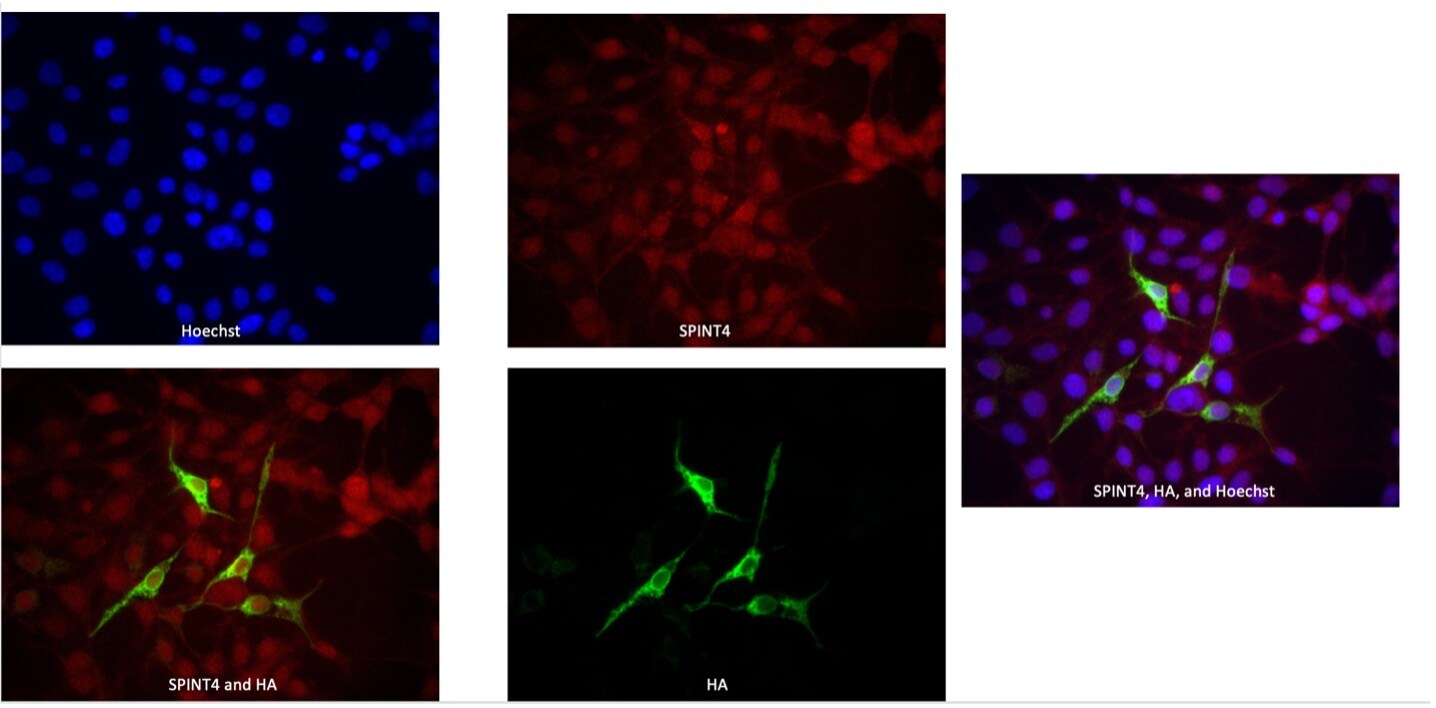
Application: ImmunocytochemistrySample Tested: plasmid transfected HEK293T cellsSpecies: HumanVerified Customer | Posted 05/10/202340x Image of SPINT4-HA 293T cells showing the non-specific binding and high background noise of NovusBio’s SPINT4 Antibody.Overview/Application Description: The specificity and signal/noise ratio of the anti-SPINT4 antibody was tested in immunofluorescence staining analyses of Human HEK293T cells expressing the SPINT4-HA protein. Signal seems to be non-specific (based on the degree of overlap of the SPINT4 and HA signal) with high background (based on the presence of signal in untransfected cells). Procedural Details: HEK293T cells were transfected with an expression plasmid encoding the SPINT4 protein fused to a C-terminal HA epitope (SPINT4-HA). Cells were fixed in 1.5% paraformaldehyde at room temperature, permeabilized with 0.5% Triton-X on ice, blocked using 10% fetal bovine serum in PBS and incubated with rabbit anti-SPINT4 antibodies (1:200 dilution) and with mouse anti-HA antibodies (1:200 dilution) for one hour at room temperature. Samples were washed with blocking buffer and incubated with Alexa Fluor 594-conjugated goat anti-rabbit antibodies (1:200 dilution) and with Alexa Fluor 488-conjugated goat anti-mouse antibodies (1:200 dilution) for one hour at room temperature, prior to washing in PBS and highlighting of nuclear DNA using Hoechst33342 for three minutes. Untransfected cells processed in the same way were used as controls. Coverslips were then mounted, and signal was visualized using a fluorescence microscope.

There are no reviews that match your criteria.
Protocols
Find general support by application which include: protocols, troubleshooting, illustrated assays, videos and webinars.
- Antigen Retrieval Protocol (PIER)
- Antigen Retrieval for Frozen Sections Protocol
- Appropriate Fixation of IHC/ICC Samples
- Cellular Response to Hypoxia Protocols
- Chromogenic IHC Staining of Formalin-Fixed Paraffin-Embedded (FFPE) Tissue Protocol
- Chromogenic Immunohistochemistry Staining of Frozen Tissue
- ClariTSA™ Fluorophore Kits
- Detection & Visualization of Antibody Binding
- Fluorescent IHC Staining of Frozen Tissue Protocol
- Graphic Protocol for Heat-induced Epitope Retrieval
- Graphic Protocol for the Preparation and Fluorescent IHC Staining of Frozen Tissue Sections
- Graphic Protocol for the Preparation and Fluorescent IHC Staining of Paraffin-embedded Tissue Sections
- Graphic Protocol for the Preparation of Gelatin-coated Slides for Histological Tissue Sections
- IHC Sample Preparation (Frozen sections vs Paraffin)
- Immunofluorescent IHC Staining of Formalin-Fixed Paraffin-Embedded (FFPE) Tissue Protocol
- Immunohistochemistry (IHC) and Immunocytochemistry (ICC) Protocols
- Immunohistochemistry Frozen Troubleshooting
- Immunohistochemistry Paraffin Troubleshooting
- Preparing Samples for IHC/ICC Experiments
- Preventing Non-Specific Staining (Non-Specific Binding)
- Primary Antibody Selection & Optimization
- Protocol for Heat-Induced Epitope Retrieval (HIER)
- Protocol for Making a 4% Formaldehyde Solution in PBS
- Protocol for VisUCyte™ HRP Polymer Detection Reagent
- Protocol for the Preparation & Fixation of Cells on Coverslips
- Protocol for the Preparation and Chromogenic IHC Staining of Frozen Tissue Sections
- Protocol for the Preparation and Chromogenic IHC Staining of Frozen Tissue Sections - Graphic
- Protocol for the Preparation and Chromogenic IHC Staining of Paraffin-embedded Tissue Sections
- Protocol for the Preparation and Chromogenic IHC Staining of Paraffin-embedded Tissue Sections - Graphic
- Protocol for the Preparation and Fluorescent IHC Staining of Frozen Tissue Sections
- Protocol for the Preparation and Fluorescent IHC Staining of Paraffin-embedded Tissue Sections
- Protocol for the Preparation of Gelatin-coated Slides for Histological Tissue Sections
- TUNEL and Active Caspase-3 Detection by IHC/ICC Protocol
- The Importance of IHC/ICC Controls
- Troubleshooting Guide: Immunohistochemistry
- View all Protocols, Troubleshooting, Illustrated assays and Webinars
Loading...