VEGF-C Antibody (107/F10) - Azide and BSA Free
Novus Biologicals | Catalog # NBP1-18626
![Immunohistochemistry: VEGF-C Antibody (107/F10) - Azide and BSA Free [NBP1-18626]](https://resources.rndsystems.com/images/products/VEGF-C-Antibody-107-F10-Immunohistochemistry-NBP1-18626-img0004.jpg "Immunohistochemistry: VEGF-C Antibody (107/F10) - Azide and BSA Free [NBP1-18626]")
Key Product Details
Species Reactivity
Validated:
Cited:
Applications
Validated:
Cited:
Label
Antibody Source
Format
Product Specifications
Immunogen
Specificity
Clonality
Host
Isotype
Scientific Data Images for VEGF-C Antibody (107/F10) - Azide and BSA Free
Immunohistochemistry: VEGF-C Antibody (107/F10) - Azide and BSA Free [NBP1-18626]
VEGF-C-Antibody-107-F10-Immunohistochemistry-NBP1-18626-img0004.jpg![Western Blot: VEGF-C Antibody (107/F10)Azide and BSA Free [NBP1-18626]](https://resources.rndsystems.com/images/products/VEGF-C-Antibody-107-F10-Western-Blot-NBP1-18626-img0001.jpg "Western Blot: VEGF-C Antibody (107/F10)Azide and BSA Free [NBP1-18626]")
Western Blot: VEGF-C Antibody (107/F10)Azide and BSA Free [NBP1-18626]
Western Blot: VEGF-C Antibody (107/F10) [NBP1-18626] - Analysis of recombinant human VEGF-C derived from insect cells using a monoclonal antibody directed against recombinant human/rat VEGF-C.![Immunocytochemistry/ Immunofluorescence: VEGF-C Antibody (107/F10) - Azide and BSA Free [NBP1-18626]](https://resources.rndsystems.com/images/products/VEGF-C-Antibody-107-F10-Immunocytochemistry-Immunofluorescence-NBP1-18626-img0003.jpg "Immunocytochemistry/ Immunofluorescence: VEGF-C Antibody (107/F10) - Azide and BSA Free [NBP1-18626]")
Immunocytochemistry/ Immunofluorescence: VEGF-C Antibody (107/F10) - Azide and BSA Free [NBP1-18626]
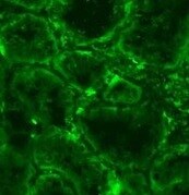
Immunocytochemistry/Immunofluorescence: VEGF-C Antibody (107/F10) [NBP1-18626] - Human myofibroblasts stained with VEGF-C antibody. ICC/IF image submitted by a verified customer review.![Immunohistochemistry-Paraffin: VEGF-C Antibody (107/F10) - Azide and BSA Free [NBP1-18626]](https://resources.rndsystems.com/images/products/VEGF-C-Antibody-107-F10-Immunohistochemistry-Paraffin-NBP1-18626-img0002.jpg "Immunohistochemistry-Paraffin: VEGF-C Antibody (107/F10) - Azide and BSA Free [NBP1-18626]")
Immunohistochemistry-Paraffin: VEGF-C Antibody (107/F10) - Azide and BSA Free [NBP1-18626]
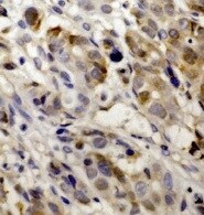
Immunohistochemistry-Paraffin: VEGF-C Antibody (107/F10) [NBP1-18626] - Human gastric cancer tissue section stained with VEGF-C antibody. IHC-P image submitted by a verified customer review. - Azide and BSA Free [NBP1-18626] -")
Immunohistochemistry: VEGF-C Antibody (107/F10) - Azide and BSA Free [NBP1-18626] -
Immunohistochemistry: VEGF-C Antibody (107/F10) - Azide and BSA Free [NBP1-18626] - Internal thoracic artery wall harvested from a patient diagnosed with double-vessel CAD showing immunohistochemical staining. Immunohistochemical staining of VEGF-C (a), CAV2 (b) & CAV3 (c) in an internal thoracic artery obtained from a 67-year-old patient who developed the early graft restenosis within 7 months after coronary artery bypass grafting. VEGF-C is expressed in smooth muscle cells of the tunica media (TM) & endothelial cells present in the tunica intima (TI). CAV2 is present exclusively in smooth muscle cells. Strong expression of CAV3 is observed in smooth muscle cells & in endothelial cells. d Negative control. TA tunica adventitia Image collected & cropped by CiteAb from the following publication (http://link.springer.com/10.1007/s00380-018-1158-9), licensed under a CC-BY license. Not internally tested by Novus Biologicals. - Azide and BSA Free [NBP1-18626] -")
Immunohistochemistry: VEGF-C Antibody (107/F10) - Azide and BSA Free [NBP1-18626] -
Immunohistochemistry: VEGF-C Antibody (107/F10) - Azide and BSA Free [NBP1-18626] - Immunohistochemical staining of a saphenous vein wall harvested from a patient diagnosed with triple-vessel CAD. Three serial histological sections of a graft wall obtained from a 71-year-old patient who developed early graft failure 11 months after coronary artery bypass grafting (the same patients as mentioned in Fig. 7). VEGF-C (a), VEGFR-3 (b), & CAV3 (c) shown to be expressed by immunohistochemical staining in smooth muscle cells of the tunica media (TM). In addition, VEGFR-3 is strongly present in endothelial cells of the tunica intima (TI). d Negative control. TA tunica adventitia Image collected & cropped by CiteAb from the following publication (http://link.springer.com/10.1007/s00380-018-1158-9), licensed under a CC-BY license. Not internally tested by Novus Biologicals. - Azide and BSA Free [NBP1-18626] -")
Immunohistochemistry: VEGF-C Antibody (107/F10) - Azide and BSA Free [NBP1-18626] -
Immunohistochemistry: VEGF-C Antibody (107/F10) - Azide and BSA Free [NBP1-18626] - Immunohistochemical staining of an internal thoracic artery & saphenous vein wall harvested from a patient with no evidence of luminal stenosis 12 months after CABG. Immunohistochemical staining of VEGF-C (a) & CAV2 (b) in an internal thoracic artery (ITA) graft & VEGFR-3 (c) in saphenous vein (SV) transplant harvested from a 65-year-old patient diagnosed with triple-vessel CAD. Note that lack of CAV2 expression within smooth muscle cells (SMC) in ITA wall is accompanied by VEGF-C positive expression in the same area. In SV tissue sample harvested from the same patient, no VEGRR-3 expression was found. d Negative control. TI tunica intima, TM tunica media, TA tunica adventitia Image collected & cropped by CiteAb from the following publication (http://link.springer.com/10.1007/s00380-018-1158-9), licensed under a CC-BY license. Not internally tested by Novus Biologicals. - Azide and BSA Free [NBP1-18626] -")
Immunohistochemistry: VEGF-C Antibody (107/F10) - Azide and BSA Free [NBP1-18626] -
Immunohistochemistry: VEGF-C Antibody (107/F10) - Azide and BSA Free [NBP1-18626] - Saphenous vein wall harvested from a patient diagnosed with double-vessel CAD showing immunohistochemical staining. VEGF-C (a) & VEGFR-3 (b) reactivity in a graft wall obtained from a 67-year-old patient who developed the early graft restenosis within 7 months after coronary artery bypass grafting (the same patient as mentioned in Fig. 6). VEGF-C is localized exclusively within individual smooth muscle cells of the tunica media (TM). Cells with positive VEGFR-3 expression are situated in endothelial cells of the tunica intima (TI). c Negative control. TA tunica adventitia Image collected & cropped by CiteAb from the following publication (http://link.springer.com/10.1007/s00380-018-1158-9), licensed under a CC-BY license. Not internally tested by Novus Biologicals. - Azide and BSA Free [NBP1-18626] -")
Immunohistochemistry: VEGF-C Antibody (107/F10) - Azide and BSA Free [NBP1-18626] -
Immunohistochemistry: VEGF-C Antibody (107/F10) - Azide and BSA Free [NBP1-18626] - Immunohistochemistry of an internal thoracic artery wall harvested from a patient diagnosed with triple-vessel CAD. Immunohistochemical staining of VEGF-C (a), CAV2 (b), & CAV3 (c) in an internal thoracic artery graft harvested from a 71-year-old patient who developed early graft failure 11 months after coronary artery bypass grafting. Intensive VEGF-C & CAV3 expression is observed in the smooth muscle cells of the tunica media (TM) & in the endothelial cells of the tunica intima (TI). The presence of immunopositive expression of CAV2 (c) is limited to smooth muscle cells of the tunica media (TM). Expression of all analyzed proteins is present in the small blood vessels localized in the tunica adventitia (TA, arrows). d Negative control Image collected & cropped by CiteAb from the following publication (http://link.springer.com/10.1007/s00380-018-1158-9), licensed under a CC-BY license. Not internally tested by Novus Biologicals.Applications for VEGF-C Antibody (107/F10) - Azide and BSA Free
ELISA
Immunocytochemistry/ Immunofluorescence
Immunohistochemistry
Western Blot
Reviewed Applications
Read 2 reviews rated 5 using NBP1-18626 in the following applications:
Formulation, Preparation, and Storage
Purification
Reconstitution
Formulation
Format
Preservative
Concentration
Shipping
Stability & Storage
Calculators
Background: VEGF-C
Long Name
Alternate Names
Entrez Gene IDs
Gene Symbol
Additional VEGF-C Products
Product Documents for VEGF-C Antibody (107/F10) - Azide and BSA Free
Certificate of Analysis
To download a Certificate of Analysis, please enter a lot or batch number in the search box below.
Product Specific Notices for VEGF-C Antibody (107/F10) - Azide and BSA Free
This product is for research use only and is not approved for use in humans or in clinical diagnosis. Primary Antibodies are guaranteed for 1 year from date of receipt.
Citations for VEGF-C Antibody (107/F10) - Azide and BSA Free
 Powered by Bioz
Customer Reviews for VEGF-C Antibody (107/F10) - Azide and BSA Free (2)
Have you used VEGF-C Antibody (107/F10) - Azide and BSA Free?
Submit a review and receive an Amazon gift card!
$25/€18/£15/$25CAN/¥2500 Yen for a review with an image
$10/€7/£6/$10CAN/¥1110 Yen for a review without an image
Submit a review
Customer Images
-
Application: ImmunocytochemistrySample Tested: MyofibroblastsSpecies: HumanVerified Customer | Posted 08/13/2021Myofibroblast immunofluorescent staining

-
Application: Immunohistochemistry-ParaffinSample Tested: gastric cancerSpecies: HumanVerified Customer | Posted 08/11/2021VEGF-C Antibody in human gastric cancer tissue.
There are no reviews that match your criteria.
Protocols
Find general support by application which include: protocols, troubleshooting, illustrated assays, videos and webinars.
- Antigen Retrieval Protocol (PIER)
- Antigen Retrieval for Frozen Sections Protocol
- Appropriate Fixation of IHC/ICC Samples
- Cellular Response to Hypoxia Protocols
- Chromogenic IHC Staining of Formalin-Fixed Paraffin-Embedded (FFPE) Tissue Protocol
- Chromogenic Immunohistochemistry Staining of Frozen Tissue
- ClariTSA™ Fluorophore Kits
- Detection & Visualization of Antibody Binding
- ELISA Sample Preparation & Collection Guide
- ELISA Troubleshooting Guide
- Fluorescent IHC Staining of Frozen Tissue Protocol
- Graphic Protocol for Heat-induced Epitope Retrieval
- Graphic Protocol for the Preparation and Fluorescent IHC Staining of Frozen Tissue Sections
- Graphic Protocol for the Preparation and Fluorescent IHC Staining of Paraffin-embedded Tissue Sections
- Graphic Protocol for the Preparation of Gelatin-coated Slides for Histological Tissue Sections
- How to Run an R&D Systems DuoSet ELISA
- How to Run an R&D Systems Quantikine ELISA
- How to Run an R&D Systems Quantikine™ QuicKit™ ELISA
- ICC Cell Smear Protocol for Suspension Cells
- ICC Immunocytochemistry Protocol Videos
- ICC for Adherent Cells
- IHC Sample Preparation (Frozen sections vs Paraffin)
- Immunocytochemistry (ICC) Protocol
- Immunocytochemistry Troubleshooting
- Immunofluorescence of Organoids Embedded in Cultrex Basement Membrane Extract
- Immunofluorescent IHC Staining of Formalin-Fixed Paraffin-Embedded (FFPE) Tissue Protocol
- Immunohistochemistry (IHC) and Immunocytochemistry (ICC) Protocols
- Immunohistochemistry Frozen Troubleshooting
- Immunohistochemistry Paraffin Troubleshooting
- Preparing Samples for IHC/ICC Experiments
- Preventing Non-Specific Staining (Non-Specific Binding)
- Primary Antibody Selection & Optimization
- Protocol for Heat-Induced Epitope Retrieval (HIER)
- Protocol for Making a 4% Formaldehyde Solution in PBS
- Protocol for VisUCyte™ HRP Polymer Detection Reagent
- Protocol for the Fluorescent ICC Staining of Cell Smears - Graphic
- Protocol for the Fluorescent ICC Staining of Cultured Cells on Coverslips - Graphic
- Protocol for the Preparation & Fixation of Cells on Coverslips
- Protocol for the Preparation and Chromogenic IHC Staining of Frozen Tissue Sections
- Protocol for the Preparation and Chromogenic IHC Staining of Frozen Tissue Sections - Graphic
- Protocol for the Preparation and Chromogenic IHC Staining of Paraffin-embedded Tissue Sections
- Protocol for the Preparation and Chromogenic IHC Staining of Paraffin-embedded Tissue Sections - Graphic
- Protocol for the Preparation and Fluorescent ICC Staining of Cells on Coverslips
- Protocol for the Preparation and Fluorescent ICC Staining of Non-adherent Cells
- Protocol for the Preparation and Fluorescent ICC Staining of Stem Cells on Coverslips
- Protocol for the Preparation and Fluorescent IHC Staining of Frozen Tissue Sections
- Protocol for the Preparation and Fluorescent IHC Staining of Paraffin-embedded Tissue Sections
- Protocol for the Preparation of Gelatin-coated Slides for Histological Tissue Sections
- Protocol for the Preparation of a Cell Smear for Non-adherent Cell ICC - Graphic
- Quantikine HS ELISA Kit Assay Principle, Alkaline Phosphatase
- Quantikine HS ELISA Kit Principle, Streptavidin-HRP Polymer
- R&D Systems Quality Control Western Blot Protocol
- Sandwich ELISA (Colorimetric) – Biotin/Streptavidin Detection Protocol
- Sandwich ELISA (Colorimetric) – Direct Detection Protocol
- TUNEL and Active Caspase-3 Detection by IHC/ICC Protocol
- The Importance of IHC/ICC Controls
- Troubleshooting Guide: ELISA
- Troubleshooting Guide: Immunohistochemistry
- Troubleshooting Guide: Western Blot Figures
- Western Blot Conditions
- Western Blot Protocol
- Western Blot Protocol for Cell Lysates
- Western Blot Troubleshooting
- Western Blot Troubleshooting Guide
- View all Protocols, Troubleshooting, Illustrated assays and Webinars
FAQs for VEGF-C Antibody (107/F10) - Azide and BSA Free
-
Q: I was wondering if you know of any blocking antibodies against IL-6 or VEGF-C that can be used in cell culture to quench these cytokines?
A:
We have no antibodies that are currently known to show blocking antibodies to VEGF-C. Please see this link to our IL-6 antibodies with blocking/neutralizing activity can be found at this link.