


Scientific images may be resized, cropped, or adjusted for brightness or contrast for presentation purposes. These updates are limited to presentation and do not affect the underlying scientific interpretation of the data. Where available, additional source data may be provided upon request.
Key Product Details
Species Reactivity
Validated:
Cited:
Predicted:
Applications
Validated:
Cited:
Label
Antibody Source
Product Specifications
Immunogen
Reactivity Notes
Specificity
Clonality
Host
Isotype
Theoretical MW
Disclaimer note: The observed molecular weight of the protein may vary from the listed predicted molecular weight due to post translational modifications, post translation cleavages, relative charges, and other experimental factors.
Scientific Data Images for VPS35 Antibody
Western Blot: VPS35 Antibody [NB100-1397]
Western Blot: VPS35 Antibody [NB100-1397] - Staining of HepG2 (A) and HEK293 (B) cell lysate (35 ug protein in RIPA buffer). Antibody at 0.03 ug/mL. Detected by chemiluminescence.
Immunocytochemistry/ Immunofluorescence: VPS35 Antibody [NB100-1397]
Immunocytochemistry/Immunofluorescence: VPS35 Antibody [NB100-1397] - Immunofluorescence analysis of paraformaldehyde fixed U2OS cells, permeabilized with 0.15% Triton. Primary incubation 1hr (10ug/ml) followed by Alexa Fluor 488 secondary antibody (2ug/ml), showing cytoplasmic/vesicle staining. The nuclear stain is DAPI (blue). Negative control: Unimmunized goat IgG (10ug/ml) followed by Alexa Fluor 488 secondary antibody (2ug/ml).
Immunohistochemistry-Paraffin: VPS35 Antibody [NB100-1397]
Immunohistochemistry-Paraffin: VPS35 Antibody [NB100-1397] - Negative Control showing staining of paraffin embedded Human Prostate, with no primary antibody.
Western Blot: VPS35 Antibody [NB100-1397]
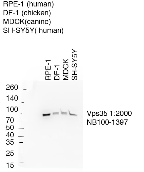
Western Blot: VPS35 Antibody [NB100-1397] - 10 ug cell lysate per lane of various cell lines as indicated, immunoblotted with vps35 antibody at 1:2000 at 4C for 20 hrs. Secondary antibody donkey anti-goat HRP at 1:5000 for 1 hr at room temp. WB image submitted by a verified customer review.
Western Blot: VPS35 Antibody [NB100-1397]
Western Blot: VPS35 Antibody [NB100-1397] - HEK293T cell lysate. WB image submitted by a verified customer review.
Western Blot: VPS35 Antibody [NB100-1397]
Western Blot: VPS35 Antibody [NB100-1397] - Staining of Human (A) Mouse (B) and Rat (C) Brain lysate (35 ug protein in RIPA buffer). antibody at 0.03 ug/mL. Detected by chemiluminescence.
Immunocytochemistry/ Immunofluorescence: VPS35 Antibody [NB100-1397]
Immunocytochemistry/Immunofluorescence: VPS35 Antibody [NB100-1397] - Confocal microscopy image of human RPE-1 cells fixed with PFA, permeablilized with 0.3% Triton X-100,then immunostained with vps35 antibody at 1:2000 overnight in 1% BSA at 4 degrees. ICC image submitted by a verified customer review.
Immunocytochemistry/ Immunofluorescence: VPS35 Antibody [NB100-1397]
Immunocytochemistry/Immunofluorescence: VPS35 Antibody [NB100-1397] - Immunofluorescence analysis of paraformaldehyde fixed HEK293 cells, permeabilized with 0.15% Triton. Primary incubation 1hr (10ug/ml) followed by Alexa Fluor 488 secondary antibody (2ug/ml), showing cytoplasmic/vesicle staining. The nuclear stain is DAPI (blue). Negative control: Unimmunized goat IgG (10ug/ml) followed by Alexa Fluor 488 secondary antibody (2ug/ml).
Immunohistochemistry-Paraffin: VPS35 Antibody [NB100-1397]
Immunohistochemistry-Paraffin: VPS35 Antibody [NB100-1397] - Staining of paraffin embedded Human Prostate. Antibody at 8 ug/mL. Heat induced antigen retrieval with citrate buffer pH 6, HRP-staining.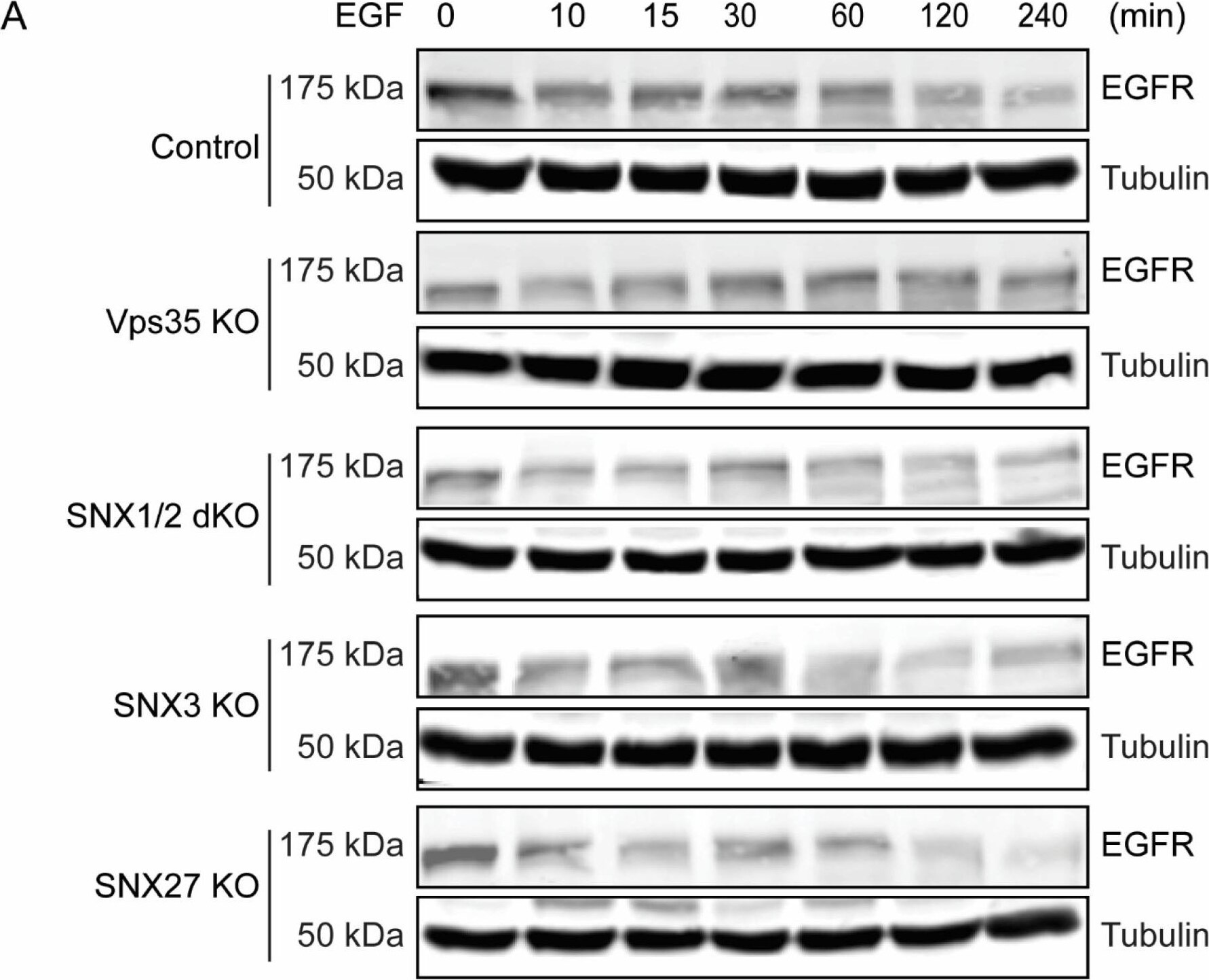
Western Blot: VPS35 Antibody [NB100-1397] -
Retromer Vps35 or SNX1/2 depletion altered EGF-stimulated EGFR degradation. (A) Serum-starved cells were treated with 100 ug/mL of EGF for different time points. At indicated time points, cells were harvested, and equal amounts of protein samples were used for immunoblotting with EGFR antibody. Representative blots from at least three independent experiments are shown. (B) EGFR levels at each time point were normalized to the EGFR level at 0 min for each cell line. Graph represents the difference of EGFR levels during EGF stimulation (means +/- SEM). Two-tailed student’s t-test indicated the difference, * p < 0.05, HeLa control vs. Vps35 KO (120 min); * p < 0.05, HeLa control vs. Vps35 KO (240 min); ** p < 0.01, HeLa control vs. SNX1/2 dKO (240 min). Image collected and cropped by CiteAb from the following open publication (https://pubmed.ncbi.nlm.nih.gov/36359754), licensed under a CC-BY license. Not internally tested by Novus Biologicals.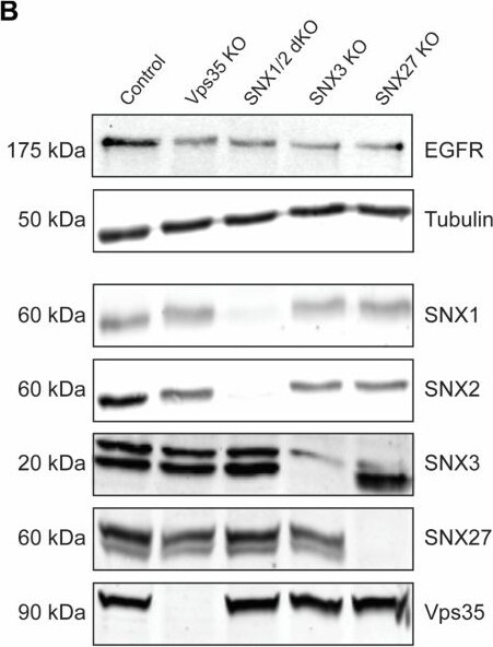
Western Blot: VPS35 Antibody [NB100-1397] -
Depletion of retromer or retromer-associated SNXs decreases EGFR protein levels. (A) Steady-state HeLa control, Vps35 KO, SNX1/2 dKO, SNX3 KO, and SNX27 KO cell monolayers grown in complete-DMEM media on coverslips were fixed in ice-cold methanol, stained with EGFR antibody, and count-stained with DAPI. Images were captured on a Leica SP8 DMi8 confocal microscope using a 60× glycerol objective (Scale Bar, 10 um). (B) Equal amounts of protein samples from HeLa control, Vps35 KO, SNX1/2 dKO, SNX3 KO, and SNX27 KO cells were subjected to SDS-PAGE and immunoblotting with the antibodies against EGFR, SNX1, SNX2, SNX3, SNX27, and Vps35, whereas tubulin served as a loading control. Representative blots from at least three independent experiments are shown. (C) The fold differences for EGFR protein levels are presented (means +/- SEM). Two-tailed student’s t-test indicated the difference between HeLa control and HeLa KO cells, ** p < 0.01, HeLa control vs. Vps35 KO; *** p < 0.001, HeLa control vs. SNX1/2 dKO; *** p < 0.001, HeLa control vs. SNX3 KO; *** p < 0.001, HeLa control vs. SNX27 KO. Image collected and cropped by CiteAb from the following open publication (https://pubmed.ncbi.nlm.nih.gov/36359754), licensed under a CC-BY license. Not internally tested by Novus Biologicals.Applications for VPS35 Antibody
Immunocytochemistry/ Immunofluorescence
Immunohistochemistry
Immunohistochemistry-Paraffin
Peptide ELISA
Western Blot
Reviewed Applications
Read 5 reviews rated 4.8 using NB100-1397 in the following applications:
Formulation, Preparation, and Storage
Purification
Formulation
Preservative
Concentration
Shipping
Stability & Storage
Background: VPS35
Long Name
Alternate Names
Gene Symbol
UniProt
Additional VPS35 Products
Product Documents for VPS35 Antibody
Certificate of Analysis
To download a Certificate of Analysis, please enter a lot or batch number in the search box below.
Product Specific Notices for VPS35 Antibody
This product is for research use only and is not approved for use in humans or in clinical diagnosis. Primary Antibodies are guaranteed for 1 year from date of receipt.
Citations for VPS35 Antibody
 Powered by Bioz
Customer Reviews for VPS35 Antibody (5)
Have you used VPS35 Antibody?
Submit a review and receive an Amazon gift card!
$25/€18/£15/$25CAN/¥2500 for a review with an image
$10/€7/£6/$10CAN/¥1110 for a review without an image
Submit a review
Customer Images
-
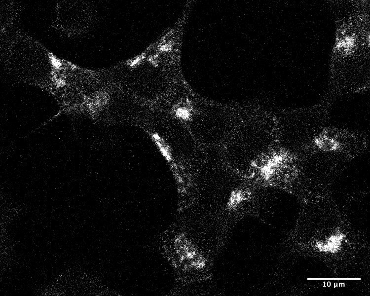
Application: ImmunofluorescenceSample Tested: INS-1Species: RatVerified Customer | Posted 10/29/202020%FCS to block, 0.5% Triton, secondary was Anti-Goat-647

-
Application: ImmunocytochemistrySample Tested: hela cellSpecies: HumanVerified Customer | Posted 05/28/2020
-
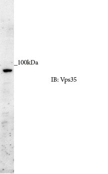
Application: Western BlotSample Tested: HEK293T cell lysateSpecies: HumanVerified Customer | Posted 05/28/2020
-
Application: Western BlotSample Tested: RPE-1, DF-1, MDCK canine kidney epithelial cell line and SH-SY5Y whole cell lysatesSpecies: Human, Chicken and CanineVerified Customer | Posted 01/07/202010ug of various cell lines indicated immunbloted with vps35 antibody 1:2000samples prepared in 1.5x lammeli buffer, 10ug of protein loaded per lane transferred onto nitrocellulose at 100V for 1.5 hours 4 degrees. blotting coditions membrane blocked in 5% milk in PBS 0.1% tween 20 (PBST) for 1 hour primary ab incubated 1:2000 in 5% milk in PBS at 4 degrees for 20 hours washed 3X with PBST secondary donkey anti goat HPR 1:5000 for 1 hour room temp washed 2x PBST, 1X PBS blot developed using ECL prime reagent (GE healthcare )
-
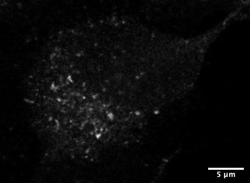
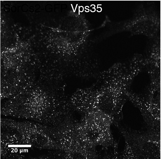
Application: ImmunocytochemistrySample Tested: RPE-1 cells and RPE-1Species: HumanVerified Customer | Posted 01/07/2020confocal RPE-1 cells fixed with PFA perm 0.3% tx100 then immunostained with vps35 ab 1:2000 overnight in 1% BSA at 4 degrees.
There are no reviews that match your criteria.
Protocols
Find general support by application which include: protocols, troubleshooting, illustrated assays, videos and webinars.
- Antigen Retrieval Protocol (PIER)
- Antigen Retrieval for Frozen Sections Protocol
- Appropriate Fixation of IHC/ICC Samples
- Cellular Response to Hypoxia Protocols
- Chromogenic IHC Staining of Formalin-Fixed Paraffin-Embedded (FFPE) Tissue Protocol
- Chromogenic Immunohistochemistry Staining of Frozen Tissue
- ClariTSA™ Fluorophore Kits
- Detection & Visualization of Antibody Binding
- ELISA Sample Preparation & Collection Guide
- ELISA Troubleshooting Guide
- Fluorescent IHC Staining of Frozen Tissue Protocol
- Graphic Protocol for Heat-induced Epitope Retrieval
- Graphic Protocol for the Preparation and Fluorescent IHC Staining of Frozen Tissue Sections
- Graphic Protocol for the Preparation and Fluorescent IHC Staining of Paraffin-embedded Tissue Sections
- Graphic Protocol for the Preparation of Gelatin-coated Slides for Histological Tissue Sections
- How to Run an R&D Systems DuoSet ELISA
- How to Run an R&D Systems Quantikine ELISA
- How to Run an R&D Systems Quantikine™ QuicKit™ ELISA
- ICC Cell Smear Protocol for Suspension Cells
- ICC Immunocytochemistry Protocol Videos
- ICC for Adherent Cells
- IHC Sample Preparation (Frozen sections vs Paraffin)
- Immunocytochemistry (ICC) Protocol
- Immunocytochemistry Troubleshooting
- Immunofluorescence of Organoids Embedded in Cultrex Basement Membrane Extract
- Immunofluorescent IHC Staining of Formalin-Fixed Paraffin-Embedded (FFPE) Tissue Protocol
- Immunohistochemistry (IHC) and Immunocytochemistry (ICC) Protocols
- Immunohistochemistry Frozen Troubleshooting
- Immunohistochemistry Paraffin Troubleshooting
- Preparing Samples for IHC/ICC Experiments
- Preventing Non-Specific Staining (Non-Specific Binding)
- Primary Antibody Selection & Optimization
- Protocol for Heat-Induced Epitope Retrieval (HIER)
- Protocol for Making a 4% Formaldehyde Solution in PBS
- Protocol for VisUCyte™ HRP Polymer Detection Reagent
- Protocol for the Fluorescent ICC Staining of Cell Smears - Graphic
- Protocol for the Fluorescent ICC Staining of Cultured Cells on Coverslips - Graphic
- Protocol for the Preparation & Fixation of Cells on Coverslips
- Protocol for the Preparation and Chromogenic IHC Staining of Frozen Tissue Sections
- Protocol for the Preparation and Chromogenic IHC Staining of Frozen Tissue Sections - Graphic
- Protocol for the Preparation and Chromogenic IHC Staining of Paraffin-embedded Tissue Sections
- Protocol for the Preparation and Chromogenic IHC Staining of Paraffin-embedded Tissue Sections - Graphic
- Protocol for the Preparation and Fluorescent ICC Staining of Cells on Coverslips
- Protocol for the Preparation and Fluorescent ICC Staining of Non-adherent Cells
- Protocol for the Preparation and Fluorescent ICC Staining of Stem Cells on Coverslips
- Protocol for the Preparation and Fluorescent IHC Staining of Frozen Tissue Sections
- Protocol for the Preparation and Fluorescent IHC Staining of Paraffin-embedded Tissue Sections
- Protocol for the Preparation of Gelatin-coated Slides for Histological Tissue Sections
- Protocol for the Preparation of a Cell Smear for Non-adherent Cell ICC - Graphic
- Quantikine HS ELISA Kit Assay Principle, Alkaline Phosphatase
- Quantikine HS ELISA Kit Principle, Streptavidin-HRP Polymer
- R&D Systems Quality Control Western Blot Protocol
- Sandwich ELISA (Colorimetric) – Biotin/Streptavidin Detection Protocol
- Sandwich ELISA (Colorimetric) – Direct Detection Protocol
- TUNEL and Active Caspase-3 Detection by IHC/ICC Protocol
- The Importance of IHC/ICC Controls
- Troubleshooting Guide: ELISA
- Troubleshooting Guide: Immunohistochemistry
- Troubleshooting Guide: Western Blot Figures
- Western Blot Conditions
- Western Blot Protocol
- Western Blot Protocol for Cell Lysates
- Western Blot Troubleshooting
- Western Blot Troubleshooting Guide
- View all Protocols, Troubleshooting, Illustrated assays and Webinars