
ZMAT1 Antibody - BSA Free
Novus Biologicals | Catalog # NBP1-81375

![Western Blot: ZMAT1 Antibody [NBP1-81375]](https://resources.rndsystems.com/images/products/ZMAT1-Antibody-Western-Blot-NBP1-81375-img0005.jpg "Western Blot: ZMAT1 Antibody [NBP1-81375]")
Loading...
Key Product Details
Validated by
Orthogonal Validation
Species Reactivity
Human
Applications
Immunohistochemistry, Immunohistochemistry-Paraffin, Western Blot
Label
Unconjugated
Antibody Source
Polyclonal Rabbit IgG
Format
BSA Free
Loading...
Product Specifications
Immunogen
This antibody was developed against Recombinant Protein corresponding to amino acids: NSRKTQDSYQNECADYINVQKARGLEAKTCFRKMEESSLETRRYREVVDSRPRHRMFEQRLPFETFRTYAAPYNISQAMEKQLPHSKKTYDSFQDELEDYIKVQKARGLDPKTCFRKMRENSVDTHGYREMVDSGP
Clonality
Polyclonal
Host
Rabbit
Isotype
IgG
Theoretical MW
75 kDa.
Disclaimer note: The observed molecular weight of the protein may vary from the listed predicted molecular weight due to post translational modifications, post translation cleavages, relative charges, and other experimental factors.
Disclaimer note: The observed molecular weight of the protein may vary from the listed predicted molecular weight due to post translational modifications, post translation cleavages, relative charges, and other experimental factors.
Scientific Data Images for ZMAT1 Antibody - BSA Free
Western Blot: ZMAT1 Antibody [NBP1-81375]
Western Blot: ZMAT1 Antibody [NBP1-81375] - Lane 1: Marker [kDa] 230, 130, 95, 72, 56, 36, 28, 17, 11. Lane 2: Human cell line RT-4. Lane 3: Human cell line U-251MG sp![Immunohistochemistry-Paraffin: ZMAT1 Antibody [NBP1-81375]](https://resources.rndsystems.com/images/products/ZMAT1-Antibody-Immunohistochemistry-Paraffin-NBP1-81375-img0008.jpg "Immunohistochemistry-Paraffin: ZMAT1 Antibody [NBP1-81375]")
![Immunohistochemistry-Paraffin: ZMAT1 Antibody [NBP1-81375]](https://resources.rndsystems.com/images/products/ZMAT1-Antibody-Immunohistochemistry-Paraffin-NBP1-81375-img0006.jpg "Immunohistochemistry-Paraffin: ZMAT1 Antibody [NBP1-81375]")
Immunohistochemistry-Paraffin: ZMAT1 Antibody [NBP1-81375]
Immunohistochemistry-Paraffin: ZMAT1 Antibody [NBP1-81375] - Staining of human epididymis shows high expression.![Immunohistochemistry-Paraffin: ZMAT1 Antibody [NBP1-81375]](https://resources.rndsystems.com/images/products/ZMAT1-Antibody-Immunohistochemistry-Paraffin-NBP1-81375-img0007.jpg "Immunohistochemistry-Paraffin: ZMAT1 Antibody [NBP1-81375]")
Immunohistochemistry-Paraffin: ZMAT1 Antibody [NBP1-81375]
Immunohistochemistry-Paraffin: ZMAT1 Antibody [NBP1-81375] - Staining of human pancreas shows low expression as expected.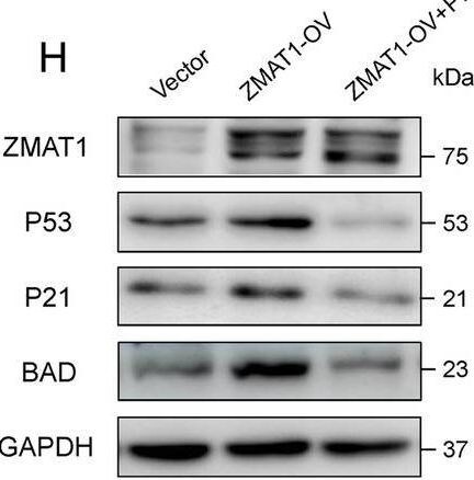
Western Blot: ZMAT1 Antibody - BSA Free [NBP1-81375] -
ZMAT1 functions in a p53-dependent manner. A Gene set enrichment analysis (GSEA) of RNA sequencing on SW1990/Vector and SW1990/ZMAT1-OV cells groups. B Heatmap of RNA sequencing showed differential expression levels of key nodes in p53-assicoated cell cycle and apoptosis pathways. C-D RT-qPCR and western blot were used to detect the p53 levels in ZMAT1 over-expressed (C) and knockdown cells (D). E Double-label immunofluorescence staining for the intracellular localization of ZMAT1 and p53 in SW1990 cells. F luciferase activity assays were performed on 293 T cells with co-transfection of pGL3-p53 with 0.01 μg, 0.05 μg, 0.1 μg, 0.5 μg and 1 μg of vector encoding ZMAT1. G SW1990/ZMAT1-OV cells were treated with Pifithrin-alpha for 24 h and the protein levels of ZMAT1, p53, p21, and BAD were analyzed by immunoblotting with the indicated antibodies. H CCK-8 assays were performed on SW1990/ZMAT1-OV cells after treating with Pifithrin-alpha for 24 h. I Colony formation assays were performed on SW1990/ZMAT1-OV cells after treating with Pifithrin-alpha for 24 h. All * P-value < 0.05, ** P-value < 0.01, *** P-value < 0.001. P-values were assessed using two-tailed t-tests and ANOVA followed by Dunnett’s tests for multiple comparison in B, C, D, E, G, J and K. All figures represent mean +/- SD from three independent experiments Image collected and cropped by CiteAb from the following open publication (https://pubmed.ncbi.nlm.nih.gov/35392973), licensed under a CC-BY license. Not internally tested by Novus Biologicals.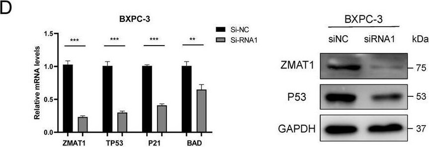
Western Blot: ZMAT1 Antibody - BSA Free [NBP1-81375] -
ZMAT1 functions in a p53-dependent manner. A Gene set enrichment analysis (GSEA) of RNA sequencing on SW1990/Vector and SW1990/ZMAT1-OV cells groups. B Heatmap of RNA sequencing showed differential expression levels of key nodes in p53-assicoated cell cycle and apoptosis pathways. C-D RT-qPCR and western blot were used to detect the p53 levels in ZMAT1 over-expressed (C) and knockdown cells (D). E Double-label immunofluorescence staining for the intracellular localization of ZMAT1 and p53 in SW1990 cells. F luciferase activity assays were performed on 293 T cells with co-transfection of pGL3-p53 with 0.01 μg, 0.05 μg, 0.1 μg, 0.5 μg and 1 μg of vector encoding ZMAT1. G SW1990/ZMAT1-OV cells were treated with Pifithrin-alpha for 24 h and the protein levels of ZMAT1, p53, p21, and BAD were analyzed by immunoblotting with the indicated antibodies. H CCK-8 assays were performed on SW1990/ZMAT1-OV cells after treating with Pifithrin-alpha for 24 h. I Colony formation assays were performed on SW1990/ZMAT1-OV cells after treating with Pifithrin-alpha for 24 h. All * P-value < 0.05, ** P-value < 0.01, *** P-value < 0.001. P-values were assessed using two-tailed t-tests and ANOVA followed by Dunnett’s tests for multiple comparison in B, C, D, E, G, J and K. All figures represent mean +/- SD from three independent experiments Image collected and cropped by CiteAb from the following open publication (https://pubmed.ncbi.nlm.nih.gov/35392973), licensed under a CC-BY license. Not internally tested by Novus Biologicals.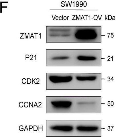
Western Blot: ZMAT1 Antibody - BSA Free [NBP1-81375] -
Over-expression of ZMAT1 arrests the cell cycle in Pancreatic Ductal Adenocarcinoma (PDAC) cells. A Gene Ontology (GO) enrichment analysis of ZMAT1 based on TCGA cohort. B Kyoto Encyclopedia of Genes and Genomes (KEGG) pathway analysis of ZMAT1 based on TCGA cohort. C-E Flow cytometry showed over-expression of ZMAT1 triggered G2/S blockade in SW1990 cells (C), while depletion of ZMAT1 increased the percentage of S phase in BXPC-3 (D) and Capan-2 cells (E). F–H Western blot showed over-expression of ZMAT1 up-regulated p21 and decreased CDK2 and CCNA2 in SW1990 cells (F), whereas ZMAT1 knockdown declined p21 and increased CDK2 and CCNA2 in BXPC-3 (G) and Capan-2 cells (H). All * P-value < 0.05, ** P-value < 0.01, *** P-value < 0.001. P-values were assessed using two-tailed t-tests in C-E. All figures represent mean +/- SD from three independent experiments Image collected and cropped by CiteAb from the following open publication (https://pubmed.ncbi.nlm.nih.gov/35392973), licensed under a CC-BY license. Not internally tested by Novus Biologicals.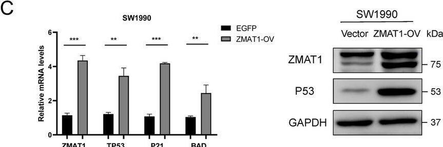
Western Blot: ZMAT1 Antibody - BSA Free [NBP1-81375] -
ZMAT1 functions in a p53-dependent manner. A Gene set enrichment analysis (GSEA) of RNA sequencing on SW1990/Vector and SW1990/ZMAT1-OV cells groups. B Heatmap of RNA sequencing showed differential expression levels of key nodes in p53-assicoated cell cycle and apoptosis pathways. C-D RT-qPCR and western blot were used to detect the p53 levels in ZMAT1 over-expressed (C) and knockdown cells (D). E Double-label immunofluorescence staining for the intracellular localization of ZMAT1 and p53 in SW1990 cells. F luciferase activity assays were performed on 293 T cells with co-transfection of pGL3-p53 with 0.01 μg, 0.05 μg, 0.1 μg, 0.5 μg and 1 μg of vector encoding ZMAT1. G SW1990/ZMAT1-OV cells were treated with Pifithrin-alpha for 24 h and the protein levels of ZMAT1, p53, p21, and BAD were analyzed by immunoblotting with the indicated antibodies. H CCK-8 assays were performed on SW1990/ZMAT1-OV cells after treating with Pifithrin-alpha for 24 h. I Colony formation assays were performed on SW1990/ZMAT1-OV cells after treating with Pifithrin-alpha for 24 h. All * P-value < 0.05, ** P-value < 0.01, *** P-value < 0.001. P-values were assessed using two-tailed t-tests and ANOVA followed by Dunnett’s tests for multiple comparison in B, C, D, E, G, J and K. All figures represent mean +/- SD from three independent experiments Image collected and cropped by CiteAb from the following open publication (https://pubmed.ncbi.nlm.nih.gov/35392973), licensed under a CC-BY license. Not internally tested by Novus Biologicals.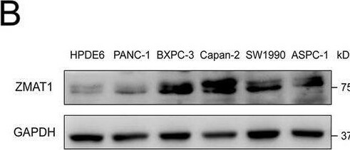
Western Blot: ZMAT1 Antibody - BSA Free [NBP1-81375] -
ZMAT1 inhibits proliferation and migration of Pancreatic Ductal Adenocarcinoma (PDAC) cells. A-B Real-time quantitative polymerase chain reaction (RT-qPCR) (A) and western blot (B) showed ZMAT1 was highly expressed in BXPC-3 and Capan-2 cells, while relatively low-expressed in SW1990 cells among PDAC cell lines. C RT-qPCR and western blot were used to detect the over-expression of ZMAT1 in SW1990 cells. D CCK-8 assays showed that ZMAT1 over-expression reduced the proliferation in SW1990 cells. E RT-qPCR and western blot were used to detect the down-regulation of ZMAT1 in BXPC-3 cells transfected with ZMAT1-siRNA. F CCK-8 assays showed that ZMAT1 down-regulation promoted the proliferation in BXPC-3 cells. G RT-qPCR and western blot were used to detect the down-regulation of ZMAT1 in Capan-2 cells transfected with ZMAT1-siRNA. H CCK-8 assays showed that ZMAT1 down-regulation promoted the proliferation in Capan-2 cells. I-K Colony formation assays showed that ZMAT1 over-expression reduced the proliferation in SW1990 cells (I), while ZMAT1 knockdown promoted the proliferation in BXPC-3 (J) and Capan-2 cells (K). L-N Transwell assays showed that ZMAT1 over-expression reduced the migration in SW1990 cells (L), while ZMAT1 knockdown promoted the migration in BXPC-3 (M) and Capan-2 cells (N). All * P-value < 0.05, ** P-value < 0.01, *** P-value < 0.001. Scale bars: 100 μm. P-values were assessed using two-tailed t-tests and ANOVA followed by Dunnett’s tests for multiple comparison in C-N. All figures represent mean +/- SD from three independent experiments Image collected and cropped by CiteAb from the following open publication (https://pubmed.ncbi.nlm.nih.gov/35392973), licensed under a CC-BY license. Not internally tested by Novus Biologicals.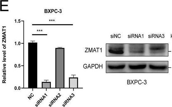
Western Blot: ZMAT1 Antibody - BSA Free [NBP1-81375] -
ZMAT1 inhibits proliferation and migration of Pancreatic Ductal Adenocarcinoma (PDAC) cells. A-B Real-time quantitative polymerase chain reaction (RT-qPCR) (A) and western blot (B) showed ZMAT1 was highly expressed in BXPC-3 and Capan-2 cells, while relatively low-expressed in SW1990 cells among PDAC cell lines. C RT-qPCR and western blot were used to detect the over-expression of ZMAT1 in SW1990 cells. D CCK-8 assays showed that ZMAT1 over-expression reduced the proliferation in SW1990 cells. E RT-qPCR and western blot were used to detect the down-regulation of ZMAT1 in BXPC-3 cells transfected with ZMAT1-siRNA. F CCK-8 assays showed that ZMAT1 down-regulation promoted the proliferation in BXPC-3 cells. G RT-qPCR and western blot were used to detect the down-regulation of ZMAT1 in Capan-2 cells transfected with ZMAT1-siRNA. H CCK-8 assays showed that ZMAT1 down-regulation promoted the proliferation in Capan-2 cells. I-K Colony formation assays showed that ZMAT1 over-expression reduced the proliferation in SW1990 cells (I), while ZMAT1 knockdown promoted the proliferation in BXPC-3 (J) and Capan-2 cells (K). L-N Transwell assays showed that ZMAT1 over-expression reduced the migration in SW1990 cells (L), while ZMAT1 knockdown promoted the migration in BXPC-3 (M) and Capan-2 cells (N). All * P-value < 0.05, ** P-value < 0.01, *** P-value < 0.001. Scale bars: 100 μm. P-values were assessed using two-tailed t-tests and ANOVA followed by Dunnett’s tests for multiple comparison in C-N. All figures represent mean +/- SD from three independent experiments Image collected and cropped by CiteAb from the following open publication (https://pubmed.ncbi.nlm.nih.gov/35392973), licensed under a CC-BY license. Not internally tested by Novus Biologicals.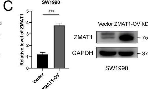
Western Blot: ZMAT1 Antibody - BSA Free [NBP1-81375] -
ZMAT1 inhibits proliferation and migration of Pancreatic Ductal Adenocarcinoma (PDAC) cells. A-B Real-time quantitative polymerase chain reaction (RT-qPCR) (A) and western blot (B) showed ZMAT1 was highly expressed in BXPC-3 and Capan-2 cells, while relatively low-expressed in SW1990 cells among PDAC cell lines. C RT-qPCR and western blot were used to detect the over-expression of ZMAT1 in SW1990 cells. D CCK-8 assays showed that ZMAT1 over-expression reduced the proliferation in SW1990 cells. E RT-qPCR and western blot were used to detect the down-regulation of ZMAT1 in BXPC-3 cells transfected with ZMAT1-siRNA. F CCK-8 assays showed that ZMAT1 down-regulation promoted the proliferation in BXPC-3 cells. G RT-qPCR and western blot were used to detect the down-regulation of ZMAT1 in Capan-2 cells transfected with ZMAT1-siRNA. H CCK-8 assays showed that ZMAT1 down-regulation promoted the proliferation in Capan-2 cells. I-K Colony formation assays showed that ZMAT1 over-expression reduced the proliferation in SW1990 cells (I), while ZMAT1 knockdown promoted the proliferation in BXPC-3 (J) and Capan-2 cells (K). L-N Transwell assays showed that ZMAT1 over-expression reduced the migration in SW1990 cells (L), while ZMAT1 knockdown promoted the migration in BXPC-3 (M) and Capan-2 cells (N). All * P-value < 0.05, ** P-value < 0.01, *** P-value < 0.001. Scale bars: 100 μm. P-values were assessed using two-tailed t-tests and ANOVA followed by Dunnett’s tests for multiple comparison in C-N. All figures represent mean +/- SD from three independent experiments Image collected and cropped by CiteAb from the following open publication (https://pubmed.ncbi.nlm.nih.gov/35392973), licensed under a CC-BY license. Not internally tested by Novus Biologicals.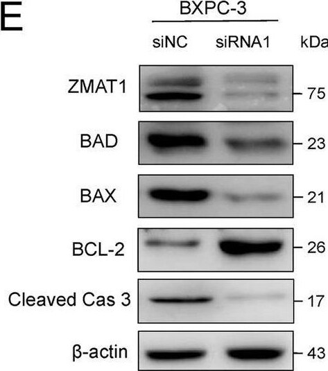
Western Blot: ZMAT1 Antibody - BSA Free [NBP1-81375] -
Over-expression of ZMAT1 promotes apoptosis in Pancreatic Ductal Adenocarcinoma (PDAC) cells. A Flow cytometry showed that over-expression of ZMAT1 increased the percentage of apoptotic SW1990 cells. B-C Flow cytometry showed that ZMAT1 deletion reduced the percentage of apoptotic SW1990 cells. D-F Western blot showed over-expression of ZMAT1 up-regulated BAD, BAX, Cleaved Caspase 3 and declined Bcl-2 in SW1990 cells (D), whereas ZMAT1 knockdown reduced BAD, BAX, Cleaved Caspase 3 and increased Bcl-2 in BXPC-3 (F) and Capan-2 cells (G). All * P-value < 0.05, ** P-value < 0.01, *** P-value < 0.001. P-values were assessed using two-tailed t-tests in C-E. All figures represent mean +/- SD from three independent experiments Image collected and cropped by CiteAb from the following open publication (https://pubmed.ncbi.nlm.nih.gov/35392973), licensed under a CC-BY license. Not internally tested by Novus Biologicals.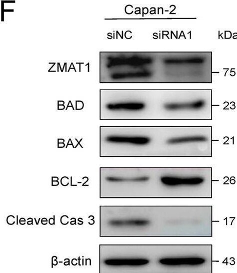
Western Blot: ZMAT1 Antibody - BSA Free [NBP1-81375] -
Over-expression of ZMAT1 promotes apoptosis in Pancreatic Ductal Adenocarcinoma (PDAC) cells. A Flow cytometry showed that over-expression of ZMAT1 increased the percentage of apoptotic SW1990 cells. B-C Flow cytometry showed that ZMAT1 deletion reduced the percentage of apoptotic SW1990 cells. D-F Western blot showed over-expression of ZMAT1 up-regulated BAD, BAX, Cleaved Caspase 3 and declined Bcl-2 in SW1990 cells (D), whereas ZMAT1 knockdown reduced BAD, BAX, Cleaved Caspase 3 and increased Bcl-2 in BXPC-3 (F) and Capan-2 cells (G). All * P-value < 0.05, ** P-value < 0.01, *** P-value < 0.001. P-values were assessed using two-tailed t-tests in C-E. All figures represent mean +/- SD from three independent experiments Image collected and cropped by CiteAb from the following open publication (https://pubmed.ncbi.nlm.nih.gov/35392973), licensed under a CC-BY license. Not internally tested by Novus Biologicals.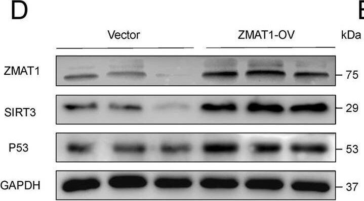
Western Blot: ZMAT1 Antibody - BSA Free [NBP1-81375] -
ZMAT1 correlates with p53 in Pancreatic Ductal Adenocarcinoma (PDAC). A BALB/c-nudes (n = 6 per group) were sacrificed 60 days after the injection and tumors dissected from respective groups were shown. B Tumor growth curves after the injection of SW1990/Vector cells and SW1990/ZMAT1-OV cells. Tumor volume was calculated every 10 days. C Tumor weight was measured in ZMAT1-OV and control groups. D The protein levels of ZMAT1, SIRT3 and p53 of tumors were analyzed by immunoblotting with the indicated antibodies. E IHC staining of ZMAT1, SIRT3, p53, Ki67 and Tunel in tumors from ZMAT1-OV and control groups. F Representative images of double-label immunofluorescence (IF) staining of ZMAT1 and p53 in 60 PDAC tissues. G IF staining showed ZMAT1 expression level highly correlated with p53 expression. H Kaplan–Meier analysis in PDAC patients grouped according to the expression levels of ZMAT1 and p53 showed that PDAC patients with high ZMAT1/high p53 expression had the longest overall survival among all the groups. I A schematic diagram for the role of the ZMAT1-SIRT3-p53 axis in regulation of cell cycle and apoptosis in PDAC. All * P-value < 0.05, ** P-value < 0.01, *** P-value < 0.001. Scale bars: 200 μm. P-values were assessed using two-tailed t-tests and ANOVA followed by Dunnett’s tests for multiple comparison in B-C. Spearman’s correlation was performed in G. Kaplan–Meier analyses and log-rank tests were conducted in H Image collected and cropped by CiteAb from the following open publication (https://pubmed.ncbi.nlm.nih.gov/35392973), licensed under a CC-BY license. Not internally tested by Novus Biologicals.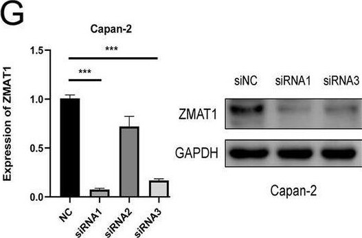
Western Blot: ZMAT1 Antibody - BSA Free [NBP1-81375] -
ZMAT1 inhibits proliferation and migration of Pancreatic Ductal Adenocarcinoma (PDAC) cells. A-B Real-time quantitative polymerase chain reaction (RT-qPCR) (A) and western blot (B) showed ZMAT1 was highly expressed in BXPC-3 and Capan-2 cells, while relatively low-expressed in SW1990 cells among PDAC cell lines. C RT-qPCR and western blot were used to detect the over-expression of ZMAT1 in SW1990 cells. D CCK-8 assays showed that ZMAT1 over-expression reduced the proliferation in SW1990 cells. E RT-qPCR and western blot were used to detect the down-regulation of ZMAT1 in BXPC-3 cells transfected with ZMAT1-siRNA. F CCK-8 assays showed that ZMAT1 down-regulation promoted the proliferation in BXPC-3 cells. G RT-qPCR and western blot were used to detect the down-regulation of ZMAT1 in Capan-2 cells transfected with ZMAT1-siRNA. H CCK-8 assays showed that ZMAT1 down-regulation promoted the proliferation in Capan-2 cells. I-K Colony formation assays showed that ZMAT1 over-expression reduced the proliferation in SW1990 cells (I), while ZMAT1 knockdown promoted the proliferation in BXPC-3 (J) and Capan-2 cells (K). L-N Transwell assays showed that ZMAT1 over-expression reduced the migration in SW1990 cells (L), while ZMAT1 knockdown promoted the migration in BXPC-3 (M) and Capan-2 cells (N). All * P-value < 0.05, ** P-value < 0.01, *** P-value < 0.001. Scale bars: 100 μm. P-values were assessed using two-tailed t-tests and ANOVA followed by Dunnett’s tests for multiple comparison in C-N. All figures represent mean +/- SD from three independent experiments Image collected and cropped by CiteAb from the following open publication (https://pubmed.ncbi.nlm.nih.gov/35392973), licensed under a CC-BY license. Not internally tested by Novus Biologicals.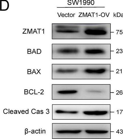
Western Blot: ZMAT1 Antibody - BSA Free [NBP1-81375] -
Over-expression of ZMAT1 promotes apoptosis in Pancreatic Ductal Adenocarcinoma (PDAC) cells. A Flow cytometry showed that over-expression of ZMAT1 increased the percentage of apoptotic SW1990 cells. B-C Flow cytometry showed that ZMAT1 deletion reduced the percentage of apoptotic SW1990 cells. D-F Western blot showed over-expression of ZMAT1 up-regulated BAD, BAX, Cleaved Caspase 3 and declined Bcl-2 in SW1990 cells (D), whereas ZMAT1 knockdown reduced BAD, BAX, Cleaved Caspase 3 and increased Bcl-2 in BXPC-3 (F) and Capan-2 cells (G). All * P-value < 0.05, ** P-value < 0.01, *** P-value < 0.001. P-values were assessed using two-tailed t-tests in C-E. All figures represent mean +/- SD from three independent experiments Image collected and cropped by CiteAb from the following open publication (https://pubmed.ncbi.nlm.nih.gov/35392973), licensed under a CC-BY license. Not internally tested by Novus Biologicals.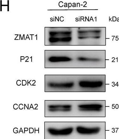
Western Blot: ZMAT1 Antibody - BSA Free [NBP1-81375] -
Over-expression of ZMAT1 arrests the cell cycle in Pancreatic Ductal Adenocarcinoma (PDAC) cells. A Gene Ontology (GO) enrichment analysis of ZMAT1 based on TCGA cohort. B Kyoto Encyclopedia of Genes and Genomes (KEGG) pathway analysis of ZMAT1 based on TCGA cohort. C-E Flow cytometry showed over-expression of ZMAT1 triggered G2/S blockade in SW1990 cells (C), while depletion of ZMAT1 increased the percentage of S phase in BXPC-3 (D) and Capan-2 cells (E). F–H Western blot showed over-expression of ZMAT1 up-regulated p21 and decreased CDK2 and CCNA2 in SW1990 cells (F), whereas ZMAT1 knockdown declined p21 and increased CDK2 and CCNA2 in BXPC-3 (G) and Capan-2 cells (H). All * P-value < 0.05, ** P-value < 0.01, *** P-value < 0.001. P-values were assessed using two-tailed t-tests in C-E. All figures represent mean +/- SD from three independent experiments Image collected and cropped by CiteAb from the following open publication (https://pubmed.ncbi.nlm.nih.gov/35392973), licensed under a CC-BY license. Not internally tested by Novus Biologicals.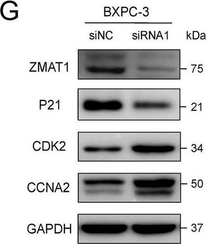
Western Blot: ZMAT1 Antibody - BSA Free [NBP1-81375] -
Over-expression of ZMAT1 arrests the cell cycle in Pancreatic Ductal Adenocarcinoma (PDAC) cells. A Gene Ontology (GO) enrichment analysis of ZMAT1 based on TCGA cohort. B Kyoto Encyclopedia of Genes and Genomes (KEGG) pathway analysis of ZMAT1 based on TCGA cohort. C-E Flow cytometry showed over-expression of ZMAT1 triggered G2/S blockade in SW1990 cells (C), while depletion of ZMAT1 increased the percentage of S phase in BXPC-3 (D) and Capan-2 cells (E). F–H Western blot showed over-expression of ZMAT1 up-regulated p21 and decreased CDK2 and CCNA2 in SW1990 cells (F), whereas ZMAT1 knockdown declined p21 and increased CDK2 and CCNA2 in BXPC-3 (G) and Capan-2 cells (H). All * P-value < 0.05, ** P-value < 0.01, *** P-value < 0.001. P-values were assessed using two-tailed t-tests in C-E. All figures represent mean +/- SD from three independent experiments Image collected and cropped by CiteAb from the following open publication (https://pubmed.ncbi.nlm.nih.gov/35392973), licensed under a CC-BY license. Not internally tested by Novus Biologicals.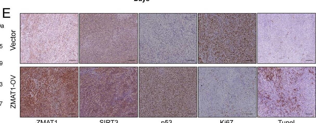
Immunohistochemistry: ZMAT1 Antibody - BSA Free [NBP1-81375] -
ZMAT1 correlates with p53 in Pancreatic Ductal Adenocarcinoma (PDAC). A BALB/c-nudes (n = 6 per group) were sacrificed 60 days after the injection and tumors dissected from respective groups were shown. B Tumor growth curves after the injection of SW1990/Vector cells and SW1990/ZMAT1-OV cells. Tumor volume was calculated every 10 days. C Tumor weight was measured in ZMAT1-OV and control groups. D The protein levels of ZMAT1, SIRT3 and p53 of tumors were analyzed by immunoblotting with the indicated antibodies. E IHC staining of ZMAT1, SIRT3, p53, Ki67 and Tunel in tumors from ZMAT1-OV and control groups. F Representative images of double-label immunofluorescence (IF) staining of ZMAT1 and p53 in 60 PDAC tissues. G IF staining showed ZMAT1 expression level highly correlated with p53 expression. H Kaplan–Meier analysis in PDAC patients grouped according to the expression levels of ZMAT1 and p53 showed that PDAC patients with high ZMAT1/high p53 expression had the longest overall survival among all the groups. I A schematic diagram for the role of the ZMAT1-SIRT3-p53 axis in regulation of cell cycle and apoptosis in PDAC. All * P-value < 0.05, ** P-value < 0.01, *** P-value < 0.001. Scale bars: 200 μm. P-values were assessed using two-tailed t-tests and ANOVA followed by Dunnett’s tests for multiple comparison in B-C. Spearman’s correlation was performed in G. Kaplan–Meier analyses and log-rank tests were conducted in H Image collected and cropped by CiteAb from the following open publication (https://pubmed.ncbi.nlm.nih.gov/35392973), licensed under a CC-BY license. Not internally tested by Novus Biologicals.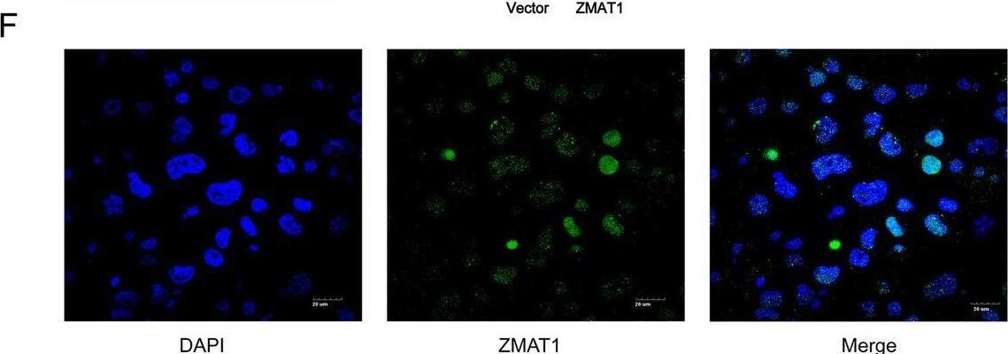
Immunocytochemistry/ Immunofluorescence: ZMAT1 Antibody - BSA Free [NBP1-81375] -
ZMAT1 functions in a p53-dependent manner. A Gene set enrichment analysis (GSEA) of RNA sequencing on SW1990/Vector and SW1990/ZMAT1-OV cells groups. B Heatmap of RNA sequencing showed differential expression levels of key nodes in p53-assicoated cell cycle and apoptosis pathways. C-D RT-qPCR and western blot were used to detect the p53 levels in ZMAT1 over-expressed (C) and knockdown cells (D). E Double-label immunofluorescence staining for the intracellular localization of ZMAT1 and p53 in SW1990 cells. F luciferase activity assays were performed on 293 T cells with co-transfection of pGL3-p53 with 0.01 μg, 0.05 μg, 0.1 μg, 0.5 μg and 1 μg of vector encoding ZMAT1. G SW1990/ZMAT1-OV cells were treated with Pifithrin-alpha for 24 h and the protein levels of ZMAT1, p53, p21, and BAD were analyzed by immunoblotting with the indicated antibodies. H CCK-8 assays were performed on SW1990/ZMAT1-OV cells after treating with Pifithrin-alpha for 24 h. I Colony formation assays were performed on SW1990/ZMAT1-OV cells after treating with Pifithrin-alpha for 24 h. All * P-value < 0.05, ** P-value < 0.01, *** P-value < 0.001. P-values were assessed using two-tailed t-tests and ANOVA followed by Dunnett’s tests for multiple comparison in B, C, D, E, G, J and K. All figures represent mean +/- SD from three independent experiments Image collected and cropped by CiteAb from the following open publication (https://pubmed.ncbi.nlm.nih.gov/35392973), licensed under a CC-BY license. Not internally tested by Novus Biologicals.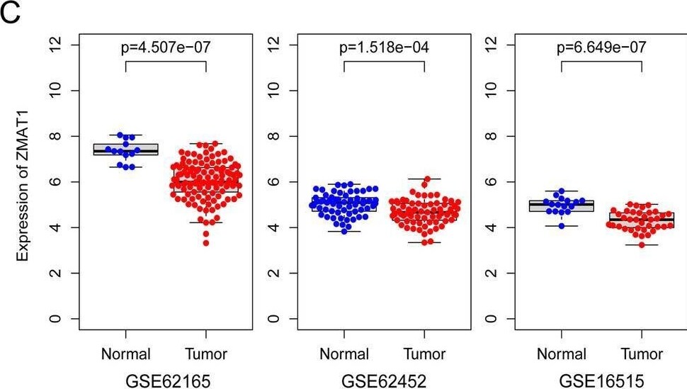
Flow Cytometry: ZMAT1 Antibody - BSA Free [NBP1-81375] -
ZMAT1 is down-regulated and correlates with unfavorable clinical characteristics and adverse outcome in Pancreatic Ductal Adenocarcinoma (PDAC). A Down-regulation of ZMAT1 in PDAC was identified in GEPIA database. B Down-regulation of ZMAT1 was identified in PDAC in Oncomine database (Pei’s dataset and Ishikawa’s dataset). C Down-regulation of ZMAT1 was identified in PDAC in three individual GEO datasets (GSE62165, GSE62452 and GSE16515). D The mRNA low-expression levels of ZMAT1 were identified in PDAC tissues and normal pancreas tissues of 25 samples. E Representative images of ZMAT1 staining in PDAC specimens and normal pancreas tissues. F Immunohistochemistry staining showed the protein levels of ZMAT1 were down-regulated in PDAC tissues. G ZMAT1 expression of PDAC was significantly correlated with differentiation, TNM stage, CA19-9 index and lymph nodes metastasis. H Multivariate Cox regression analyses showed low expression of ZMAT1 was independent risk factor for overall survival (OS) and disease-free survival (DFS) of 122 PDAC patients from validation cohort. I-J Kaplan–Meier analyses showed PDAC patients with high expression of ZMAT1 had superior OS and DFS than those with low expression in both TCGA cohort (I) and validation cohort (J). T, tumor; N, normal; OS, overall survival; DFS, disease-free survival. CA19-9, carbohydrate antigen 19–9. All * P-value < 0.05, ** P-value < 0.01, *** P-value < 0.001. Scale bars: 200 μm. P-values were determined by Non-parametric Mann–Whitney U-test in A-C. P-values were assessed by two-tailed t-tests in D and F. P-values were determined by χ2 tests or Fisher’s exact tests in G. The Hazard Ratios (HR) and P-values by the log-rank (Mantel-Cox) test are calculated in H-J Image collected and cropped by CiteAb from the following open publication (https://pubmed.ncbi.nlm.nih.gov/35392973), licensed under a CC-BY license. Not internally tested by Novus Biologicals.Applications for ZMAT1 Antibody - BSA Free
Application
Recommended Usage
Immunohistochemistry
1:50 - 1:200
Immunohistochemistry-Paraffin
1:50 - 1:200
Western Blot
0.04-0.4 ug/ml
Application Notes
For IHC-Paraffin, HIER pH 6 retrieval is recommended.
Formulation, Preparation, and Storage
Purification
Affinity purified
Formulation
PBS (pH 7.2) and 40% Glycerol
Format
BSA Free
Preservative
0.02% Sodium Azide
Concentration
Concentrations vary lot to lot. See vial label for concentration. If unlisted please contact technical services.
Shipping
The product is shipped with polar packs. Upon receipt, store it immediately at the temperature recommended below.
Stability & Storage
Store at 4C short term. Aliquot and store at -20C long term. Avoid freeze-thaw cycles.
Background: ZMAT1
Alternate Names
KIAA1789, zinc finger, matrin type 1, zinc finger, matrin-type 1
Entrez Gene IDs
84460 (Human)
Gene Symbol
ZMAT1
UniProt
Additional ZMAT1 Products
Product Documents for ZMAT1 Antibody - BSA Free
Certificate of Analysis
To download a Certificate of Analysis, please enter a lot or batch number in the search box below.
Product Specific Notices for ZMAT1 Antibody - BSA Free
This product is for research use only and is not approved for use in humans or in clinical diagnosis. Primary Antibodies are guaranteed for 1 year from date of receipt.
Citations for ZMAT1 Antibody - BSA Free
 Powered by Bioz
Customer Reviews for ZMAT1 Antibody - BSA Free
There are currently no reviews for this product. Be the first to review ZMAT1 Antibody - BSA Free and earn rewards!
Have you used ZMAT1 Antibody - BSA Free?
Submit a review and receive an Amazon gift card!
$25/€18/£15/$25CAN/¥2500 for a review with an image
$10/€7/£6/$10CAN/¥1110 for a review without an image
Submit a review
Protocols
Find general support by application which include: protocols, troubleshooting, illustrated assays, videos and webinars.
- Antigen Retrieval Protocol (PIER)
- Antigen Retrieval for Frozen Sections Protocol
- Appropriate Fixation of IHC/ICC Samples
- Cellular Response to Hypoxia Protocols
- Chromogenic IHC Staining of Formalin-Fixed Paraffin-Embedded (FFPE) Tissue Protocol
- Chromogenic Immunohistochemistry Staining of Frozen Tissue
- ClariTSA™ Fluorophore Kits
- Detection & Visualization of Antibody Binding
- Fluorescent IHC Staining of Frozen Tissue Protocol
- Graphic Protocol for Heat-induced Epitope Retrieval
- Graphic Protocol for the Preparation and Fluorescent IHC Staining of Frozen Tissue Sections
- Graphic Protocol for the Preparation and Fluorescent IHC Staining of Paraffin-embedded Tissue Sections
- Graphic Protocol for the Preparation of Gelatin-coated Slides for Histological Tissue Sections
- IHC Sample Preparation (Frozen sections vs Paraffin)
- Immunofluorescent IHC Staining of Formalin-Fixed Paraffin-Embedded (FFPE) Tissue Protocol
- Immunohistochemistry (IHC) and Immunocytochemistry (ICC) Protocols
- Immunohistochemistry Frozen Troubleshooting
- Immunohistochemistry Paraffin Troubleshooting
- Preparing Samples for IHC/ICC Experiments
- Preventing Non-Specific Staining (Non-Specific Binding)
- Primary Antibody Selection & Optimization
- Protocol for Heat-Induced Epitope Retrieval (HIER)
- Protocol for Making a 4% Formaldehyde Solution in PBS
- Protocol for VisUCyte™ HRP Polymer Detection Reagent
- Protocol for the Preparation & Fixation of Cells on Coverslips
- Protocol for the Preparation and Chromogenic IHC Staining of Frozen Tissue Sections
- Protocol for the Preparation and Chromogenic IHC Staining of Frozen Tissue Sections - Graphic
- Protocol for the Preparation and Chromogenic IHC Staining of Paraffin-embedded Tissue Sections
- Protocol for the Preparation and Chromogenic IHC Staining of Paraffin-embedded Tissue Sections - Graphic
- Protocol for the Preparation and Fluorescent IHC Staining of Frozen Tissue Sections
- Protocol for the Preparation and Fluorescent IHC Staining of Paraffin-embedded Tissue Sections
- Protocol for the Preparation of Gelatin-coated Slides for Histological Tissue Sections
- R&D Systems Quality Control Western Blot Protocol
- TUNEL and Active Caspase-3 Detection by IHC/ICC Protocol
- The Importance of IHC/ICC Controls
- Troubleshooting Guide: Immunohistochemistry
- Troubleshooting Guide: Western Blot Figures
- Western Blot Conditions
- Western Blot Protocol
- Western Blot Protocol for Cell Lysates
- Western Blot Troubleshooting
- Western Blot Troubleshooting Guide
- View all Protocols, Troubleshooting, Illustrated assays and Webinars
Loading...