
Extracellular Vesicle Analysis with Simple Western Technology
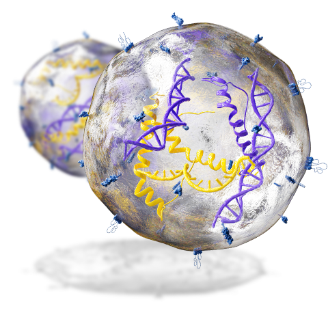
Extracellular vesicles (EVs) and exosomes have been shown to facilitate intercellular communication and are involved in numerous important cellular & disease processes, making them an exciting potential source for biomarker discovery and for liquid biopsy analysis. While EV protein and exosome characterization is important, analyzing protein expression in intact EVs can be a challenge due to the limited amount of material and low expression levels of many EVs proteins.
Simple Western automated western analysis systems are hands-free automated capillary electrophoresis (CE)-based platforms which offer high sensitivity and eliminate many of the tedious, error-prone steps of traditional western blotting, making them particularly well-suited for extracellular vesicle analysis. The systems automate protein separation and immunodetection and offer the following advantages for extracellular vesicle analysis:
- Low sample volume: Only 3 µL starting volume required, enabling you to utilize <5% of your EV sample volume for protein expression characterization and the rest for other isolation or characterization methods (e.g., functional testing, nucleic acid isolation, electron microscopy, nanoparticle tracking, etc.)
- Industry-leading sensitivity: chemiluminescence or fluorescence detection
- Multiplexed protein analysis and RePlex™ sequential immunoassays or total protein detection
- Fully quantitative data with built-in total protein normalization in the same capillary
- Hands-free automation of up to 96 samples in one 3-hour run
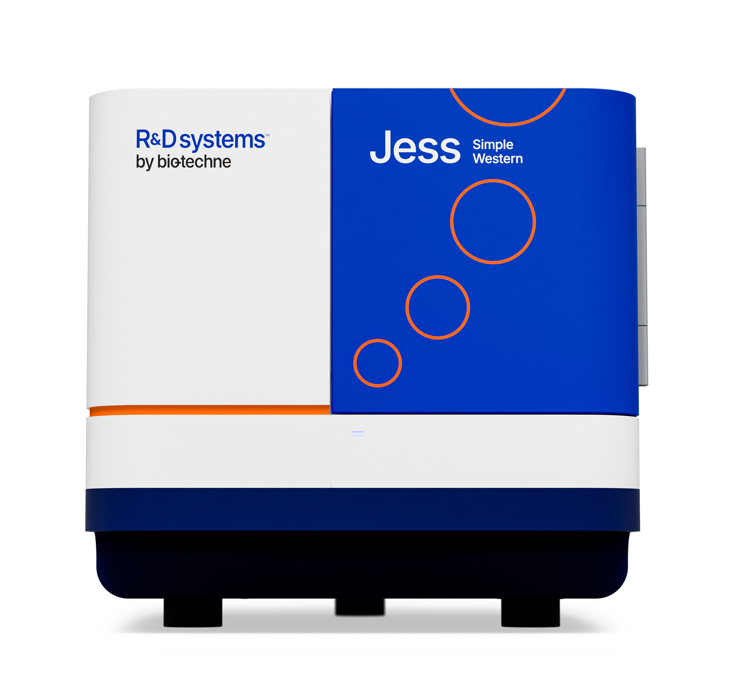
Dr. Sanooj Soni and his team at Imperial College London use Simple Western’s Jess™ to revolutionize how they study Extracellular Vesicles — tiny messengers that influence how target cells and organs respond to injury and infection in their critically ill patients.
With small and scarce EV samples from critically ill patients, Traditional Western Blotting lacked the sensitivity and reproducibility this team desperately needed to advance their research.
With fully automated, sensitive and fast analysis on Jess, Dr Soni reports that “We've been able to expand the scope of research significantly. This opens up whole new avenues and possibilities for biomarker discovery and therapeutic signaling. We’re now investing in more complex signaling pathways and protein markers that were previously beyond our reach. Being confident in the consistency of our data while reducing hands-on time is invaluable. Jess is a huge boost to our productivity."
With Jess on board, Dr Soni’s team is bridging the gap between discovery and clinical impact, by developing strategies for faster recovery, fewer long-term disabilities, and most importantly for saving lives.
Fast, Validated EV Protein Analysis Workflow
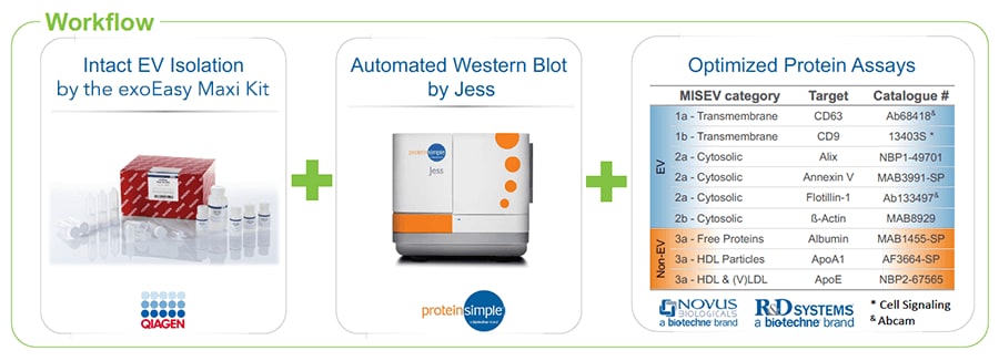
We have partnered with QIAGEN to develop and validate a streamlined workflow for exosome isolation and protein analysis, designed to tackle common challenges in extracellular vesicle (EV) research. This workflow combines QIAGEN’s exoEasy Maxi Kit for high-purity EV isolation with Simple Western technology for automated, high-sensitivity western analysis.
By utilizing this integrated approach, researchers can analyze protein expression in intact EV samples more efficiently. The cumbersome, time-consuming ultracentrifugation and precipitation-based methods for EV isolation are replaced with a simple column-based system, while the traditional western blotting process is automated for reproducibility and improved sensitivity.
This validated workflow offers several advantages for EV researchers:
- Simplified and standardized process: Facilitates protein analysis in EV samples with minimal manual intervention.
- High sensitivity and low sample requirements: Enables detection of low-abundance EV proteins using conventional antibodies, even with limited sample availability.
- Applications across EV research workflows: Supports exosome biomarker discovery, characterization studies, and liquid biopsy analysis.
By reducing complexity and improving sensitivity, this combined solution accelerates research aimed at understanding exosome function, identifying disease biomarkers, and advancing translational applications.
High Performance Workflow for Extracellular Vesicle Analysis
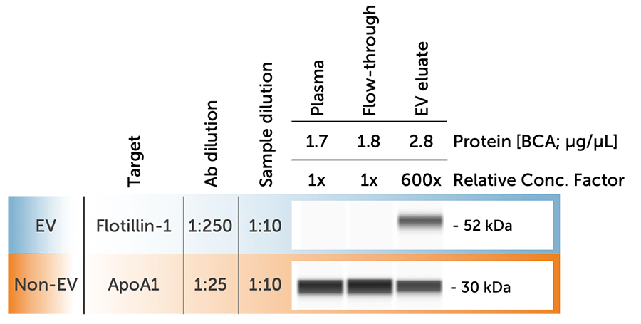
EV Protein Expression Across Plasma Fractions Using the exoEasy Maxi Kit and Simple Western
EV eluate of plasma (3 µL) compared to unprocessed plasma and the unbound fraction found in the column flow-through. EV marker Flotillin-1 (Ab133497; Abcam) is not detectable in neat plasma but is detectable in EV eluate, while HDL-marker ApoA1 (AF3664-SP; R&D Systems) is depleted in EV eluate compared to plasma and flow-through fraction.
Detection of MISEV recommended EV Protein-markers using the validated workflow
The limited amount of material and the diverse methods for isolation of extracellular vesicles (EV) pose unique challenges to proper characterization of experimental EV preparations. The “Minimal Information for Studies of Extracellular Vesicles” (MISEV) guidelines recommend characterizing preparations for both trans-membrane-, cytosolic- and contaminating non-EV proteins. However, compliance with these guidelines can be a considerable effort due to lack of easy and robust analytical protocols and the time consuming and user variable nature of standard western blotting protocols. Below we used a simple method for isolation of EVs and Simple Western Technology for automated protein separation and immunodetection of MISEV-recommended proteins. The total EVs were isolated by affinity-membrane spin columns from pre-filtered 0.5-4 mL plasma or 2-20 mL urine, respectively. Intact vesicles were eluted and the EV-depleted biofluid fraction was collected from the flow-through. A small fraction (4 μL) was analyzed by a simple western blot workflow providing automated capillary electrophoresis-based protein separation and immunodetection, characterizing each fraction for presence or absence of MISEV-recommended proteins. A range of specific antibodies were identified and the EV fractions were shown to be enriched in EV-proteins, whereas contaminating non-EV proteins were significantly reduced. Isolation of EVs was necessary to allow detection of the low abundant EV protein markers, whereas non-EV proteins were readily detectable both in the neat biofluids and in the EV-depleted flow-through. We characterized the effect of washing on the purity of EV isolates and defined the dynamic range of the workflow using titrations of input volume of both plasma and urine EV isolations. In conclusion, Simple western blotting protocols were established for quality control of isolated EVs in accordance with MISEV-guidelines. EVs isolated using affinity-membrane spin columns were shown to be enriched in EV markers and depleted for non-EV proteins.
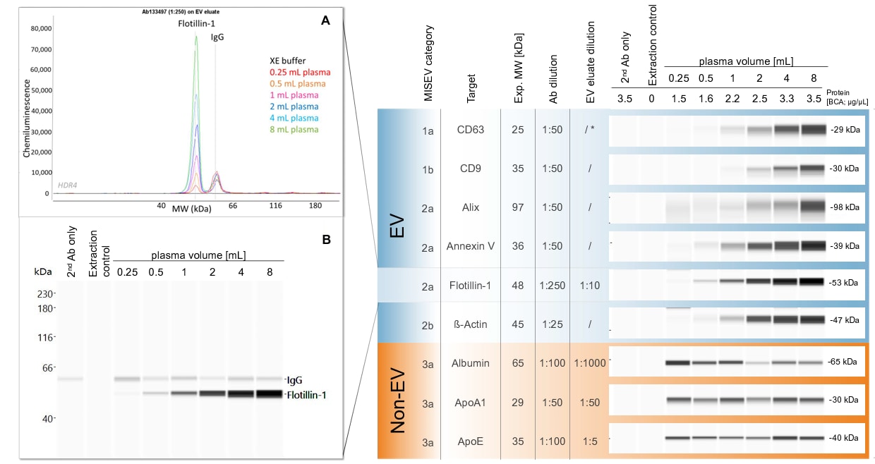
Immunodetected Flotillin-1 signal in 1:10 diluted human plasma derived EV eluate shown as [A] Electropherogram data and [B] Lane view data at expected MW of ca. 50 kDa. Flotillin-1 signal increases with input volume while background signal from plasma-contained antibodies (IgG) stays at the same level and can be excluded via secondary antibody-only-control.
Virtual Blot-Like Image (HDR4) overview of all immunodetected proteins in plasma-derived EV eluate ordered by MISEV classification and specific settings. For CD63 protein (marked with *) a deglycosylation reaction (PNGaseF, NEB) was essential to receive a band at the expected MW. Increasing human plasma input lead to a dose-response in protein assay signal for immunodetected EV proteins (MISEV category 1-2), but not for contaminants (MISEV category 3).
Exosomes: Contents by Cell Type and Process

Protocol: Examining Exosomes on Simple Western

Validated Antibodies for Extracellular Research

The exoEasy Maxi Kit for Exosome Isolation
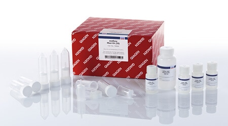
The exoEasy Maxi Kit from QIAGEN utilizes membrane affinity spin columns and specialized buffers to efficiently isolate and purify EVs (including exosomes) from plasma, serum, cell culture supernatant and other biological samples.
- Consistent isolation of intact exosomes and other EVs
- Suitable for downstream functional testing including physical and biochemical analysis of EVs
- Fast, 25-minute workflow
- Process multiple samples in parallel using a simple spin-column
- Accommodates large sample volumes including up to 4 mL of plasma or serum or 32 mL of cell culture supernatant
ProteiNext: Extracellular Vesicles Symposium
Discover ProteiNext Symposium, an annual event dedicated to advancing protein analysis. In 2024 the focus was on Extracellular Vesicles (EVs) with insights from 10+ leading scientists, addressing key topics like sample preparation, protein characterization, and data normalization. Watch the recordings below to explore innovative solutions and methods driving progress in extracellular vesicle research.