14-3-3 Antibody - BSA Free
Novus Biologicals | Catalog # NBP2-27202
![Western Blot: 14-3-3 Antibody [NBP2-27202]](https://resources.rndsystems.com/images/products/14-3-3-Antibody-Western-Blot-NBP2-27202-img0001.jpg "Western Blot: 14-3-3 Antibody [NBP2-27202]")
Loading...
Key Product Details
Species Reactivity
Validated:
Human, Mouse, Rat, Porcine, Canine, Chicken, Chinese Hamster, Feline, Opossum, Primate
Cited:
Human
Predicted:
Amphibian (100%), Bovine (94%), Monkey (100%), Orangutan (100%), Reptile (94%), Zebrafish (94%). Backed by our 100% Guarantee.
Applications
Western Blot, Immunocytochemistry/ Immunofluorescence, Simple Western
Label
Unconjugated
Antibody Source
Polyclonal Rabbit IgG
Format
BSA Free
Loading...
Product Specifications
Immunogen
A portion of amino acids 125-175 from human 14-3-3 was used as the immunogen for this antibody.
Reactivity Notes
The amino acid sequence used as immunogen for this antibody is 100% homologous in Frog, Crab-eating Macaque.
Clonality
Polyclonal
Host
Rabbit
Isotype
IgG
Scientific Data Images for 14-3-3 Antibody - BSA Free
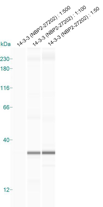
Western Blot: 14-3-3 Antibody [NBP2-27202]
Western Blot: 14-3-3 Antibody [NBP2-27202] - analysis of 14-3-3 using 14-3-3 antibody. Human Brain tissue lysate in the 1) absence and 2) presence of immunizing peptide, 3) mouse brain, and 4) rat brain lysate probed with 2 ug/ml of 14-3-3 antibody. I goat anti-rabbit Ig HRP secondary antibody and PicoTect ECL substrate solution were used for this test.![Immunocytochemistry/ Immunofluorescence: 14-3-3 Antibody [NBP2-27202]](https://resources.rndsystems.com/images/products/14-3-3-Antibody-Immunocytochemistry-Immunofluorescence-NBP2-27202-img0004.jpg "Immunocytochemistry/ Immunofluorescence: 14-3-3 Antibody [NBP2-27202]")
Immunocytochemistry/ Immunofluorescence: 14-3-3 Antibody [NBP2-27202]
Immunocytochemistry/Immunofluorescence: 14-3-3 Antibody [NBP2-27202] - A431 cells were fixed for 10 minutes using 10% formalin and then permeabilized for 5 minutes using 1X PBS + 0.05% Triton-X100. The cells were incubated with anti-14-3-3 at 2 ug/ml overnight at 4C and detected with an anti-rabbit Dylight 488 (Green) at a 1:500 dilution. Alpha tubulin (DM1A) NB100-690 was used as a co-stain at a 1:1000 dilution and detected with an anti-mouse Dylight 550 (Red) at a 1:500 dilution. Nuclei were counterstained with DAPI (Blue). Cells were imaged using a 40X objective.![Simple Western: 14-3-3 Antibody [NBP2-27202]](https://resources.rndsystems.com/images/products/14-3-3-Antibody-Simple-Western-NBP2-27202-img0002.jpg "Simple Western: 14-3-3 Antibody [NBP2-27202]")
Simple Western: 14-3-3 Antibody [NBP2-27202]
Simple Western: 14-3-3 Antibody [NBP2-27202] - Simple Western lane view shows a specific band for 14-3-3 Gamma in 0.5 mg/ml of Human Brain lysate. This experiment was performed under reducing conditions using the 12-230 kDa separation system.![Simple Western: 14-3-3 Antibody [NBP2-27202]](https://resources.rndsystems.com/images/products/14-3-3-Antibody-Simple-Western-NBP2-27202-img0003.jpg "Simple Western: 14-3-3 Antibody [NBP2-27202]")
Simple Western: 14-3-3 Antibody [NBP2-27202]
Simple Western: 14-3-3 Antibody [NBP2-27202] - Analysis in mouse brain lysate. Image from verified customer review.Applications for 14-3-3 Antibody - BSA Free
Application
Recommended Usage
Immunocytochemistry/ Immunofluorescence
2-5 ug/ml
Simple Western
1:50
Western Blot
2-4ug/ml
Application Notes
In Simple Western only 10 - 15 uL of the recommended dilution is used per data point.
See Simple Western Antibody Database for Simple Western validation: Tested in Human Brain lysate 0.5 mg/mL, separated by Size, antibody dilution of 1:50, apparent MW was 33 kDa.
See Simple Western Antibody Database for Simple Western validation: Tested in Human Brain lysate 0.5 mg/mL, separated by Size, antibody dilution of 1:50, apparent MW was 33 kDa.
Reviewed Applications
Read 1 review rated 5 using NBP2-27202 in the following applications:
Formulation, Preparation, and Storage
Purification
Protein A purified
Formulation
PBS
Format
BSA Free
Preservative
0.05% Sodium Azide
Concentration
0.5 mg/ml
Shipping
The product is shipped with polar packs. Upon receipt, store it immediately at the temperature recommended below.
Stability & Storage
Store at 4C short term. Aliquot and store at -20C long term. Avoid freeze-thaw cycles.
Background: 14-3-3
Additional 14-3-3 Products
Product Documents for 14-3-3 Antibody - BSA Free
Certificate of Analysis
To download a Certificate of Analysis, please enter a lot or batch number in the search box below.
Product Specific Notices for 14-3-3 Antibody - BSA Free
This product is for research use only and is not approved for use in humans or in clinical diagnosis. Primary Antibodies are guaranteed for 1 year from date of receipt.
Related Research Areas
Citations for 14-3-3 Antibody - BSA Free
 Powered by Bioz
Customer Reviews for 14-3-3 Antibody - BSA Free (1)
5 out of 5
1 Customer Rating
Have you used 14-3-3 Antibody - BSA Free?
Submit a review and receive an Amazon gift card!
$25/€18/£15/$25CAN/¥2500 Yen for a review with an image
$10/€7/£6/$10CAN/¥1110 Yen for a review without an image
Submit a review
Customer Images
Showing
1
-
1 of
1 review
Showing All
Filter By:
-
Application: Simple WesternSample Tested: whole brain homogenates from miceSpecies: MouseVerified Customer | Posted 01/21/2016Simple Western: 14-3-3 expression in mouse brain lysate

There are no reviews that match your criteria.
Protocols
Find general support by application which include: protocols, troubleshooting, illustrated assays, videos and webinars.
- Appropriate Fixation of IHC/ICC Samples
- Cellular Response to Hypoxia Protocols
- ClariTSA™ Fluorophore Kits
- Detection & Visualization of Antibody Binding
- ICC Cell Smear Protocol for Suspension Cells
- ICC Immunocytochemistry Protocol Videos
- ICC for Adherent Cells
- Immunocytochemistry (ICC) Protocol
- Immunocytochemistry Troubleshooting
- Immunofluorescence of Organoids Embedded in Cultrex Basement Membrane Extract
- Immunohistochemistry (IHC) and Immunocytochemistry (ICC) Protocols
- Preparing Samples for IHC/ICC Experiments
- Preventing Non-Specific Staining (Non-Specific Binding)
- Primary Antibody Selection & Optimization
- Protocol for VisUCyte™ HRP Polymer Detection Reagent
- Protocol for the Fluorescent ICC Staining of Cell Smears - Graphic
- Protocol for the Fluorescent ICC Staining of Cultured Cells on Coverslips - Graphic
- Protocol for the Preparation and Fluorescent ICC Staining of Cells on Coverslips
- Protocol for the Preparation and Fluorescent ICC Staining of Non-adherent Cells
- Protocol for the Preparation and Fluorescent ICC Staining of Stem Cells on Coverslips
- Protocol for the Preparation of a Cell Smear for Non-adherent Cell ICC - Graphic
- R&D Systems Quality Control Western Blot Protocol
- TUNEL and Active Caspase-3 Detection by IHC/ICC Protocol
- The Importance of IHC/ICC Controls
- Troubleshooting Guide: Western Blot Figures
- Western Blot Conditions
- Western Blot Protocol
- Western Blot Protocol for Cell Lysates
- Western Blot Troubleshooting
- Western Blot Troubleshooting Guide
- View all Protocols, Troubleshooting, Illustrated assays and Webinars
Loading...