5-MethylCytosine Antibody (15H61) - BSA Free
Novus Biologicals | Catalog # NBP2-59166
![Immunocytochemistry/ Immunofluorescence: 5-MethylCytosine Antibody (15H61) [NBP2-59166]](https://resources.rndsystems.com/images/products/5-MethylCytosine-Antibody-C-15200003-Immunofluorescence-NBP2-59166-img0005.jpg "Immunocytochemistry/ Immunofluorescence: 5-MethylCytosine Antibody (15H61) [NBP2-59166]")
Loading...
Key Product Details
Validated by
Biological Validation
Species Reactivity
Human, Mouse, Drosophila
Applications
Immunocytochemistry/ Immunofluorescence, Methylated DNA Immunoprecipitation, Dot Blot, In-situ Hybridization
Label
Unconjugated
Antibody Source
Monoclonal Mouse IgG1 Clone # 15H61
Format
BSA Free
Loading...
Product Specifications
Immunogen
The exact immunogen is propietary information.
Clonality
Monoclonal
Host
Mouse
Isotype
IgG1
Description
Monoclonal antibody raised in mouse against 5-mC (5-methylcytosine) conjugated to BSA.
Scientific Data Images for 5-MethylCytosine Antibody (15H61) - BSA Free
Immunocytochemistry/ Immunofluorescence: 5-MethylCytosine Antibody (15H61) [NBP2-59166]
Immunocytochemistry/Immunofluorescence: 5-MethylCytosine Antibody (C.15200003) [NBP2-59166] - Human osteosarcoma (U2OS) cells were stained with the monoclonal antibody against 5-mC. Cells were fixed with 2.5% PFA in PBS for 30 minutes, permeabilized with 0.5% Triton X-100 for 1 hour and treated with 2N HCl for 1 hour followed by 2 x 5 minutes with 0.1 M borate buffer to depurinate the DNA. After blocking with PBS containing 0.1% Triton X-100 and 1% BSA, the cells were immunofluorescently labelled with the 5-mC antibody diluted 1:500 in blocking solution, followed by a goat anti-mouse antibody conjugated to Alexa488. Figure 3A: cells were immunofluorescently labelled with the 5-mC antibody after incubation of the antibody with 50 uM mCTP (left) or with DAPI (right). Figure 3B: staining of the cells with the 5-mC antibody after incubation of the antibody with 50 uM hmCTP and with DAPI.![Immunocytochemistry/ Immunofluorescence: 5-MethylCytosine Antibody (15H61) [NBP2-59166]](https://resources.rndsystems.com/images/products/5-MethylCytosine-Antibody-C-15200003-Immunofluorescence-NBP2-59166-img0004.jpg "Immunocytochemistry/ Immunofluorescence: 5-MethylCytosine Antibody (15H61) [NBP2-59166]")
Immunocytochemistry/ Immunofluorescence: 5-MethylCytosine Antibody (15H61) [NBP2-59166]
Immunocytochemistry/Immunofluorescence: 5-MethylCytosine Antibody (C.15200003) [NBP2-59166] - HeLa cells were stained with the antibody against 5-mC and with DAPI. Cells were fixed with 4% formaldehyde for 10 minutes and blocked with PBS/Triton X-100 containing 1% BSA. The cells were immunofluorescently labelled with the 5-mC antibody (middle) diluted 1:1000 in blocking solution followed by an anti-mouse antibody conjugated to Alexa594. The left panel shows staining of the nuclei with DAPI. A merge of the two stainings is shown on the right.![In-situ Hybridization: 5-MethylCytosine Antibody (15H61) [NBP2-59166]](https://resources.rndsystems.com/images/products/5-MethylCytosine-Antibody-C-15200003-Immunofluorescence-NBP2-59166-img0002.jpg "In-situ Hybridization: 5-MethylCytosine Antibody (15H61) [NBP2-59166]")
In-situ Hybridization: 5-MethylCytosine Antibody (15H61) [NBP2-59166]
In-situ Hybridization: 5-MethylCytosine Antibody (C.15200003) [NBP2-59166] - To detect methylated chromosomal regions, FISH was performed on metaphase chromosomes from HeLa cells using the antibody against 5-mC. The cells were blocked in metaphase by treatment with colcemid (0.1 ug/mL) for 1 - 2 hours, fixed overnight at -20C with ethanol/glacial acetic acid and treated with 2N HCl for 30 minutes at room temperature. Subsequently, the cells were blocked with PBS containing 1% BSA and 0.1% Triton X-100 and stained with the 5-mC antibody (left) diluted 1:1,000 in blocking solution, followed by an anti-mouse antibody conjugated to Alexa594. The middle panel shows staining of the chromosomes with DAPI. A merge of the two stainings is shown on the right.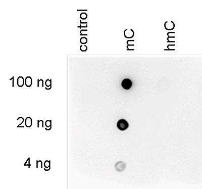
Dot Blot: 5-MethylCytosine Antibody (C.15200003) [NBP2-59166] - To demonstrate the specificity of the antibody against 5-mC, a Dot blot analysis was performed using hmC, mC and C controls. 100 to 4 ng (equivalent of 5 to 0.2 pmol of C-bases) of the controls were spotted on a membrane (Amersham Hybond-N+). The antibody was used at a dilution of 1:600. Dot blot shows a high specificity of the antibody for the methylated control.
Applications for 5-MethylCytosine Antibody (15H61) - BSA Free
Application
Recommended Usage
Dot Blot
1:600
Immunocytochemistry/ Immunofluorescence
1:1000
In-situ Hybridization
1:200 - 1:1000
Methylated DNA Immunoprecipitation
1 - 2 ug/IP
Formulation, Preparation, and Storage
Purification
Protein A purified
Formulation
PBS
Format
BSA Free
Preservative
0.05% Sodium Azide
Concentration
Please see the vial label for concentration. If unlisted please contact technical services.
Shipping
The product is shipped with polar packs. Upon receipt, store it immediately at the temperature recommended below.
Stability & Storage
Store at -20C. Avoid freeze-thaw cycles.
Background: 5-MethylCytosine
Alternate Names
5-mC, 5-Methyl Cytosine, Cytosine (5-Methyl)
Additional 5-MethylCytosine Products
Product Documents for 5-MethylCytosine Antibody (15H61) - BSA Free
Certificate of Analysis
To download a Certificate of Analysis, please enter a lot or batch number in the search box below.
Product Specific Notices for 5-MethylCytosine Antibody (15H61) - BSA Free
This product is for research use only and is not approved for use in humans or in clinical diagnosis. Primary Antibodies are guaranteed for 1 year from date of receipt.
Customer Reviews for 5-MethylCytosine Antibody (15H61) - BSA Free
There are currently no reviews for this product. Be the first to review 5-MethylCytosine Antibody (15H61) - BSA Free and earn rewards!
Have you used 5-MethylCytosine Antibody (15H61) - BSA Free?
Submit a review and receive an Amazon gift card!
$25/€18/£15/$25CAN/¥2500 Yen for a review with an image
$10/€7/£6/$10CAN/¥1110 Yen for a review without an image
Submit a review
Protocols
Find general support by application which include: protocols, troubleshooting, illustrated assays, videos and webinars.
- Appropriate Fixation of IHC/ICC Samples
- Cellular Response to Hypoxia Protocols
- ClariTSA™ Fluorophore Kits
- Detection & Visualization of Antibody Binding
- ICC Cell Smear Protocol for Suspension Cells
- ICC Immunocytochemistry Protocol Videos
- ICC for Adherent Cells
- Immunocytochemistry (ICC) Protocol
- Immunocytochemistry Troubleshooting
- Immunofluorescence of Organoids Embedded in Cultrex Basement Membrane Extract
- Immunohistochemistry (IHC) and Immunocytochemistry (ICC) Protocols
- Immunoprecipitation Protocol
- Preparing Samples for IHC/ICC Experiments
- Preventing Non-Specific Staining (Non-Specific Binding)
- Primary Antibody Selection & Optimization
- Protocol for VisUCyte™ HRP Polymer Detection Reagent
- Protocol for the Fluorescent ICC Staining of Cell Smears - Graphic
- Protocol for the Fluorescent ICC Staining of Cultured Cells on Coverslips - Graphic
- Protocol for the Preparation and Fluorescent ICC Staining of Cells on Coverslips
- Protocol for the Preparation and Fluorescent ICC Staining of Non-adherent Cells
- Protocol for the Preparation and Fluorescent ICC Staining of Stem Cells on Coverslips
- Protocol for the Preparation of a Cell Smear for Non-adherent Cell ICC - Graphic
- TUNEL and Active Caspase-3 Detection by IHC/ICC Protocol
- The Importance of IHC/ICC Controls
- View all Protocols, Troubleshooting, Illustrated assays and Webinars
Loading...