53BP1 Antibody (1285A) - Azide and BSA Free
Novus Biologicals | Catalog # MAB18771
Recombinant Monoclonal Antibody
![Western Blot: 53BP1 Antibody (1285A)Azide and BSA Free [MAB18771]](https://resources.rndsystems.com/images/products/53BP1-Antibody-1285A-Western-Blot-MAB18771-img0002.jpg "Western Blot: 53BP1 Antibody (1285A)Azide and BSA Free [MAB18771]")
Key Product Details
Species Reactivity
Validated:
Human
Predicted:
Bovine (92%), Canine (90%), Chimpanzee (96%), Feline (90%), Orangutan (96%), Porcine (90%), Primate (96%), Rabbit (90%), Sheep (90%). Backed by our 100% Guarantee.
Applications
Western Blot, Immunocytochemistry
Label
Unconjugated
Antibody Source
Monoclonal Rabbit IgG Clone # 1285A
Format
Azide and BSA Free
Loading...
Product Specifications
Immunogen
Recombinant monoclonal 53BP1 Antibody (1285A) was made to amino acids 350 - 400 of human 53BP1 (NP_005648.1).
Reactivity Notes
Predicted cross-reactivity based on sequence identity: Chimpanzee (96%), Gorilla (96%), Orangutan (96%), Gibbon (94%), Bovine (92%), Marmoset (92%), Canine (90%), Feline (90%), Porcine (90%), Rabbit (90%), Sheep (90%)
Clonality
Monoclonal
Host
Rabbit
Isotype
IgG
Description
>90% by SDS PAGE
Scientific Data Images for 53BP1 Antibody (1285A) - Azide and BSA Free
Western Blot: 53BP1 Antibody (1285A)Azide and BSA Free [MAB18771]
Western Blot: 53BP1 Antibody (1285A) [MAB18771] - Analysis of HEK293 and ZR-75 cell lysates with 1 ug/ml of h53BP1 Monoclonal Antibody followed by HRP-conjugated Anti-Rabbit IgG Antibody. The molecular weight was observed for h53BP1 at approximately 350 kDa (as indicated), and the theoretical molecular weight of the whole endogenous protein is 214 kDa. This experiment was conducted under reducing conditions and using Immunoblot Buffer Group 1.![Immunocytochemistry: 53BP1 Antibody (1285A) - Azide and BSA Free [MAB18771]](https://resources.rndsystems.com/images/products/53BP1-Antibody-1285A-Immunocytochemistry-MAB18771-img0001.jpg "Immunocytochemistry: 53BP1 Antibody (1285A) - Azide and BSA Free [MAB18771]")
Immunocytochemistry: 53BP1 Antibody (1285A) - Azide and BSA Free [MAB18771]
Immunocytochemistry: 53BP1 Antibody (1285A) [MAB18771] - 53BP1 was detected in immersion fixed HeLa cell using 53BP1 Monoclonal Antibody at 3 ug/ml dilution. Cells were stained using the NorthernLights 557-conjugated Anti-Rabbit IgG Secondary Antibody (red) and counterstained with DAPI (blue). Specific staining was localized to nuclei.Applications for 53BP1 Antibody (1285A) - Azide and BSA Free
Application
Recommended Usage
Immunocytochemistry
Optimal dilutions of this antibody should be experimentally determined.
Western Blot
Optimal dilutions of this antibody should be experimentally determined.
Reviewed Applications
Read 1 review rated 5 using MAB18771 in the following applications:
Formulation, Preparation, and Storage
Purification
Protein A or G purified
Formulation
PBS
Format
Azide and BSA Free
Preservative
No Preservative
Concentration
1.0 mg/ml
Shipping
The product is shipped with polar packs. Upon receipt, store it immediately at the temperature recommended below.
Stability & Storage
Store at -20C. Avoid freeze-thaw cycles.
Background: 53BP1
References
1.Henry, E., Souissi-Sahraoui, I., Deynoux, M., Lefevre, A., Barroca, V., Campalans, A.,... Arcangeli, M. L. (2019). Human hematopoietic stem/progenitor cells display ROS-dependent long-term hematopoietic defects after exposure to low dose of ionizing radiations. Haematologica. doi:10.3324/haematol.2019.226936
2.Janoshazi, A. K., Horton, J. K., Zhao, M. L., Prasad, R., Scappini, E. L., Tucker, C. J., & Wilson, S. H. (2020). Shining light on the response to repair intermediates in DNA of living cells. DNA Repair (Amst), 85, 102749. doi:10.1016/j.dnarep.2019.102749
Long Name
p53 Binding Protein 1
Alternate Names
p202, TP53BP1
Gene Symbol
TP53BP1
Additional 53BP1 Products
Product Documents for 53BP1 Antibody (1285A) - Azide and BSA Free
Certificate of Analysis
To download a Certificate of Analysis, please enter a lot or batch number in the search box below.
Product Specific Notices for 53BP1 Antibody (1285A) - Azide and BSA Free
This product is for research use only and is not approved for use in humans or in clinical diagnosis. Primary Antibodies are guaranteed for 1 year from date of receipt.
Related Research Areas
Customer Reviews for 53BP1 Antibody (1285A) - Azide and BSA Free (1)
5 out of 5
1 Customer Rating
Have you used 53BP1 Antibody (1285A) - Azide and BSA Free?
Submit a review and receive an Amazon gift card!
$25/€18/£15/$25CAN/¥2500 Yen for a review with an image
$10/€7/£6/$10CAN/¥1110 Yen for a review without an image
Submit a review
Customer Images
Showing
1
-
1 of
1 review
Showing All
Filter By:
-
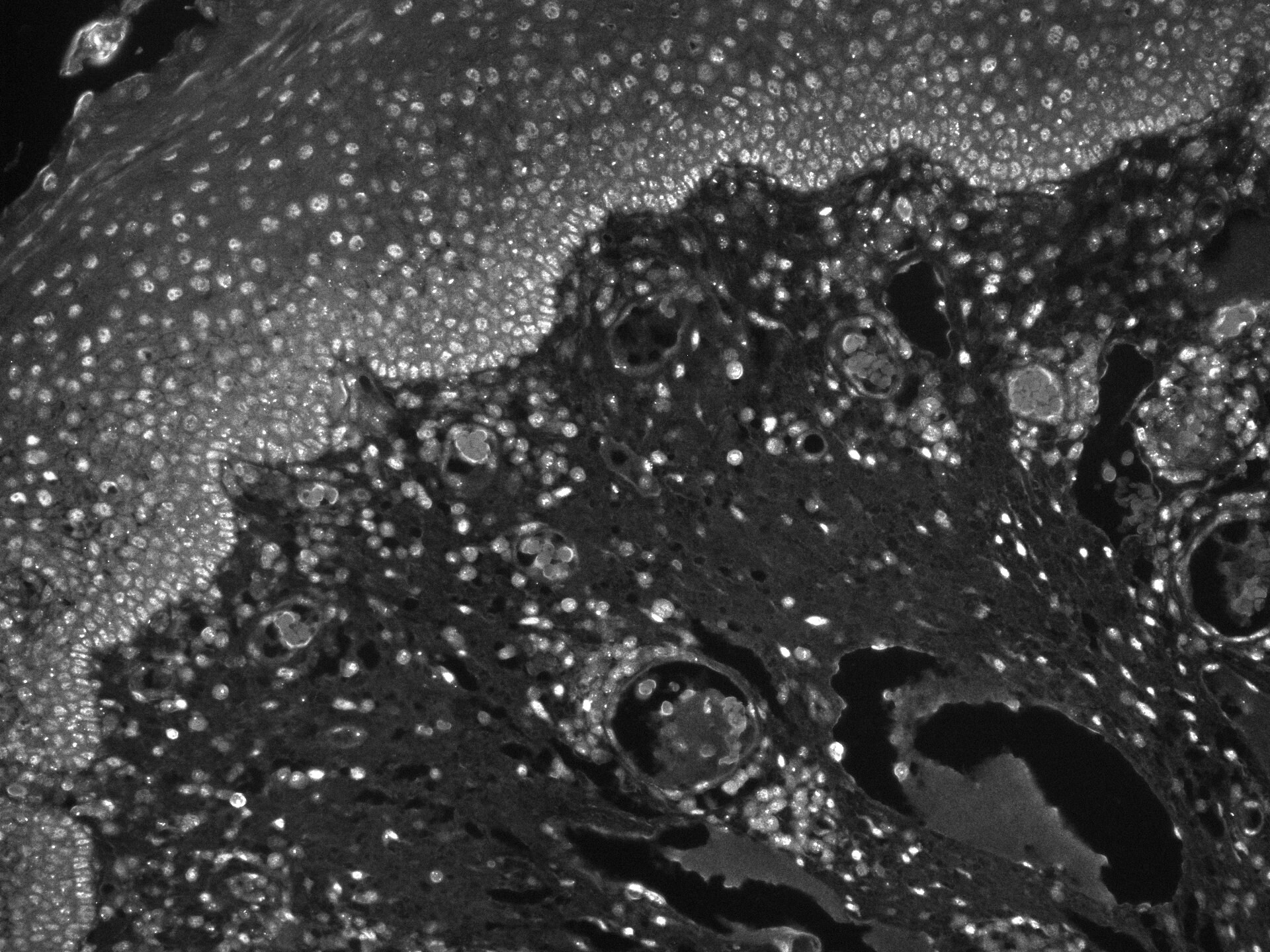
Application: Immunohistochemistry-ParaffinSample Tested: human tonsilSpecies: HumanVerified Customer | Posted 04/03/202353BP1 in Tonsil tissue section
 Bio-Techne ResponseThis review was submitted through the legacy Novus Innovators Program, reflecting a new species or application tested on a primary antibody.
There are no reviews that match your criteria.
Protocols
Find general support by application which include: protocols, troubleshooting, illustrated assays, videos and webinars.
- Appropriate Fixation of IHC/ICC Samples
- Cellular Response to Hypoxia Protocols
- ClariTSA™ Fluorophore Kits
- Detection & Visualization of Antibody Binding
- ICC Cell Smear Protocol for Suspension Cells
- ICC Immunocytochemistry Protocol Videos
- ICC for Adherent Cells
- Immunocytochemistry (ICC) Protocol
- Immunocytochemistry Troubleshooting
- Immunofluorescence of Organoids Embedded in Cultrex Basement Membrane Extract
- Immunohistochemistry (IHC) and Immunocytochemistry (ICC) Protocols
- Preparing Samples for IHC/ICC Experiments
- Preventing Non-Specific Staining (Non-Specific Binding)
- Primary Antibody Selection & Optimization
- Protocol for VisUCyte™ HRP Polymer Detection Reagent
- Protocol for the Fluorescent ICC Staining of Cell Smears - Graphic
- Protocol for the Fluorescent ICC Staining of Cultured Cells on Coverslips - Graphic
- Protocol for the Preparation and Fluorescent ICC Staining of Cells on Coverslips
- Protocol for the Preparation and Fluorescent ICC Staining of Non-adherent Cells
- Protocol for the Preparation and Fluorescent ICC Staining of Stem Cells on Coverslips
- Protocol for the Preparation of a Cell Smear for Non-adherent Cell ICC - Graphic
- R&D Systems Quality Control Western Blot Protocol
- TUNEL and Active Caspase-3 Detection by IHC/ICC Protocol
- The Importance of IHC/ICC Controls
- Troubleshooting Guide: Western Blot Figures
- Western Blot Conditions
- Western Blot Protocol
- Western Blot Protocol for Cell Lysates
- Western Blot Troubleshooting
- Western Blot Troubleshooting Guide
- View all Protocols, Troubleshooting, Illustrated assays and Webinars
FAQs for 53BP1 Antibody (1285A) - Azide and BSA Free
Showing
1
-
2 of
2 FAQs
Showing All
-
Q: Hello I want to ask you for an advice in case of antibodies. In our research we focus on DNA repair foci and use your Novus antibodies for phosphorylated histone H2AX (monoclonal mouse) and protein 53BP1(polyclonal rabbit). Now we want to use antobody for phosphorylated 53BP1 protein to increase specifity and I want to ask you which from your NOVUS antibodies will be the best for us.
A:
We currently stock just one antibody to phosphorylated 53BP1, which you can see at the following link: NB100-1803. This is guaranteed for detection of 53BP1 (pSer25) in human and mouse samples by WB, FLOW, ICC/IF,IHC-P, IP and PLA. As this antibody is a rabbit polyclonal, you would be unable to stain for both 53BP1 and its phosphorylated isoform in the same sample, unless you used an antibody from a different host for detection of total 53BP1 or used directly conjugated primaries. Our mouse monoclonal to 53BP1, with catalogue number NBP2-25028, is validated for detection of the human protein by WB, FLOW and IHC. Unfortunately we do not currently stock any other antibodies to phosphorylated 53BP1.
-
Q: We're looking for anti53BP1 antibody covalently labeled with a fluorofor preferabley Cy3. Do you make it?
A:
We sell 53BP1 antibodies available conjugated to DyLight 488, 550 (very similar in spectrum to Cy3) and 650. You may also purchase the unlabeled antibodies and conjugate them to the fluorophores of your choice. The catalog numbers that you may be interested in are: NB100-904R, NB100-304R, NB100-305R.
-
Q: Hello I want to ask you for an advice in case of antibodies. In our research we focus on DNA repair foci and use your Novus antibodies for phosphorylated histone H2AX (monoclonal mouse) and protein 53BP1(polyclonal rabbit). Now we want to use antobody for phosphorylated 53BP1 protein to increase specifity and I want to ask you which from your NOVUS antibodies will be the best for us.
A:
We currently stock just one antibody to phosphorylated 53BP1, which you can see at the following link: NB100-1803. This is guaranteed for detection of 53BP1 (pSer25) in human and mouse samples by WB, FLOW, ICC/IF,IHC-P, IP and PLA. As this antibody is a rabbit polyclonal, you would be unable to stain for both 53BP1 and its phosphorylated isoform in the same sample, unless you used an antibody from a different host for detection of total 53BP1 or used directly conjugated primaries. Our mouse monoclonal to 53BP1, with catalogue number NBP2-25028, is validated for detection of the human protein by WB, FLOW and IHC. Unfortunately we do not currently stock any other antibodies to phosphorylated 53BP1.
-
Q: We're looking for anti53BP1 antibody covalently labeled with a fluorofor preferabley Cy3. Do you make it?
A:
We sell 53BP1 antibodies available conjugated to DyLight 488, 550 (very similar in spectrum to Cy3) and 650. You may also purchase the unlabeled antibodies and conjugate them to the fluorophores of your choice. The catalog numbers that you may be interested in are: NB100-904R, NB100-304R, NB100-305R.
Loading...