 by Immunohistochemistry")
Loading...
Key Product Details
Species Reactivity
Validated:
Multi-Species
Cited:
Human, Mouse, Rat, Drosophila, Insect - Drosophila, Transgenic Mouse
Applications
Validated:
Immunohistochemistry, ELISA, Immunocytochemistry
Cited:
Immunohistochemistry, Immunohistochemistry-Frozen, Western Blot, Flow Cytometry, Immunocytochemistry, Chromatin Immunoprecipitation (ChIP), Bioassay, Co-Immunoprecipitation, Comet Assay
Label
Unconjugated
Antibody Source
Monoclonal Mouse IgG2B Clone # 15A3
Loading...
Product Specifications
Immunogen
8-oxo-dG-conjugated-KLH
Specificity
This mouse monoclonal antibody specifically binds to 8-hydroxy-2'-
deoxyguanosine within DNA in H
Clonality
Monoclonal
Host
Mouse
Isotype
IgG2B
Scientific Data Images for 8-oxo-dG Antibody (15A3)
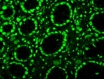
Detection of Rat 8-oxo-dG 8-oxo-dG Antibody (15A3) by Immunohistochemistry
Expression of apoptotic factors in denervated muscle 2 weeks after denervation.(A) Expression of Bcl-2, Bad, and Bax in denervated muscle subjected to different treatment (B) Quantitative analysis of apoptotic markers in different treatment groups (C) The expression of 8-oxo-dG in normal, control, ES, and AFS group. (D) The representative of western blot analysis of 8-oxo-dG related to different treatment (n = 2) (F) Quantitative analysis of caspase-3 in different treatment groups Bar length = 100 μm. R = right, L = left, N = 6 for each group, * p<0.05 and **p<0.01indicated a statistical difference compared to the control group; # p<0.05 indicated a statistical difference as compared to the ES group. Image collected and cropped by CiteAb from the following publication (https://pubmed.ncbi.nlm.nih.gov/25945496), licensed under a CC-BY license. Not internally tested by R&D Systems. by Western Blot")
Detection of Rat 8-oxo-dG 8-oxo-dG Antibody (15A3) by Western Blot
Expression of apoptotic factors in denervated muscle 2 weeks after denervation.(A) Expression of Bcl-2, Bad, and Bax in denervated muscle subjected to different treatment (B) Quantitative analysis of apoptotic markers in different treatment groups (C) The expression of 8-oxo-dG in normal, control, ES, and AFS group. (D) The representative of western blot analysis of 8-oxo-dG related to different treatment (n = 2) (F) Quantitative analysis of caspase-3 in different treatment groups Bar length = 100 μm. R = right, L = left, N = 6 for each group, * p<0.05 and **p<0.01indicated a statistical difference compared to the control group; # p<0.05 indicated a statistical difference as compared to the ES group. Image collected and cropped by CiteAb from the following publication (https://pubmed.ncbi.nlm.nih.gov/25945496), licensed under a CC-BY license. Not internally tested by R&D Systems.
Detection of Rat 8-oxo-dG by Western Blot
Expression of apoptotic factors in denervated muscle 2 weeks after denervation.(A) Expression of Bcl-2, Bad, and Bax in denervated muscle subjected to different treatment (B) Quantitative analysis of apoptotic markers in different treatment groups (C) The expression of 8-oxo-dG in normal, control, ES, and AFS group. (D) The representative of western blot analysis of 8-oxo-dG related to different treatment (n = 2) (F) Quantitative analysis of caspase-3 in different treatment groups Bar length = 100 μm. R = right, L = left, N = 6 for each group, * p<0.05 and **p<0.01indicated a statistical difference compared to the control group; # p<0.05 indicated a statistical difference as compared to the ES group. Image collected and cropped by CiteAb from the following open publication (https://pubmed.ncbi.nlm.nih.gov/25945496), licensed under a CC-BY license. Not internally tested by R&D Systems.
Detection of Rat 8-oxo-dG by Immunocytochemistry/ Immunofluorescence
Expression of apoptotic factors in denervated muscle 2 weeks after denervation.(A) Expression of Bcl-2, Bad, and Bax in denervated muscle subjected to different treatment (B) Quantitative analysis of apoptotic markers in different treatment groups (C) The expression of 8-oxo-dG in normal, control, ES, and AFS group. (D) The representative of western blot analysis of 8-oxo-dG related to different treatment (n = 2) (F) Quantitative analysis of caspase-3 in different treatment groups Bar length = 100 μm. R = right, L = left, N = 6 for each group, * p<0.05 and **p<0.01indicated a statistical difference compared to the control group; # p<0.05 indicated a statistical difference as compared to the ES group. Image collected and cropped by CiteAb from the following open publication (https://pubmed.ncbi.nlm.nih.gov/25945496), licensed under a CC-BY license. Not internally tested by R&D Systems.Applications for 8-oxo-dG Antibody (15A3)
Application
Recommended Usage
ELISA
Empirical determination will be required for optimal results.
Immunocytochemistry
1:250 dilution
Sample:
Sample:
H2O2 treated MCF 10A human breast epithelial cell line
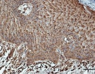
Immunohistochemistry
1:250 dilution
Sample: Paraffin embedded rat thymus tissue
Sample: Paraffin embedded rat thymus tissue
Reviewed Applications
Read 4 reviews rated 4.8 using 4354-MC-050 in the following applications:
Formulation, Preparation, and Storage
Purification
Protein A or G purified from ascites
Formulation
This antibody is provided as purified immunoglobulin from mouse ascites
at 0.5 mg/ml in 1X PBS containing 0.1% sodium azide, 50% glycerol.
Shipping
The product is shipped with dry ice or equivalent. Upon receipt, store it immediately at the temperature recommended below.
Stability & Storage
Store the unopened product at -20 to -70 °C. Use a manual defrost freezer and avoid repeated freeze-thaw cycles. Do not use past expiration date.
Background: 8-oxo-dG
Long Name
8-Hydroxyguanine
Alternate Names
8-OHG, 8oxodG
Additional 8-oxo-dG Products
Product Documents for 8-oxo-dG Antibody (15A3)
Certificate of Analysis
To download a Certificate of Analysis, please enter a lot or batch number in the search box below.
Note: Certificate of Analysis not available for kit components.
Product Specific Notices for 8-oxo-dG Antibody (15A3)
For research use only
Related Research Areas
Citations for 8-oxo-dG Antibody (15A3)
 Powered by Bioz
Customer Reviews for 8-oxo-dG Antibody (15A3) (4)
4.8 out of 5
4 Customer Ratings
Have you used 8-oxo-dG Antibody (15A3)?
Submit a review and receive an Amazon gift card!
$25/€18/£15/$25CAN/¥2500 Yen for a review with an image
$10/€7/£6/$10CAN/¥1110 Yen for a review without an image
Submit a review
Customer Images
Showing
1
-
4 of
4 reviews
Showing All
Filter By:
-
Application: ImmunohistochemistrySample Tested: Stomach tissueSpecies: MouseVerified Customer | Posted 01/03/2022

-
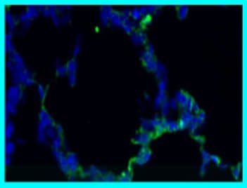
Application: Immunocytochemistry/ImmunofluorescenceSample Tested: Lung tissueSpecies: MouseVerified Customer | Posted 10/28/2021Blue- DAPI; Green:8-oxo-dG
-
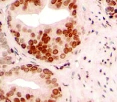
Application: ImmunohistochemistrySample Tested: prostate carcinomaSpecies: HumanVerified Customer | Posted 08/11/2021
-
Application: ImmunohistochemistrySample Tested: Skin tissueSpecies: HumanVerified Customer | Posted 12/28/2017
There are no reviews that match your criteria.
Protocols
Find general support by application which include: protocols, troubleshooting, illustrated assays, videos and webinars.
- Antigen Retrieval Protocol (PIER)
- Antigen Retrieval for Frozen Sections Protocol
- Appropriate Fixation of IHC/ICC Samples
- Cellular Response to Hypoxia Protocols
- Chromogenic IHC Staining of Formalin-Fixed Paraffin-Embedded (FFPE) Tissue Protocol
- Chromogenic Immunohistochemistry Staining of Frozen Tissue
- ClariTSA™ Fluorophore Kits
- Detection & Visualization of Antibody Binding
- ELISA Sample Preparation & Collection Guide
- ELISA Troubleshooting Guide
- Fluorescent IHC Staining of Frozen Tissue Protocol
- Graphic Protocol for Heat-induced Epitope Retrieval
- Graphic Protocol for the Preparation and Fluorescent IHC Staining of Frozen Tissue Sections
- Graphic Protocol for the Preparation and Fluorescent IHC Staining of Paraffin-embedded Tissue Sections
- Graphic Protocol for the Preparation of Gelatin-coated Slides for Histological Tissue Sections
- How to Run an R&D Systems DuoSet ELISA
- How to Run an R&D Systems Quantikine ELISA
- How to Run an R&D Systems Quantikine™ QuicKit™ ELISA
- ICC Cell Smear Protocol for Suspension Cells
- ICC Immunocytochemistry Protocol Videos
- ICC for Adherent Cells
- IHC Sample Preparation (Frozen sections vs Paraffin)
- Immunocytochemistry (ICC) Protocol
- Immunocytochemistry Troubleshooting
- Immunofluorescence of Organoids Embedded in Cultrex Basement Membrane Extract
- Immunofluorescent IHC Staining of Formalin-Fixed Paraffin-Embedded (FFPE) Tissue Protocol
- Immunohistochemistry (IHC) and Immunocytochemistry (ICC) Protocols
- Immunohistochemistry Frozen Troubleshooting
- Immunohistochemistry Paraffin Troubleshooting
- Preparing Samples for IHC/ICC Experiments
- Preventing Non-Specific Staining (Non-Specific Binding)
- Primary Antibody Selection & Optimization
- Protocol for Heat-Induced Epitope Retrieval (HIER)
- Protocol for Making a 4% Formaldehyde Solution in PBS
- Protocol for VisUCyte™ HRP Polymer Detection Reagent
- Protocol for the Fluorescent ICC Staining of Cell Smears - Graphic
- Protocol for the Fluorescent ICC Staining of Cultured Cells on Coverslips - Graphic
- Protocol for the Preparation & Fixation of Cells on Coverslips
- Protocol for the Preparation and Chromogenic IHC Staining of Frozen Tissue Sections
- Protocol for the Preparation and Chromogenic IHC Staining of Frozen Tissue Sections - Graphic
- Protocol for the Preparation and Chromogenic IHC Staining of Paraffin-embedded Tissue Sections
- Protocol for the Preparation and Chromogenic IHC Staining of Paraffin-embedded Tissue Sections - Graphic
- Protocol for the Preparation and Fluorescent ICC Staining of Cells on Coverslips
- Protocol for the Preparation and Fluorescent ICC Staining of Non-adherent Cells
- Protocol for the Preparation and Fluorescent ICC Staining of Stem Cells on Coverslips
- Protocol for the Preparation and Fluorescent IHC Staining of Frozen Tissue Sections
- Protocol for the Preparation and Fluorescent IHC Staining of Paraffin-embedded Tissue Sections
- Protocol for the Preparation of Gelatin-coated Slides for Histological Tissue Sections
- Protocol for the Preparation of a Cell Smear for Non-adherent Cell ICC - Graphic
- Quantikine HS ELISA Kit Assay Principle, Alkaline Phosphatase
- Quantikine HS ELISA Kit Principle, Streptavidin-HRP Polymer
- Sandwich ELISA (Colorimetric) – Biotin/Streptavidin Detection Protocol
- Sandwich ELISA (Colorimetric) – Direct Detection Protocol
- TUNEL and Active Caspase-3 Detection by IHC/ICC Protocol
- The Importance of IHC/ICC Controls
- Troubleshooting Guide: ELISA
- Troubleshooting Guide: Immunohistochemistry
- View all Protocols, Troubleshooting, Illustrated assays and Webinars
Loading...