A. aeolicus BPL/BioID2 Antibody (SS QD1) - BSA Free
Novus Biologicals | Catalog # NBP2-59940
![Western Blot: A. aeolicus BPL/BioID2 Antibody (SS QD1)BSA Free [NBP2-59940]](https://resources.rndsystems.com/images/products/A-aeolicus-BPL-BioID2-Antibody-SS-QD1-Western-Blot-NBP2-59940-img0004.jpg "Western Blot: A. aeolicus BPL/BioID2 Antibody (SS QD1)BSA Free [NBP2-59940]")
Key Product Details
Species Reactivity
Bacteria
Applications
Validated:
Western Blot, Immunocytochemistry/ Immunofluorescence, Simple Western
Cited:
Immunocytochemistry/ Immunofluorescence
Label
Unconjugated
Antibody Source
Monoclonal Mouse IgG2 kappa Clone # SS QD1
Format
BSA Free
Loading...
Product Specifications
Immunogen
GST fused to A. aeolicus BPL/BioID2
Reactivity Notes
Aquifex aeolicus
Clonality
Monoclonal
Host
Mouse
Isotype
IgG2 kappa
Theoretical MW
27 kDa.
Disclaimer note: The observed molecular weight of the protein may vary from the listed predicted molecular weight due to post translational modifications, post translation cleavages, relative charges, and other experimental factors.
Disclaimer note: The observed molecular weight of the protein may vary from the listed predicted molecular weight due to post translational modifications, post translation cleavages, relative charges, and other experimental factors.
Scientific Data Images for A. aeolicus BPL/BioID2 Antibody (SS QD1) - BSA Free
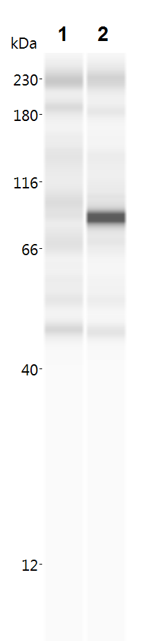
Western Blot: A. aeolicus BPL/BioID2 Antibody (SS QD1)BSA Free [NBP2-59940]
Western Blot: A. aeolicus BPL/BioID2 Antibody (SS QD1) [NBP2-59940] - Analysis in total cell lysate of parental HeLa cell line (lane 1) or HeLa polyclonal cell line expressing BioID2 tagged to TorsinA delta E302/3 (6) without (lane 2) or with induced expression (lane 3) were transferred to a nitrocellulose membrane and blotted for BPL R40G/BioID2 with SS QD1 antibody. The predicted molecular weight of BioID2-TorsinA delta E302/3 fusion protein is 65 kDa.![Immunocytochemistry/ Immunofluorescence: A. aeolicus BPL/BioID2 Antibody (SS QD1) - BSA Free [NBP2-59940]](https://resources.rndsystems.com/images/products/A-aeolicus-BPL-BioID2-Antibody-SS-QD1-Immunocytochemistry-Immunofluorescence-NBP2-59940-img0003.jpg "Immunocytochemistry/ Immunofluorescence: A. aeolicus BPL/BioID2 Antibody (SS QD1) - BSA Free [NBP2-59940]")
Immunocytochemistry/ Immunofluorescence: A. aeolicus BPL/BioID2 Antibody (SS QD1) - BSA Free [NBP2-59940]
Immunocytochemistry/Immunofluorescence: A. aeolicus BPL/BioID2 Antibody (SS QD1) [NBP2-59940] - Staining of HeLa cell line expressing doxycycline (dox) - inducible BioID2 tagged to TorsinA delta E302/3 with SS QD1 monoclonal antibody. Uninduced (-dox) or induced (+dox) cells were fixed in (A) cold methanol or (B) 4% PFA and stained with anti-BPL R40G/BioID2 SS QD1 antibody (in green). The nuclei were counter-stained with Hoechst.![Simple Western: A. aeolicus BPL/BioID2 Antibody (SS QD1)BSA Free [NBP2-59940]](https://resources.rndsystems.com/images/products/A-aeolicus-BPL-BioID2-Antibody-SS-QD1-Simple-Western-NBP2-59940-img0005.jpg "Simple Western: A. aeolicus BPL/BioID2 Antibody (SS QD1)BSA Free [NBP2-59940]")
Simple Western: A. aeolicus BPL/BioID2 Antibody (SS QD1)BSA Free [NBP2-59940]
Simple Western: A. aeolicus BPL/BioID2 Antibody (SS QD1) [NBP2-59940] - Lane 1: lysate from untransfected HEK293 cells. Lane 2: lysate from HEK293 cells transfected with BioID2-PTEN. A specific signal for BioID2-PTEN is visible at ~94kDa.Applications for A. aeolicus BPL/BioID2 Antibody (SS QD1) - BSA Free
Application
Recommended Usage
Immunocytochemistry/ Immunofluorescence
1:10 - 1:500
Western Blot
1:100 - 1:2000
Application Notes
Positive Control: Cells overexpressing A. aeolicus BPL/BioID2 construct.
See Simple Western Antibody Database for Simple Western validation: separated by Size, apparent MW was 94 kDa
See Simple Western Antibody Database for Simple Western validation: separated by Size, apparent MW was 94 kDa
Reviewed Applications
Read 1 review rated 4 using NBP2-59940 in the following applications:
Formulation, Preparation, and Storage
Purification
Protein A purified
Formulation
PBS
Format
BSA Free
Preservative
0.02% Sodium Azide
Concentration
1 mg/ml
Shipping
The product is shipped with polar packs. Upon receipt, store it immediately at the temperature recommended below.
Stability & Storage
Store at 4C short term. Aliquot and store at -20C long term. Avoid freeze-thaw cycles.
Product Documents for A. aeolicus BPL/BioID2 Antibody (SS QD1) - BSA Free
Certificate of Analysis
To download a Certificate of Analysis, please enter a lot or batch number in the search box below.
Product Specific Notices for A. aeolicus BPL/BioID2 Antibody (SS QD1) - BSA Free
This product is for research use only and is not approved for use in humans or in clinical diagnosis. Primary Antibodies are guaranteed for 1 year from date of receipt.
Citations for A. aeolicus BPL/BioID2 Antibody (SS QD1) - BSA Free
 Powered by Bioz
Customer Reviews for A. aeolicus BPL/BioID2 Antibody (SS QD1) - BSA Free (1)
4 out of 5
1 Customer Rating
Have you used A. aeolicus BPL/BioID2 Antibody (SS QD1) - BSA Free?
Submit a review and receive an Amazon gift card!
$25/€18/£15/$25CAN/¥2500 Yen for a review with an image
$10/€7/£6/$10CAN/¥1110 Yen for a review without an image
Submit a review
Customer Images
Showing
1
-
1 of
1 review
Showing All
Filter By:
-
Application: Simple WesternSample Tested: HEK293 cells transfected with BioID2-PTENSpecies: HumanVerified Customer | Posted 11/15/20181- lysate from untransfected HEK293 cells 2- lysate from HEK293 cells transfected with BioID2-PTEN A specific signal for BioID2-PTEN is visible at ~94kDa.For Simple Western (see review image): Lysates were diluted to 0.3125mg/mL before combining with 5x Fluorescent Master Mix and subjecting samples to Simple Western using default assay parameters (Protein Simple). Primary antibody to BioID2 (NBP2-59940) that was used in assay was diluted to 5ug/mL (1:200) in Antibody Diluent 2 (Protein Simple). Product review code: SW-1WB

There are no reviews that match your criteria.
Protocols
Find general support by application which include: protocols, troubleshooting, illustrated assays, videos and webinars.
- Appropriate Fixation of IHC/ICC Samples
- Cellular Response to Hypoxia Protocols
- ClariTSA™ Fluorophore Kits
- Detection & Visualization of Antibody Binding
- ICC Cell Smear Protocol for Suspension Cells
- ICC Immunocytochemistry Protocol Videos
- ICC for Adherent Cells
- Immunocytochemistry (ICC) Protocol
- Immunocytochemistry Troubleshooting
- Immunofluorescence of Organoids Embedded in Cultrex Basement Membrane Extract
- Immunohistochemistry (IHC) and Immunocytochemistry (ICC) Protocols
- Preparing Samples for IHC/ICC Experiments
- Preventing Non-Specific Staining (Non-Specific Binding)
- Primary Antibody Selection & Optimization
- Protocol for VisUCyte™ HRP Polymer Detection Reagent
- Protocol for the Fluorescent ICC Staining of Cell Smears - Graphic
- Protocol for the Fluorescent ICC Staining of Cultured Cells on Coverslips - Graphic
- Protocol for the Preparation and Fluorescent ICC Staining of Cells on Coverslips
- Protocol for the Preparation and Fluorescent ICC Staining of Non-adherent Cells
- Protocol for the Preparation and Fluorescent ICC Staining of Stem Cells on Coverslips
- Protocol for the Preparation of a Cell Smear for Non-adherent Cell ICC - Graphic
- R&D Systems Quality Control Western Blot Protocol
- TUNEL and Active Caspase-3 Detection by IHC/ICC Protocol
- The Importance of IHC/ICC Controls
- Troubleshooting Guide: Western Blot Figures
- Western Blot Conditions
- Western Blot Protocol
- Western Blot Protocol for Cell Lysates
- Western Blot Troubleshooting
- Western Blot Troubleshooting Guide
- View all Protocols, Troubleshooting, Illustrated assays and Webinars
Loading...