Acetyl-Lysine Antibody (1C6)
Novus Biologicals | Catalog # NB100-74339
![Western Blot: Acetyl-Lysine Antibody (1C6) [NB100-74339]](https://resources.rndsystems.com/images/products/Acetylated-Lysine-Antibody-1C6-Western-Blot-NB100-74339-img0004.jpg "Western Blot: Acetyl-Lysine Antibody (1C6) [NB100-74339]")
Loading...
Key Product Details
Validated by
Biological Validation
Species Reactivity
Validated:
Non-species specific
Cited:
Human, Mouse
Applications
Validated:
Western Blot, Immunocytochemistry/ Immunofluorescence, Immunoprecipitation, Chromatin Immunoprecipitation (ChIP), Dot Blot
Cited:
Western Blot
Label
Unconjugated
Antibody Source
Monoclonal Mouse IgG1 Clone # 1C6
Loading...
Product Specifications
Immunogen
Synthetic peptide sequence surrounding the acetylated lysine 9 of histone H3.
Clonality
Monoclonal
Host
Mouse
Isotype
IgG1
Scientific Data Images for Acetyl-Lysine Antibody (1C6)
Western Blot: Acetyl-Lysine Antibody (1C6) [NB100-74339]
Western Blot: Acetyl-Lysine Antibody (1C6) [NB100-74339] - Analysis of Acetyl-Lysine in HeLA cells.![Immunocytochemistry/ Immunofluorescence: Acetyl-Lysine Antibody (1C6) [NB100-74339]](https://resources.rndsystems.com/images/products/Acetylated-Lysine-Antibody-1C6-Immunocytochemistry-Immunofluorescence-NB100-74339-img0007.jpg "Immunocytochemistry/ Immunofluorescence: Acetyl-Lysine Antibody (1C6) [NB100-74339]")
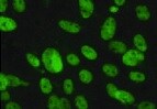
Immunocytochemistry/ Immunofluorescence: Acetyl-Lysine Antibody (1C6) [NB100-74339]
Immunocytochemistry/Immunofluorescence: Acetyl-Lysine Antibody (1C6) [NB100-74339] - Analysis of lysine acetylated proteins (green) in HeLa cells left untreated (left panels) or treated with 0.3uM Trichostatin A for 16 hours (right panels).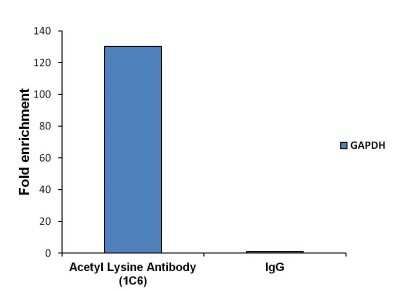
Chromatin Immunoprecipitation: Acetyl-Lysine Antibody (1C6) [NB100-74339] - Analysis of chromatin proteins with Acetyl-Lysine was performed using cross-linked chromatin from 4-10^6 LNCaP cells. Immunoprecipitation was performed using the Magnetic ChIP kit with 10ug of an Acetyl-Lysine monoclonal antibody. Quantitation of immunoprecipitated GAPDH promoter sequence is presented as fold enrichment of the acetyl lysine monoclonal antibody versus non-specific IgG.
![Western Blot: Acetyl-Lysine Antibody (1C6) [NB100-74339]](https://resources.rndsystems.com/images/products/Acetylated-Lysine-Antibody-1C6-Western-Blot-NB100-74339-img0002.jpg "Western Blot: Acetyl-Lysine Antibody (1C6) [NB100-74339]")
Western Blot: Acetyl-Lysine Antibody (1C6) [NB100-74339]
Western Blot: Acetyl-Lysine Antibody (1C6) [NB100-74339] - Analysis of Acetyl-Lysine was performed by loading various amounts of acetylated BSA or non-acetylated BSA per well and 10ul of PageRuler Prestained Protein Ladder onto a 4-20% Tris-HCl polyacrylamide gel. Proteins were transferred to a PVDF membrane and blocked with 5% BSA/TBST for at least 1 hour. The membrane was probed with an Acetyl-Lysine monoclonal antibody at a dilution of 1:1000 overnight at 4C on a rocking platform, washed in TBS-0.1%Tween-20, and probed with a goat anti-mouse IgG-HRP secondary antibody at a dilution of 1:20,000 for 1 hour. Chemiluminescent detection was performed using SuperSignal West Pico.![Western Blot: Acetyl-Lysine Antibody (1C6) [NB100-74339]](https://resources.rndsystems.com/images/products/Acetylated-Lysine-Antibody-1C6-Western-Blot-NB100-74339-img0003.jpg "Western Blot: Acetyl-Lysine Antibody (1C6) [NB100-74339]")
Western Blot: Acetyl-Lysine Antibody (1C6) [NB100-74339]
Western Blot: Acetyl-Lysine Antibody (1C6) [NB100-74339] - Analysis of lysine acetylated proteins from cells left untreated (DMSO only) or cells treated with 0.3uM or 3uM of Trichostatin A (TSA) for 16 hours was performed by loading 50 ug of the indicated whole cell lysates per well and 10ul of PageRuler Prestained Protein Ladder onto a 4-20% Tris-HCl polyacrylamide gel. Proteins were transferred to a PVDF membrane and blocked with 5% BSA/TBST for at least 1 hour. The membrane was probed with an Acetyl-Lysine monoclonal antibody at a dilution of 1:1000 overnight at 4C on a rocking platform, washed in TBS-0.1%Tween-20, and probed with a goat anti-mouse IgG-HRP secondary antibody at a dilution of 1:20,000 for 1 hour. Chemiluminescent detection was performed using SuperSignal West Pico.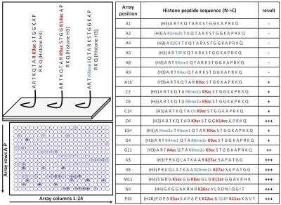
Dot Blot: Acetyl-Lysine Antibody (1C6) [NB100-74339] - Specific detection of lysine acetylated histones using an Acetyl-Lysine monoclonal antibody. Histone peptides containing 384 different modification combinations spotted in duplicate on a peptide array were used according to the manufacturer instructions. A schematic and table with representative results (positive detection in red, negative in blue) are shown. Result interpretation and quantification were performed using the manufacturer software. Chemiluminescent detection was performed.
![Immunoprecipitation: Acetyl-Lysine Antibody (1C6) [NB100-74339]](https://resources.rndsystems.com/images/products/Acetylated-Lysine-Antibody-1C6-Immunoprecipitation-NB100-74339-img0005.jpg "Immunoprecipitation: Acetyl-Lysine Antibody (1C6) [NB100-74339]")
Immunoprecipitation: Acetyl-Lysine Antibody (1C6) [NB100-74339]
Immunoprecipitation: Acetyl-Lysine Antibody (1C6) [NB100-74339] - Immunoprecipitation of acetylated alpha-Tubulin was performed.3uM or 3uM Trichostatin A (TSA) for 16 hours. Antigen-antibody complexes were formed by incubating 500ug of lysate with 3ug of an Acetyl-Lysine monoclonal antibody overnight on a rocking platform at 4C. The immune complexes were captured on 50ul Protein A/G Agarose at a dilution of 1:1000 overnight rotating at 4C, washed in TBST, and probed with Clean-blot IP Detection Reagent at a dilution of 1:2000 for at least 1 hour.![Immunoprecipitation: Acetyl-Lysine Antibody (1C6) [NB100-74339]](https://resources.rndsystems.com/images/products/Acetylated-Lysine-Antibody-1C6-Immunoprecipitation-NB100-74339-img0006.jpg "Immunoprecipitation: Acetyl-Lysine Antibody (1C6) [NB100-74339]")
Immunoprecipitation: Acetyl-Lysine Antibody (1C6) [NB100-74339]
Immunoprecipitation: Acetyl-Lysine Antibody (1C6) [NB100-74339] - Analysis of acetylated Histone H3 (Lys9) was performed using whole cell lysates from cells left untreated (DMSO only) or cells treated with 0.3uM or 3uM Trichostatin A (TSA) for 16 hours. Antigen-antibody complexes were formed by incubating 500ug of the indicated lysate with 3ug of an Acetyl-Lysine mAb overnight on a rocking platform at 4C. The immune complexes were captured on 50ul Protein A/G Agarose, washed extensively, and eluted with 5X Lane Marker Reducing Sample Buffer. Samples were resolved on a 4-20% Tris-HCl polyacrylamide gel, transferred to a PVDF membrane, and blocked with 5% BSA/TBS-0.1%Tween for at least 1 hr. The membrane was probed with an Acetyl-Histone H3 (Lys9) mAb at a dilution of 1:1000 overnight rotating at 4C, washed in TBST, and probed with Clean-blot IP Detection Reagent at a dilution of 1:2000 for at least 1 hour. Chemiluminescent detection was performed using SuperSignal West Pico.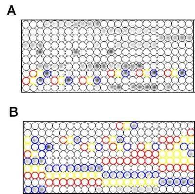
Dot Blot: Acetyl-Lysine Antibody (1C6) [NB100-74339] - Specific detection of lysine acetylated histones using an Acetyl-Lysine monoclonal antibody. Histone peptides containing 384 different modification combinations spotted in duplicate on a peptide array were used according to the manufacturer's instructions (Active Motif). Representative results showing specific detection of acetylated Histone H4 K20 (blue circles, panel A) and H3 K9 (blue circles, panel B). Note that Histone H4 K20me2/K20me3 (red and yellow circles, panel A) and H3 K9me2/K9m3 (red and yellow circles, panel B) are not detected by the Acetyl-Lysine monoclonal antibody.
Applications for Acetyl-Lysine Antibody (1C6)
Application
Recommended Usage
Chromatin Immunoprecipitation (ChIP)
10 ug
Dot Blot
1:100 - 1:2000
Immunocytochemistry/ Immunofluorescence
1:100 - 1:500
Immunoprecipitation
3 ug
Western Blot
1:500 - 1:2000
Reviewed Applications
Read 2 reviews rated 5 using NB100-74339 in the following applications:
Formulation, Preparation, and Storage
Purification
Protein G purified
Formulation
PBS, 1 mg/mL BSA
Preservative
0.05% Sodium Azide
Concentration
1 mg/ml
Shipping
The product is shipped with polar packs. Upon receipt, store it immediately at the temperature recommended below.
Stability & Storage
Store at -20C. Avoid freeze-thaw cycles.
Background: Acetyl-Lysine
Alternate Names
acetyl Lysine, AC-LYS-OH, Lysine, N-acetyl lysine
Additional Acetyl-Lysine Products
Product Documents for Acetyl-Lysine Antibody (1C6)
Certificate of Analysis
To download a Certificate of Analysis, please enter a lot or batch number in the search box below.
Product Specific Notices for Acetyl-Lysine Antibody (1C6)
This product is for research use only and is not approved for use in humans or in clinical diagnosis. Primary Antibodies are guaranteed for 1 year from date of receipt.
Citations for Acetyl-Lysine Antibody (1C6)
 Powered by Bioz
Customer Reviews for Acetyl-Lysine Antibody (1C6) (2)
5 out of 5
2 Customer Ratings
Have you used Acetyl-Lysine Antibody (1C6)?
Submit a review and receive an Amazon gift card!
$25/€18/£15/$25CAN/¥2500 Yen for a review with an image
$10/€7/£6/$10CAN/¥1110 Yen for a review without an image
Submit a review
Customer Images
Showing
1
-
2 of
2 reviews
Showing All
Filter By:
-
Application: ImmunocytochemistrySample Tested: Hepatic stellate cellsSpecies: MouseVerified Customer | Posted 11/05/2021Hepatic stellate cellsPrimary antibody dilution 1:100

-
Application: Western BlotSample Tested: SplenocytesSpecies: MouseVerified Customer | Posted 01/14/2016
There are no reviews that match your criteria.
Protocols
Find general support by application which include: protocols, troubleshooting, illustrated assays, videos and webinars.
- Appropriate Fixation of IHC/ICC Samples
- Cellular Response to Hypoxia Protocols
- ChIP Protocol Video
- Chromatin Immunoprecipitation (ChIP) Protocol
- Chromatin Immunoprecipitation Protocol
- ClariTSA™ Fluorophore Kits
- Detection & Visualization of Antibody Binding
- ICC Cell Smear Protocol for Suspension Cells
- ICC Immunocytochemistry Protocol Videos
- ICC for Adherent Cells
- Immunocytochemistry (ICC) Protocol
- Immunocytochemistry Troubleshooting
- Immunofluorescence of Organoids Embedded in Cultrex Basement Membrane Extract
- Immunohistochemistry (IHC) and Immunocytochemistry (ICC) Protocols
- Immunoprecipitation Protocol
- Preparing Samples for IHC/ICC Experiments
- Preventing Non-Specific Staining (Non-Specific Binding)
- Primary Antibody Selection & Optimization
- Protocol for VisUCyte™ HRP Polymer Detection Reagent
- Protocol for the Fluorescent ICC Staining of Cell Smears - Graphic
- Protocol for the Fluorescent ICC Staining of Cultured Cells on Coverslips - Graphic
- Protocol for the Preparation and Fluorescent ICC Staining of Cells on Coverslips
- Protocol for the Preparation and Fluorescent ICC Staining of Non-adherent Cells
- Protocol for the Preparation and Fluorescent ICC Staining of Stem Cells on Coverslips
- Protocol for the Preparation of a Cell Smear for Non-adherent Cell ICC - Graphic
- R&D Systems Quality Control Western Blot Protocol
- TUNEL and Active Caspase-3 Detection by IHC/ICC Protocol
- The Importance of IHC/ICC Controls
- Troubleshooting Guide: Western Blot Figures
- Western Blot Conditions
- Western Blot Protocol
- Western Blot Protocol for Cell Lysates
- Western Blot Troubleshooting
- Western Blot Troubleshooting Guide
- View all Protocols, Troubleshooting, Illustrated assays and Webinars
Loading...