AGER Antibody (JF0975)
Novus Biologicals | Catalog # NBP2-67095
Recombinant Monoclonal Antibody
![Western Blot: AGER Antibody (JF0975) [NBP2-67095]](https://resources.rndsystems.com/images/products/AGER-Antibody-JF0975-Western-Blot-NBP2-67095-img0005.jpg "Western Blot: AGER Antibody (JF0975) [NBP2-67095]")
Loading...
Key Product Details
Species Reactivity
Human, Mouse, Rat
Applications
Immunohistochemistry, Immunohistochemistry-Paraffin, Western Blot, Flow Cytometry
Label
Unconjugated
Antibody Source
Recombinant Monoclonal Rabbit IgG Clone # JF0975 expressed in HEK293
Loading...
Product Specifications
Immunogen
Synthetic peptide within human AGER aa 350-390. (SwissProt: Q15109 Human; SwissProt: Q62151 Mouse; SwissProt: Q63495 Rat)
Localization
Cell membrane, Secreted.
Clonality
Monoclonal
Host
Rabbit
Isotype
IgG
Scientific Data Images for AGER Antibody (JF0975)
Western Blot: AGER Antibody (JF0975) [NBP2-67095]
Western Blot: AGER Antibody (JF0975) [NBP2-67095] - Analysis of RAGE on mouse lung lysates using anti-RAGE antibody at 1/1,000 dilution.![Immunocytochemistry/ Immunofluorescence: AGER Antibody (JF0975) [NBP2-67095]](https://resources.rndsystems.com/images/products/AGER-Antibody-JF0975-Immunocytochemistry-Immunofluorescence-NBP2-67095-img0003.jpg "Immunocytochemistry/ Immunofluorescence: AGER Antibody (JF0975) [NBP2-67095]")
Immunocytochemistry/ Immunofluorescence: AGER Antibody (JF0975) [NBP2-67095]
Immunocytochemistry/Immunofluorescence: AGER Antibody (JF0975) [NBP2-67095] - Staining RAGE in MCF-7 cells (red). The nuclear counter stain is DAPI (blue). Cells were fixed in paraformaldehyde, permeabilised with 0.25% Triton X100/PBS.![Immunohistochemistry-Paraffin: AGER Antibody (JF0975) [NBP2-67095]](https://resources.rndsystems.com/images/products/AGER-Antibody-JF0975-Immunohistochemistry-Paraffin-NBP2-67095-img0006.jpg "Immunohistochemistry-Paraffin: AGER Antibody (JF0975) [NBP2-67095]")
Immunohistochemistry-Paraffin: AGER Antibody (JF0975) [NBP2-67095]
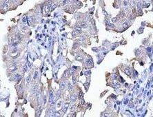
Immunohistochemistry-Paraffin: AGER Antibody (JF0975) [NBP2-67095] - Mouse lung cancer tissue stained with AGER antibody. IHC-P image submitted by a verified customer review.![Flow Cytometry: AGER Antibody (JF0975) [NBP2-67095]](https://resources.rndsystems.com/images/products/AGER-Antibody-JF0975-Flow-Cytometry-NBP2-67095-img0001.jpg "Flow Cytometry: AGER Antibody (JF0975) [NBP2-67095]")
Flow Cytometry: AGER Antibody (JF0975) [NBP2-67095]
Flow Cytometry: AGER Antibody (JF0975) [NBP2-67095] - Analysis of Jurkat cells with RAGE antibody at 1/50 dilution (red) compared with an unlabelled control (cells without incubation with primary antibody; black). Alexa Fluor 488-conjugated goat anti rabbit IgG was used as the secondary antibody.![Immunocytochemistry/ Immunofluorescence: AGER Antibody (JF0975) [NBP2-67095]](https://resources.rndsystems.com/images/products/AGER-Antibody-JF0975-Immunocytochemistry-Immunofluorescence-NBP2-67095-img0002.jpg "Immunocytochemistry/ Immunofluorescence: AGER Antibody (JF0975) [NBP2-67095]")
Immunocytochemistry/ Immunofluorescence: AGER Antibody (JF0975) [NBP2-67095]
Immunocytochemistry/Immunofluorescence: AGER Antibody (JF0975) [NBP2-67095] - Staining RAGE in A431 cells (red). The nuclear counter stain is DAPI (blue). Cells were fixed in paraformaldehyde, permeabilised with 0.25% Triton X100/PBS.![Immunohistochemistry-Paraffin: AGER Antibody (JF0975) [NBP2-67095]](https://resources.rndsystems.com/images/products/AGER-Antibody-JF0975-Immunohistochemistry-Paraffin-NBP2-67095-img0004.jpg "Immunohistochemistry-Paraffin: AGER Antibody (JF0975) [NBP2-67095]")
Immunohistochemistry-Paraffin: AGER Antibody (JF0975) [NBP2-67095]
Immunohistochemistry-Paraffin: AGER Antibody (JF0975) [NBP2-67095] - Analysis of paraffin-embedded mouse lung tissue using anti-RAGE antibody. Counter stained with hematoxylin.![AGER Antibody (JF0975) Western Blot: AGER Antibody (JF0975) [NBP2-67095]](https://resources.rndsystems.com/images/products/nbp2-67095_-western-blot-639204586675332384.jpg "Western Blot: AGER Antibody (JF0975) [NBP2-67095]")
Western Blot: AGER Antibody (JF0975) [NBP2-67095]
Western blot analysis on different lysates with NBP2-67095 at 1/5,000 dilution. Lane 1: Mouse lung tissue lysate Lane 2: Rat lung tissue lysate Lysates/proteins at 20 µg/Lane. Predicted band size: 43 kDa Observed band size: 43/50 kDa Exposure time: 24 seconds; 4-20% SDS-PAGE gel. Proteins were transferred to a PVDF membrane and blocked with 5% NFDM/TBST for 1 hour at room temperature. NBP2-67095 at 1/5,000 dilution was used in 5% NFDM/TBST at 4℃ overnight. Goat Anti-Rabbit IgG [HRP] Secondary Antibody at 1/50,000 dilution was used for 1 hour at room temperature.Applications for AGER Antibody (JF0975)
Application
Recommended Usage
Flow Cytometry
1:50-1:100
Immunohistochemistry-Paraffin
1:50-1:200
Western Blot
1:500-1:2000
Reviewed Applications
Read 1 review rated 5 using NBP2-67095 in the following applications:
Flow Cytometry Panel Builder
Bio-Techne Knows Flow Cytometry
Save time and reduce costly mistakes by quickly finding compatible reagents using the Panel Builder Tool.
Advanced Features
- Spectra Viewer - Custom analysis of spectra from multiple fluorochromes
- Spillover Popups - Visualize the spectra of individual fluorochromes
- Antigen Density Selector - Match fluorochrome brightness with antigen density

Formulation, Preparation, and Storage
Purification
Protein A purified
Formulation
TBS (pH7.4), 0.05% BSA, 40% Glycerol
Preservative
0.05% Sodium Azide
Concentration
1 mg/ml
Shipping
The product is shipped with polar packs. Upon receipt, store it immediately at the temperature recommended below.
Stability & Storage
Store at 4C short term. Aliquot and store at -20C long term. Avoid freeze-thaw cycles.
Background: RAGE/AGER
Long Name
Receptor for Advanced Glycation End Products
Alternate Names
AGER, SCARJ1
Gene Symbol
AGER
Additional RAGE/AGER Products
Product Documents for AGER Antibody (JF0975)
Certificate of Analysis
To download a Certificate of Analysis, please enter a lot or batch number in the search box below.
Product Specific Notices for AGER Antibody (JF0975)
This product is for research use only and is not approved for use in humans or in clinical diagnosis. Primary Antibodies are guaranteed for 1 year from date of receipt.
Customer Reviews for AGER Antibody (JF0975) (1)
5 out of 5
1 Customer Rating
Have you used AGER Antibody (JF0975)?
Submit a review and receive an Amazon gift card!
$25/€18/£15/$25CAN/¥2500 Yen for a review with an image
$10/€7/£6/$10CAN/¥1110 Yen for a review without an image
Submit a review
Customer Images
Showing
1
-
1 of
1 review
Showing All
Filter By:
-
Application: Immunohistochemistry-ParaffinSample Tested: Lung cancer tissueSpecies: MouseVerified Customer | Posted 12/07/2021Lung cancer tissue

There are no reviews that match your criteria.
Protocols
Find general support by application which include: protocols, troubleshooting, illustrated assays, videos and webinars.
- 7-Amino Actinomycin D (7-AAD) Cell Viability Flow Cytometry Protocol
- Antigen Retrieval Protocol (PIER)
- Antigen Retrieval for Frozen Sections Protocol
- Appropriate Fixation of IHC/ICC Samples
- Cellular Response to Hypoxia Protocols
- Chromogenic IHC Staining of Formalin-Fixed Paraffin-Embedded (FFPE) Tissue Protocol
- Chromogenic Immunohistochemistry Staining of Frozen Tissue
- ClariTSA™ Fluorophore Kits
- Detection & Visualization of Antibody Binding
- Extracellular Membrane Flow Cytometry Protocol
- Flow Cytometry Protocol for Cell Surface Markers
- Flow Cytometry Protocol for Staining Membrane Associated Proteins
- Flow Cytometry Staining Protocols
- Flow Cytometry Troubleshooting Guide
- Fluorescent IHC Staining of Frozen Tissue Protocol
- Graphic Protocol for Heat-induced Epitope Retrieval
- Graphic Protocol for the Preparation and Fluorescent IHC Staining of Frozen Tissue Sections
- Graphic Protocol for the Preparation and Fluorescent IHC Staining of Paraffin-embedded Tissue Sections
- Graphic Protocol for the Preparation of Gelatin-coated Slides for Histological Tissue Sections
- IHC Sample Preparation (Frozen sections vs Paraffin)
- Immunofluorescent IHC Staining of Formalin-Fixed Paraffin-Embedded (FFPE) Tissue Protocol
- Immunohistochemistry (IHC) and Immunocytochemistry (ICC) Protocols
- Immunohistochemistry Frozen Troubleshooting
- Immunohistochemistry Paraffin Troubleshooting
- Intracellular Flow Cytometry Protocol Using Alcohol (Methanol)
- Intracellular Flow Cytometry Protocol Using Detergents
- Intracellular Nuclear Staining Flow Cytometry Protocol Using Detergents
- Intracellular Staining Flow Cytometry Protocol Using Alcohol Permeabilization
- Intracellular Staining Flow Cytometry Protocol Using Detergents to Permeabilize Cells
- Preparing Samples for IHC/ICC Experiments
- Preventing Non-Specific Staining (Non-Specific Binding)
- Primary Antibody Selection & Optimization
- Propidium Iodide Cell Viability Flow Cytometry Protocol
- Protocol for Heat-Induced Epitope Retrieval (HIER)
- Protocol for Liperfluo
- Protocol for Making a 4% Formaldehyde Solution in PBS
- Protocol for VisUCyte™ HRP Polymer Detection Reagent
- Protocol for the Characterization of Human Th22 Cells
- Protocol for the Characterization of Human Th9 Cells
- Protocol for the Preparation & Fixation of Cells on Coverslips
- Protocol for the Preparation and Chromogenic IHC Staining of Frozen Tissue Sections
- Protocol for the Preparation and Chromogenic IHC Staining of Frozen Tissue Sections - Graphic
- Protocol for the Preparation and Chromogenic IHC Staining of Paraffin-embedded Tissue Sections
- Protocol for the Preparation and Chromogenic IHC Staining of Paraffin-embedded Tissue Sections - Graphic
- Protocol for the Preparation and Fluorescent IHC Staining of Frozen Tissue Sections
- Protocol for the Preparation and Fluorescent IHC Staining of Paraffin-embedded Tissue Sections
- Protocol for the Preparation of Gelatin-coated Slides for Histological Tissue Sections
- Protocol: Annexin V and PI Staining by Flow Cytometry
- Protocol: Annexin V and PI Staining for Apoptosis by Flow Cytometry
- R&D Systems Quality Control Western Blot Protocol
- TUNEL and Active Caspase-3 Detection by IHC/ICC Protocol
- The Importance of IHC/ICC Controls
- Troubleshooting Guide: Fluorokine Flow Cytometry Kits
- Troubleshooting Guide: Immunohistochemistry
- Troubleshooting Guide: Western Blot Figures
- Western Blot Conditions
- Western Blot Protocol
- Western Blot Protocol for Cell Lysates
- Western Blot Troubleshooting
- Western Blot Troubleshooting Guide
- View all Protocols, Troubleshooting, Illustrated assays and Webinars


