alpha Tubulin 4a Antibody (B-5-1-2) - BSA Free
Novus Biologicals | Catalog # NB120-11304
![Western Blot: alpha Tubulin 4a Antibody (B-5-1-2)BSA Free [NB120-11304]](https://resources.rndsystems.com/images/products/alpha-Tubulin-4a-Antibody-B-5-1-2-BSA-Free-Western-Blot-NB120-11304-img0004.jpg "Western Blot: alpha Tubulin 4a Antibody (B-5-1-2)BSA Free [NB120-11304]")
Key Product Details
Species Reactivity
Validated:
Human, Mouse, Rat, Bovine, Chicken, Primate
Cited:
Human, Mouse
Applications
Validated:
Western Blot, Immunocytochemistry/ Immunofluorescence, Simple Western, Immunoprecipitation, Microarray
Cited:
Western Blot
Label
Unconjugated
Antibody Source
Monoclonal Mouse IgG1 Clone # B-5-1-2
Format
BSA Free
Loading...
Product Specifications
Immunogen
Sarkosyl-resistant filaments from Stronglycentrotus purpuratus (sea urchin).
Reactivity Notes
Cross-reacts with African Green Monkey, Chlamydomonas reinhardtii, kangaroo rat and Sea Urchin. Not yet tested in other species. Please note that this antibody is reactive to Mouse and derived from the same host, Mouse. Additional Mouse on Mouse blocking steps may be required for IHC and ICC experiments. Please contact Technical Support for more information.
Localization
Cytoplasmic
Specificity
Recognizes an epitope located at the C-terminal end of the alpha-tubulin isoform in a variety of organisms.
Clonality
Monoclonal
Host
Mouse
Isotype
IgG1
Theoretical MW
~50 kDa.
Disclaimer note: The observed molecular weight of the protein may vary from the listed predicted molecular weight due to post translational modifications, post translation cleavages, relative charges, and other experimental factors.
Disclaimer note: The observed molecular weight of the protein may vary from the listed predicted molecular weight due to post translational modifications, post translation cleavages, relative charges, and other experimental factors.
Scientific Data Images for alpha Tubulin 4a Antibody (B-5-1-2) - BSA Free
Western Blot: alpha Tubulin 4a Antibody (B-5-1-2)BSA Free [NB120-11304]
Western Blot: alpha Tubulin 4a Antibody (B-5-1-2) - BSA Free [NB120-11304] - Cell line lysates were separated on SDS-PAGE and probed with Monoclonal Anti-alpha-Tubulin Clone: B-5-1-2. The antibody was developed using Goat Anti-Mouse IgG-Peroxidase and a chemiluminescent substrate. Lanes: 1.) HeLa, 2.) JURKAT, 3.) COS7, 4.) NIH-3T3, 5.) PC-12, 6.) RAT2, 7.) CHO, 8.) MDBK, 9.) MDCK![Immunocytochemistry/ Immunofluorescence: alpha Tubulin 4a Antibody (B-5-1-2) - BSA Free [NB120-11304]](https://resources.rndsystems.com/images/products/alpha-Tubulin-4a-Antibody-B-5-1-2-Immunofluorescence-NB120-11304-img0002.jpg "Immunocytochemistry/ Immunofluorescence: alpha Tubulin 4a Antibody (B-5-1-2) - BSA Free [NB120-11304]")
Immunocytochemistry/ Immunofluorescence: alpha Tubulin 4a Antibody (B-5-1-2) - BSA Free [NB120-11304]
Immunocytochemistry/Immunofluorescence: alpha Tubulin 4a Antibody (B-5-1-2) [NB120-11304] - Staining of HEK293T cells transfected with a recombinant protein HA tagged, detected with Anti-HA Mouse primary antibody and Anti-Mouse Alexa543 secondary antibody. Microtubules detected with Anti-alpha-tubulin Mouse primary antibody and Anti-Mouse Alexa488 secondary antibody.![Immunocytochemistry/ Immunofluorescence: alpha Tubulin 4a Antibody (B-5-1-2) - BSA Free [NB120-11304]](https://resources.rndsystems.com/images/products/alpha-Tubulin-4a-Antibody-B-5-1-2-Immunofluorescence-NB120-11304-img0001.jpg "Immunocytochemistry/ Immunofluorescence: alpha Tubulin 4a Antibody (B-5-1-2) - BSA Free [NB120-11304]")
Immunocytochemistry/ Immunofluorescence: alpha Tubulin 4a Antibody (B-5-1-2) - BSA Free [NB120-11304]
Immunocytochemistry/Immunofluorescence: alpha Tubulin 4a Antibody (B-5-1-2) [NB120-11304] - CFB cells were fixed and permeabilized with methanol followed by acetone. Fixed cells were stained with Monoclonal Anti-alpha-Tubulin antibody produced in mouse clone No. B-5-1-2. The antibody was developed using Anti-Mouse IgG (Fab specific)-FITC antibody produced in goat. Cells were counterstained with DAPI (blue) to stain nuclei.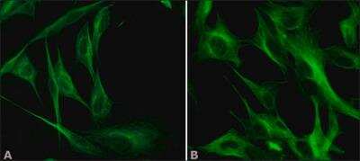
alpha Tubulin 4a Antibody (B-5-1-2) - BSA Free [NB120-11304] - Chicken fibroblasts cells were fixed and permeabilized with methanol followed by acetone. Fixed cells were stained with 1 ug/mL Anti-alpha-Tubulin antibody, Mouse monoclonal, Clone: DM1A (A). The antibody was developed using 1:40 Goat Anti-Mouse IgG (Fab specific)-FITC antibody and 1 ug/mL Monoclonal Anti-alpha-Tubulin antibody produced in Mouse, Clone: B-5-1-2 (B). The antibody was developed using 1:40 Goat Anti-Mouse IgG (Fab specific)-FITC antibodyResults:Two Anti-alpha-Tubulin antibodies, T6199 (A) and T6074 (B) target different regions of alpha-Tubulin show similar staining profiles between the two antibodies, demonstrating Independent Antibody Verification.
Applications for alpha Tubulin 4a Antibody (B-5-1-2) - BSA Free
Application
Recommended Usage
Immunocytochemistry/ Immunofluorescence
0.5-1 ug/ml
Immunoprecipitation
1:10-1:500
Western Blot
0.25-0.5 ug/ml
Application Notes
ICC: Concentration is recommended using cultured chicken fibroblasts (CFB). WB: Concentration is recommended using a total cell extract of the human foreskin fibroblast cell line (FS11). Predicted molecular weight: 50 kDa.
See Simple Western Antibody Database for Simple Western validation: tested in Caco2 cell lysates; antibody dilution of 1:50; separated by size
See Simple Western Antibody Database for Simple Western validation: tested in Caco2 cell lysates; antibody dilution of 1:50; separated by size
Formulation, Preparation, and Storage
Purification
Protein A or G purified
Formulation
10mM PBS (pH 7.4)
Format
BSA Free
Preservative
0.09% Sodium Azide
Concentration
2.0 mg/ml
Shipping
The product is shipped with polar packs. Upon receipt, store it immediately at the temperature recommended below.
Stability & Storage
Store at 4C short term. Aliquot and store at -20C long term. Avoid freeze-thaw cycles.
Background: alpha Tubulin 4a
Long Name
Tubulin alpha-4A chain
Alternate Names
Alpha-tubulin 1, TUBA1, TUBA4A, Tubulin H2-alpha
Gene Symbol
TUBA4A
Additional alpha Tubulin 4a Products
Product Documents for alpha Tubulin 4a Antibody (B-5-1-2) - BSA Free
Certificate of Analysis
To download a Certificate of Analysis, please enter a lot or batch number in the search box below.
Product Specific Notices for alpha Tubulin 4a Antibody (B-5-1-2) - BSA Free
This product is for research use only and is not approved for use in humans or in clinical diagnosis. Primary Antibodies are guaranteed for 1 year from date of receipt.
Citations for alpha Tubulin 4a Antibody (B-5-1-2) - BSA Free
 Powered by Bioz
Customer Reviews for alpha Tubulin 4a Antibody (B-5-1-2) - BSA Free
There are currently no reviews for this product. Be the first to review alpha Tubulin 4a Antibody (B-5-1-2) - BSA Free and earn rewards!
Have you used alpha Tubulin 4a Antibody (B-5-1-2) - BSA Free?
Submit a review and receive an Amazon gift card!
$25/€18/£15/$25CAN/¥2500 Yen for a review with an image
$10/€7/£6/$10CAN/¥1110 Yen for a review without an image
Submit a review
Protocols
Find general support by application which include: protocols, troubleshooting, illustrated assays, videos and webinars.
- Appropriate Fixation of IHC/ICC Samples
- Cellular Response to Hypoxia Protocols
- ClariTSA™ Fluorophore Kits
- Detection & Visualization of Antibody Binding
- ICC Cell Smear Protocol for Suspension Cells
- ICC Immunocytochemistry Protocol Videos
- ICC for Adherent Cells
- Immunocytochemistry (ICC) Protocol
- Immunocytochemistry Troubleshooting
- Immunofluorescence of Organoids Embedded in Cultrex Basement Membrane Extract
- Immunohistochemistry (IHC) and Immunocytochemistry (ICC) Protocols
- Immunoprecipitation Protocol
- Preparing Samples for IHC/ICC Experiments
- Preventing Non-Specific Staining (Non-Specific Binding)
- Primary Antibody Selection & Optimization
- Protocol for VisUCyte™ HRP Polymer Detection Reagent
- Protocol for the Fluorescent ICC Staining of Cell Smears - Graphic
- Protocol for the Fluorescent ICC Staining of Cultured Cells on Coverslips - Graphic
- Protocol for the Preparation and Fluorescent ICC Staining of Cells on Coverslips
- Protocol for the Preparation and Fluorescent ICC Staining of Non-adherent Cells
- Protocol for the Preparation and Fluorescent ICC Staining of Stem Cells on Coverslips
- Protocol for the Preparation of a Cell Smear for Non-adherent Cell ICC - Graphic
- R&D Systems Quality Control Western Blot Protocol
- TUNEL and Active Caspase-3 Detection by IHC/ICC Protocol
- The Importance of IHC/ICC Controls
- Troubleshooting Guide: Western Blot Figures
- Western Blot Conditions
- Western Blot Protocol
- Western Blot Protocol for Cell Lysates
- Western Blot Troubleshooting
- Western Blot Troubleshooting Guide
- View all Protocols, Troubleshooting, Illustrated assays and Webinars
Loading...