Key Product Details
Species Reactivity
Human, Mouse, Rat
Applications
Immunohistochemistry, Immunohistochemistry-Paraffin, Western Blot
Label
HRP
Antibody Source
Monoclonal Mouse IgG1 Clone # 961258
Loading...
Product Specifications
Immunogen
This alpha Tubulin Antibody (961258) was developed against alpha Tubulin.
Clonality
Monoclonal
Host
Mouse
Isotype
IgG1
Scientific Data Images for alpha Tubulin Antibody (961258) [HRP]
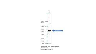
Western Blot: alpha Tubulin Antibody (961258) [HRP] [MAB93441H] - Western Blot using MAB93441. Image from customer review.
Applications for alpha Tubulin Antibody (961258) [HRP]
Application
Recommended Usage
Immunohistochemistry
Optimal dilutions of this antibody should be experimentally determined.
Immunohistochemistry-Paraffin
Optimal dilutions of this antibody should be experimentally determined.
Western Blot
Optimal dilutions of this antibody should be experimentally determined.
Application Notes
Optimal dilution of this antibody should be experimentally determined.
Reviewed Applications
Read 1 review rated 5 using MAB93441H in the following applications:
Formulation, Preparation, and Storage
Purification
Protein A or G purified
Formulation
PBS
Preservative
No Preservative
Concentration
Please see the vial label for concentration. If unlisted please contact technical services.
Shipping
The product is shipped with polar packs. Upon receipt, store it immediately at the temperature recommended below.
Stability & Storage
Store at 4C in the dark.
Background: alpha Tubulin
Tyrosine ligase adds a C-terminal tyrosine to monomeric alpha tubulin. Assembled microtubules can again be detyrosinated by a cytoskeleton associated carboxypeptidase. Detyrosinated alpha tubulin is referred to as Glu-tubulin. Another post-translational modification of detyrosinated alpha tubulin is C-terminal polyglutamylation which is characteristic for microtubules in neuronal cells and the mitotic spindle.
Like GAPDH and beta-actin, alpha/beta tubulin is often used as a loading control in immunoblot applications (1). Alpha/beta tubulin is also good for counterstaining microtubules in immunofluorescence (2).
References
1. Hannen, R., Selmansberger, M., Hauswald, M., Pagenstecher, A., Nist, A., Stiewe, T.,... Bartsch, J. W. (2019). Comparative Transcriptomic Analysis of Temozolomide Resistant Primary GBM Stem-Like Cells and Recurrent GBM Identifies Up-Regulation of the Carbonic Anhydrase CA2 Gene as Resistance Factor. Cancers (Basel), 11(7). doi:10.3390/cancers11070921
2. Nel, M., Joubert, A. M., Dohle, W., Potter, B. V., & Theron, A. E. (2018). Modes of cell death induced by tetrahydroisoquinoline-based analogs in MDA-MB-231 breast and A549 lung cancer cell lines. Drug Des Devel Ther, 12, 1881-1904. doi:10.2147/dddt.S152718
Long Name
Tubulin Alpha 1a
Alternate Names
Alpha-Tubulin 3, B-ALPHA-1, LIS3, TUBA1A, TUBA3, Tubulin B-Alpha-1
Gene Symbol
TUBA1A
Additional alpha Tubulin Products
Product Documents for alpha Tubulin Antibody (961258) [HRP]
Certificate of Analysis
To download a Certificate of Analysis, please enter a lot or batch number in the search box below.
Product Specific Notices for alpha Tubulin Antibody (961258) [HRP]
This product is for research use only and is not approved for use in humans or in clinical diagnosis. Primary Antibodies are guaranteed for 1 year from date of receipt.
Related Research Areas
Customer Reviews for alpha Tubulin Antibody (961258) [HRP] (1)
5 out of 5
1 Customer Rating
Have you used alpha Tubulin Antibody (961258) [HRP]?
Submit a review and receive an Amazon gift card!
$25/€18/£15/$25CAN/¥2500 Yen for a review with an image
$10/€7/£6/$10CAN/¥1110 Yen for a review without an image
Submit a review
Customer Images
![alpha Tubulin Antibody (961258) [HRP] MAB93441H](https://resources.rndsystems.com/images/reviews/review_mab93441h_53821_0_0.jpg)
Showing
1
-
1 of
1 review
Showing All
Filter By:
-
Application: Western BlotSample Tested: Human cancer cellSpecies: HumanVerified Customer | Posted 10/02/2020Super strong and clean antibody for loading control. It saves my time in western blot assays because no secondary antibody needed. Highly recommended.
![alpha Tubulin Antibody (961258) [HRP] MAB93441H](data:image/png;base64,R0lGODlhAQABAAD/ACwAAAAAAQABAAACADs=)
There are no reviews that match your criteria.
Protocols
Find general support by application which include: protocols, troubleshooting, illustrated assays, videos and webinars.
- Antigen Retrieval Protocol (PIER)
- Antigen Retrieval for Frozen Sections Protocol
- Appropriate Fixation of IHC/ICC Samples
- Cellular Response to Hypoxia Protocols
- Chromogenic IHC Staining of Formalin-Fixed Paraffin-Embedded (FFPE) Tissue Protocol
- Chromogenic Immunohistochemistry Staining of Frozen Tissue
- ClariTSA™ Fluorophore Kits
- Detection & Visualization of Antibody Binding
- Fluorescent IHC Staining of Frozen Tissue Protocol
- Graphic Protocol for Heat-induced Epitope Retrieval
- Graphic Protocol for the Preparation and Fluorescent IHC Staining of Frozen Tissue Sections
- Graphic Protocol for the Preparation and Fluorescent IHC Staining of Paraffin-embedded Tissue Sections
- Graphic Protocol for the Preparation of Gelatin-coated Slides for Histological Tissue Sections
- IHC Sample Preparation (Frozen sections vs Paraffin)
- Immunofluorescent IHC Staining of Formalin-Fixed Paraffin-Embedded (FFPE) Tissue Protocol
- Immunohistochemistry (IHC) and Immunocytochemistry (ICC) Protocols
- Immunohistochemistry Frozen Troubleshooting
- Immunohistochemistry Paraffin Troubleshooting
- Preparing Samples for IHC/ICC Experiments
- Preventing Non-Specific Staining (Non-Specific Binding)
- Primary Antibody Selection & Optimization
- Protocol for Heat-Induced Epitope Retrieval (HIER)
- Protocol for Making a 4% Formaldehyde Solution in PBS
- Protocol for VisUCyte™ HRP Polymer Detection Reagent
- Protocol for the Preparation & Fixation of Cells on Coverslips
- Protocol for the Preparation and Chromogenic IHC Staining of Frozen Tissue Sections
- Protocol for the Preparation and Chromogenic IHC Staining of Frozen Tissue Sections - Graphic
- Protocol for the Preparation and Chromogenic IHC Staining of Paraffin-embedded Tissue Sections
- Protocol for the Preparation and Chromogenic IHC Staining of Paraffin-embedded Tissue Sections - Graphic
- Protocol for the Preparation and Fluorescent IHC Staining of Frozen Tissue Sections
- Protocol for the Preparation and Fluorescent IHC Staining of Paraffin-embedded Tissue Sections
- Protocol for the Preparation of Gelatin-coated Slides for Histological Tissue Sections
- R&D Systems Quality Control Western Blot Protocol
- TUNEL and Active Caspase-3 Detection by IHC/ICC Protocol
- The Importance of IHC/ICC Controls
- Troubleshooting Guide: Immunohistochemistry
- Troubleshooting Guide: Western Blot Figures
- Western Blot Conditions
- Western Blot Protocol
- Western Blot Protocol for Cell Lysates
- Western Blot Troubleshooting
- Western Blot Troubleshooting Guide
- View all Protocols, Troubleshooting, Illustrated assays and Webinars
Loading...