APLP-2 Antibody - BSA Free
Novus Biologicals | Catalog # NBP1-89029
![Immunocytochemistry/ Immunofluorescence: APLP-2 Antibody [NBP1-89029]](https://resources.rndsystems.com/images/products/APLP-2-Antibody-Immunocytochemistry-Immunofluorescence-NBP1-89029-img0005.jpg "Immunocytochemistry/ Immunofluorescence: APLP-2 Antibody [NBP1-89029]")
Loading...
Key Product Details
Species Reactivity
Validated:
Human
Cited:
Mouse
Predicted:
Mouse (93%), Rat (95%). Backed by our 100% Guarantee.
Applications
Validated:
Immunohistochemistry-Paraffin, Immunocytochemistry/ Immunofluorescence
Cited:
Western Blot
Label
Unconjugated
Antibody Source
Polyclonal Rabbit IgG
Format
BSA Free
Loading...
Product Specifications
Immunogen
This antibody was developed against Recombinant Protein corresponding to amino acids: PYVAQEIQEEIDELLQEQRADMDQFTASISETPVDVRVSSEESEEIPPFHPFHPFPALPENEDTQPELYHPMK
Clonality
Polyclonal
Host
Rabbit
Isotype
IgG
Theoretical MW
87 kDa.
Disclaimer note: The observed molecular weight of the protein may vary from the listed predicted molecular weight due to post translational modifications, post translation cleavages, relative charges, and other experimental factors.
Disclaimer note: The observed molecular weight of the protein may vary from the listed predicted molecular weight due to post translational modifications, post translation cleavages, relative charges, and other experimental factors.
Scientific Data Images for APLP-2 Antibody - BSA Free
Immunocytochemistry/ Immunofluorescence: APLP-2 Antibody [NBP1-89029]
Immunocytochemistry/Immunofluorescence: APLP-2 Antibody [NBP1-89029] - Staining of human cell line U-2 OS shows localization to vesicles. Antibody staining is shown in green.![APLP-2 Antibody - BSA Free Immunohistochemistry-Paraffin: APLP-2 Antibody [NBP1-89029]](https://resources.rndsystems.com/images/products/nbp1-89029_-immunohistochemistry-paraffin-639173123471511906.jpg "Immunohistochemistry-Paraffin: APLP-2 Antibody [NBP1-89029]")
Immunohistochemistry-Paraffin: APLP-2 Antibody [NBP1-89029]
Staining of human testis shows strong cytoplasmic granular positivity in cells in seminiferous ducts.![APLP-2 Antibody - BSA Free Immunohistochemistry-Paraffin: APLP-2 Antibody [NBP1-89029]](https://resources.rndsystems.com/images/products/nbp1-89029_-immunohistochemistry-paraffin-639173124737519656.jpg "Immunohistochemistry-Paraffin: APLP-2 Antibody [NBP1-89029]")
Immunohistochemistry-Paraffin: APLP-2 Antibody [NBP1-89029]
Staining of human kidney shows strong cytoplasmic granular positivity in cells in tubules.![APLP-2 Antibody - BSA Free Immunohistochemistry-Paraffin: APLP-2 Antibody [NBP1-89029]](https://resources.rndsystems.com/images/products/nbp1-89029_-immunohistochemistry-paraffin-639173125146756173.jpg "Immunohistochemistry-Paraffin: APLP-2 Antibody [NBP1-89029]")
Immunohistochemistry-Paraffin: APLP-2 Antibody [NBP1-89029]
Staining of human placenta shows strong cytoplasmic granular positivity in trophoblastic cells.![APLP-2 Antibody - BSA Free Immunohistochemistry-Paraffin: APLP-2 Antibody [NBP1-89029]](https://resources.rndsystems.com/images/products/nbp1-89029_-immunohistochemistry-paraffin-639173132642858836.jpg "Immunohistochemistry-Paraffin: APLP-2 Antibody [NBP1-89029]")
Immunohistochemistry-Paraffin: APLP-2 Antibody [NBP1-89029]
Staining of human duodenum shows strong cytoplasmic granular positivity in glandular cells.Applications for APLP-2 Antibody - BSA Free
Application
Recommended Usage
Immunocytochemistry/ Immunofluorescence
0.25-2 ug/ml
Immunohistochemistry-Paraffin
1:50 - 1:200
Application Notes
For IHC-Paraffin, HIER pH 6 retrieval is recommended. ICC/IF Fixation Permeabilization: Use PFA/Triton X-100.
Reviewed Applications
Read 1 review rated 5 using NBP1-89029 in the following applications:
Formulation, Preparation, and Storage
Purification
Affinity purified
Formulation
PBS (pH 7.2) and 40% Glycerol
Format
BSA Free
Preservative
0.02% Sodium Azide
Concentration
Concentrations vary lot to lot. See vial label for concentration. If unlisted please contact technical services.
Shipping
The product is shipped with polar packs. Upon receipt, store it immediately at the temperature recommended below.
Stability & Storage
Store at 4C short term. Aliquot and store at -20C long term. Avoid freeze-thaw cycles.
Background: APLP-2
Long Name
Amyloid Beta [A4] Precursor-like Protein 2
Alternate Names
APLP2, APPH, APPL2, CDEBP, WALPLP2, YWK-II
Entrez Gene IDs
334 (Human)
Gene Symbol
APLP2
UniProt
Additional APLP-2 Products
Product Documents for APLP-2 Antibody - BSA Free
Certificate of Analysis
To download a Certificate of Analysis, please enter a lot or batch number in the search box below.
Product Specific Notices for APLP-2 Antibody - BSA Free
This product is for research use only and is not approved for use in humans or in clinical diagnosis. Primary Antibodies are guaranteed for 1 year from date of receipt.
Citations for APLP-2 Antibody - BSA Free
 Powered by Bioz
Customer Reviews for APLP-2 Antibody - BSA Free (1)
5 out of 5
1 Customer Rating
Have you used APLP-2 Antibody - BSA Free?
Submit a review and receive an Amazon gift card!
$25/€18/£15/$25CAN/¥2500 Yen for a review with an image
$10/€7/£6/$10CAN/¥1110 Yen for a review without an image
Submit a review
Customer Images
Showing
1
-
1 of
1 review
Showing All
Filter By:
-
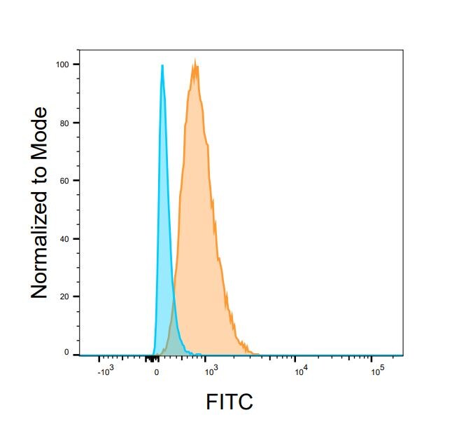
Application: Flow CytometrySample Tested: U937 human histiocytic lymphoma cell lineSpecies: HumanVerified Customer | Posted 10/17/2023U937 cells were surface stained with 2ug/mL APLP2 antibody (orange) and matched isotype control (blue) for 30 minutes at 4 degrees Celsius prior to secondary antibody stain. Isotype control and secondary antibodies were conjugated to FITC.
 Bio-Techne ResponseThis review was submitted through the legacy Novus Innovators Program, reflecting a new species or application tested on a primary antibody.
There are no reviews that match your criteria.
Protocols
Find general support by application which include: protocols, troubleshooting, illustrated assays, videos and webinars.
- Antigen Retrieval Protocol (PIER)
- Antigen Retrieval for Frozen Sections Protocol
- Appropriate Fixation of IHC/ICC Samples
- Cellular Response to Hypoxia Protocols
- Chromogenic IHC Staining of Formalin-Fixed Paraffin-Embedded (FFPE) Tissue Protocol
- Chromogenic Immunohistochemistry Staining of Frozen Tissue
- ClariTSA™ Fluorophore Kits
- Detection & Visualization of Antibody Binding
- Fluorescent IHC Staining of Frozen Tissue Protocol
- Graphic Protocol for Heat-induced Epitope Retrieval
- Graphic Protocol for the Preparation and Fluorescent IHC Staining of Frozen Tissue Sections
- Graphic Protocol for the Preparation and Fluorescent IHC Staining of Paraffin-embedded Tissue Sections
- Graphic Protocol for the Preparation of Gelatin-coated Slides for Histological Tissue Sections
- ICC Cell Smear Protocol for Suspension Cells
- ICC Immunocytochemistry Protocol Videos
- ICC for Adherent Cells
- IHC Sample Preparation (Frozen sections vs Paraffin)
- Immunocytochemistry (ICC) Protocol
- Immunocytochemistry Troubleshooting
- Immunofluorescence of Organoids Embedded in Cultrex Basement Membrane Extract
- Immunofluorescent IHC Staining of Formalin-Fixed Paraffin-Embedded (FFPE) Tissue Protocol
- Immunohistochemistry (IHC) and Immunocytochemistry (ICC) Protocols
- Immunohistochemistry Frozen Troubleshooting
- Immunohistochemistry Paraffin Troubleshooting
- Preparing Samples for IHC/ICC Experiments
- Preventing Non-Specific Staining (Non-Specific Binding)
- Primary Antibody Selection & Optimization
- Protocol for Heat-Induced Epitope Retrieval (HIER)
- Protocol for Making a 4% Formaldehyde Solution in PBS
- Protocol for VisUCyte™ HRP Polymer Detection Reagent
- Protocol for the Fluorescent ICC Staining of Cell Smears - Graphic
- Protocol for the Fluorescent ICC Staining of Cultured Cells on Coverslips - Graphic
- Protocol for the Preparation & Fixation of Cells on Coverslips
- Protocol for the Preparation and Chromogenic IHC Staining of Frozen Tissue Sections
- Protocol for the Preparation and Chromogenic IHC Staining of Frozen Tissue Sections - Graphic
- Protocol for the Preparation and Chromogenic IHC Staining of Paraffin-embedded Tissue Sections
- Protocol for the Preparation and Chromogenic IHC Staining of Paraffin-embedded Tissue Sections - Graphic
- Protocol for the Preparation and Fluorescent ICC Staining of Cells on Coverslips
- Protocol for the Preparation and Fluorescent ICC Staining of Non-adherent Cells
- Protocol for the Preparation and Fluorescent ICC Staining of Stem Cells on Coverslips
- Protocol for the Preparation and Fluorescent IHC Staining of Frozen Tissue Sections
- Protocol for the Preparation and Fluorescent IHC Staining of Paraffin-embedded Tissue Sections
- Protocol for the Preparation of Gelatin-coated Slides for Histological Tissue Sections
- Protocol for the Preparation of a Cell Smear for Non-adherent Cell ICC - Graphic
- TUNEL and Active Caspase-3 Detection by IHC/ICC Protocol
- The Importance of IHC/ICC Controls
- Troubleshooting Guide: Immunohistochemistry
- View all Protocols, Troubleshooting, Illustrated assays and Webinars
Loading...