ATF3 Antibody - BSA Free
Novus Biologicals | Catalog # NBP1-85816
![Immunocytochemistry/ Immunofluorescence: ATF3 Antibody [NBP1-85816]](https://resources.rndsystems.com/images/products/ATF3-Antibody-Immunocytochemistry-Immunofluorescence-NBP1-85816-img0012.jpg "Immunocytochemistry/ Immunofluorescence: ATF3 Antibody [NBP1-85816]")
Loading...
Key Product Details
Species Reactivity
Validated:
Human, Mouse
Cited:
Human, Mouse, Rat
Predicted:
Rat (92%). Backed by our 100% Guarantee.
Applications
Validated:
Immunohistochemistry-Paraffin, Immunocytochemistry/ Immunofluorescence
Cited:
Immunohistochemistry, Immunohistochemistry-Paraffin, Immunohistochemistry-Frozen, Western Blot, Immunocytochemistry/ Immunofluorescence, Chemotaxis, IF/IHC
Label
Unconjugated
Antibody Source
Polyclonal Rabbit IgG
Format
BSA Free
Loading...
Product Specifications
Immunogen
This antibody was developed against Recombinant Protein corresponding to amino acids: MMLQHPGQVSASEVSASAIVPCLSPPGSLVFEDFANLTPFVKEELRFAIQNKHLCHRMSSALESVTVSDRPLGVSITKAEVAPEEDERKKRRRERNKIAAAKCRNKKKEKTEC
Clonality
Polyclonal
Host
Rabbit
Isotype
IgG
Scientific Data Images for ATF3 Antibody - BSA Free
Immunocytochemistry/ Immunofluorescence: ATF3 Antibody [NBP1-85816]
Immunocytochemistry/Immunofluorescence: ATF3 Antibody [NBP1-85816] - Staining of human cell line A-431 shows localization to nucleus and nucleoli. Antibody staining is shown in green.![ATF3 Antibody - BSA Free Immunohistochemistry-Paraffin: ATF3 Antibody - BSA Free [NBP1-85816]](https://resources.rndsystems.com/images/products/nbp1-85816_rabbit-polyclonal-atf3-antibody-23420251573932.jpg "Immunohistochemistry-Paraffin: ATF3 Antibody - BSA Free [NBP1-85816]")
Immunohistochemistry-Paraffin: ATF3 Antibody - BSA Free [NBP1-85816]
Staining of mouse dorsal root ganglion shows strong nuclear immunoreactivity in single neurons.![ATF3 Antibody - BSA Free Immunohistochemistry-Paraffin: ATF3 Antibody - BSA Free [NBP1-85816]](https://resources.rndsystems.com/images/products/nbp1-85816_rabbit-polyclonal-atf3-antibody-23420251592520.jpg "Immunohistochemistry-Paraffin: ATF3 Antibody - BSA Free [NBP1-85816]")
Immunohistochemistry-Paraffin: ATF3 Antibody - BSA Free [NBP1-85816]
Staining of human skeletal muscle shows very weak nucleus-cytoplasmic positivity in myocytes.![ATF3 Antibody - BSA Free Immunocytochemistry/ Immunofluorescence: ATF3 Antibody - BSA Free [NBP1-85816]](https://resources.rndsystems.com/images/products/nbp1-85816_-immunocytochemistry-immunofluorescence-639192267705482787.jpg "Immunocytochemistry/ Immunofluorescence: ATF3 Antibody - BSA Free [NBP1-85816]")
Immunocytochemistry/ Immunofluorescence: ATF3 Antibody - BSA Free [NBP1-85816]
Staining of human cell line A-431 shows localization to nucleus & nucleoli.![ATF3 Antibody - BSA Free Immunohistochemistry-Paraffin: ATF3 Antibody - BSA Free [NBP1-85816]](https://resources.rndsystems.com/images/products/nbp1-85816_-immunohistochemistry-paraffin-639192267705801840.jpg "Immunohistochemistry-Paraffin: ATF3 Antibody - BSA Free [NBP1-85816]")
Immunohistochemistry-Paraffin: ATF3 Antibody - BSA Free [NBP1-85816]
Staining of human fallopian tube shows moderate nuclear positivity in a subset of glandular cells with weak cytoplasmic positivity.![ATF3 Antibody - BSA Free Immunohistochemistry-Paraffin: ATF3 Antibody - BSA Free [NBP1-85816]](https://resources.rndsystems.com/images/products/nbp1-85816_-immunohistochemistry-paraffin-639192267706111772.jpg "Immunohistochemistry-Paraffin: ATF3 Antibody - BSA Free [NBP1-85816]")
Immunohistochemistry-Paraffin: ATF3 Antibody - BSA Free [NBP1-85816]
Staining of human placenta shows strong nuclear positivity in trophoblastic cells with weak cytoplasmic positivity.![ATF3 Antibody - BSA Free Immunohistochemistry-Paraffin: ATF3 Antibody - BSA Free [NBP1-85816]](https://resources.rndsystems.com/images/products/nbp1-85816_-immunohistochemistry-paraffin-639192267709098476.jpg "Immunohistochemistry-Paraffin: ATF3 Antibody - BSA Free [NBP1-85816]")
Immunohistochemistry-Paraffin: ATF3 Antibody - BSA Free [NBP1-85816]
Staining of human skeletal muscle shows very weak nucleus-cytoplasmic positivity in myocytes.![ATF3 Antibody - BSA Free Immunohistochemistry-Paraffin: ATF3 Antibody - BSA Free [NBP1-85816]](https://resources.rndsystems.com/images/products/nbp1-85816_-immunohistochemistry-paraffin-639192267707267296.jpg "Immunohistochemistry-Paraffin: ATF3 Antibody - BSA Free [NBP1-85816]")
Immunohistochemistry-Paraffin: ATF3 Antibody - BSA Free [NBP1-85816]
Staining of human urinary bladder shows moderate nuclear positivity in epithelial cells with weak cytoplasmic positivity.![ATF3 Antibody - BSA Free Immunohistochemistry-Paraffin: ATF3 Antibody - BSA Free [NBP1-85816]](https://resources.rndsystems.com/images/products/nbp1-85816_-immunohistochemistry-paraffin-639192267708688329.jpg "Immunohistochemistry-Paraffin: ATF3 Antibody - BSA Free [NBP1-85816]")
Immunohistochemistry-Paraffin: ATF3 Antibody - BSA Free [NBP1-85816]
Staining of mouse dorsal root ganglion shows strong nuclear immunoreactivity in single neurons.Applications for ATF3 Antibody - BSA Free
Application
Recommended Usage
Immunocytochemistry/ Immunofluorescence
0.25-2 ug/ml
Immunohistochemistry-Paraffin
1:200 - 1:500
Application Notes
IHC-Paraffin, HIER pH 6 retrieval is recommended. ICC/IF, Fixation Permeabilization: Use PFA/Triton X-100.
Reviewed Applications
Read 3 reviews rated 4.3 using NBP1-85816 in the following applications:
Formulation, Preparation, and Storage
Purification
Affinity purified
Formulation
PBS (pH 7.2) and 40% Glycerol
Format
BSA Free
Preservative
0.02% Sodium Azide
Concentration
Concentrations vary lot to lot. See vial label for concentration. If unlisted please contact technical services.
Shipping
The product is shipped with polar packs. Upon receipt, store it immediately at the temperature recommended below.
Stability & Storage
Store at 4C short term. Aliquot and store at -20C long term. Avoid freeze-thaw cycles.
Background: ATF3
Alternate Names
activating transcription factor 3cAMP-dependent transcription factor ATF-3, ATF3deltaZip2, ATF3deltaZip2c, ATF3deltaZip3, cyclic AMP-dependent transcription factor ATF-3
Gene Symbol
ATF3
Additional ATF3 Products
Product Documents for ATF3 Antibody - BSA Free
Certificate of Analysis
To download a Certificate of Analysis, please enter a lot or batch number in the search box below.
Product Specific Notices for ATF3 Antibody - BSA Free
This product is for research use only and is not approved for use in humans or in clinical diagnosis. Primary Antibodies are guaranteed for 1 year from date of receipt.
Citations for ATF3 Antibody - BSA Free
 Powered by Bioz
Customer Reviews for ATF3 Antibody - BSA Free (3)
4.3 out of 5
3 Customer Ratings
Have you used ATF3 Antibody - BSA Free?
Submit a review and receive an Amazon gift card!
$25/€18/£15/$25CAN/¥2500 Yen for a review with an image
$10/€7/£6/$10CAN/¥1110 Yen for a review without an image
Submit a review
Customer Images
Showing
1
-
3 of
3 reviews
Showing All
Filter By:
-
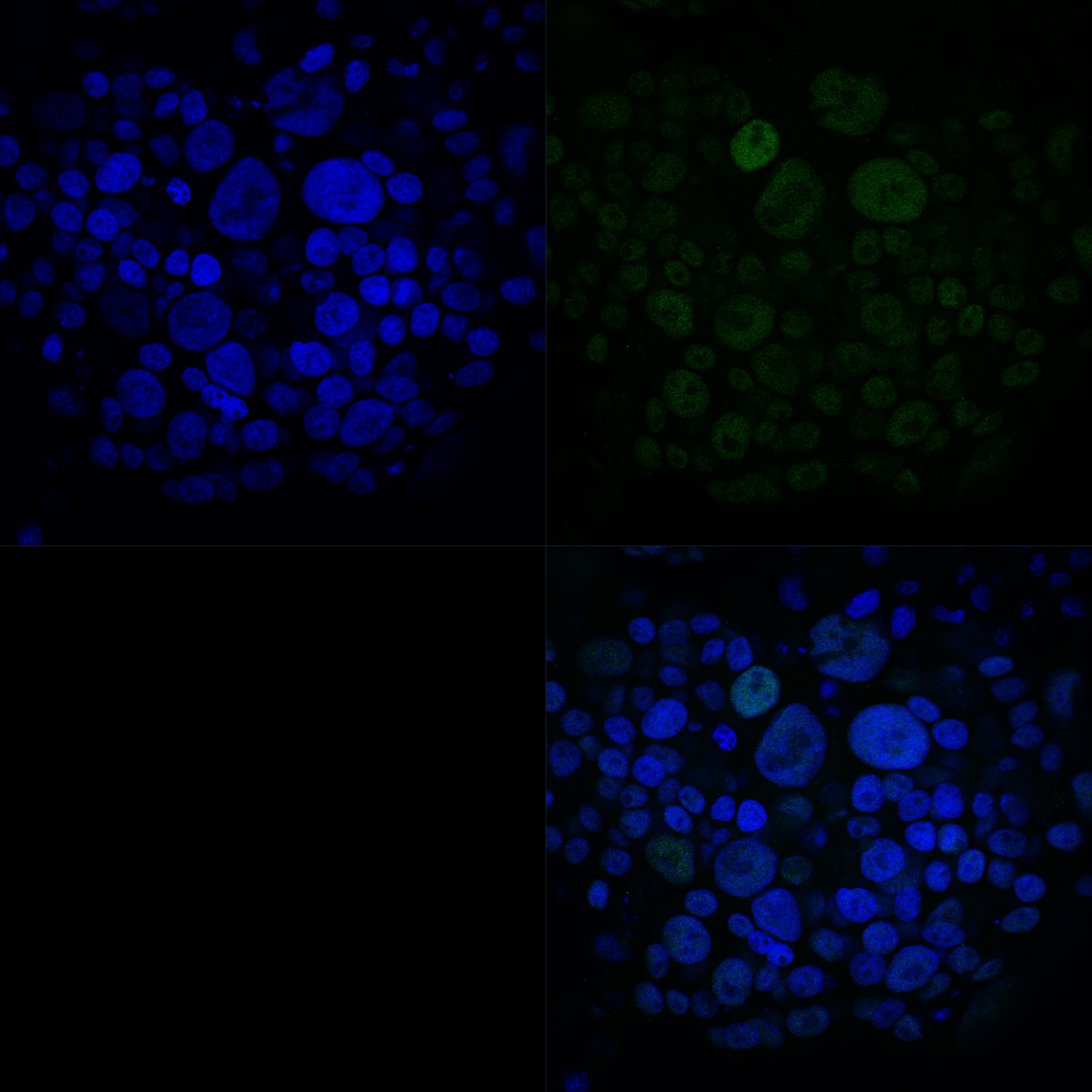
Application: Indirect ImmunofluorescenceSample Tested: Cell cultureSpecies: HumanVerified Customer | Posted 05/03/2022ATF3 antibody in indirect immunofluorescence staining of human tumor cells using a green-fluorescent secondary antibody showing nuclear localization. Nuclei stained with DAPI (blue).

-
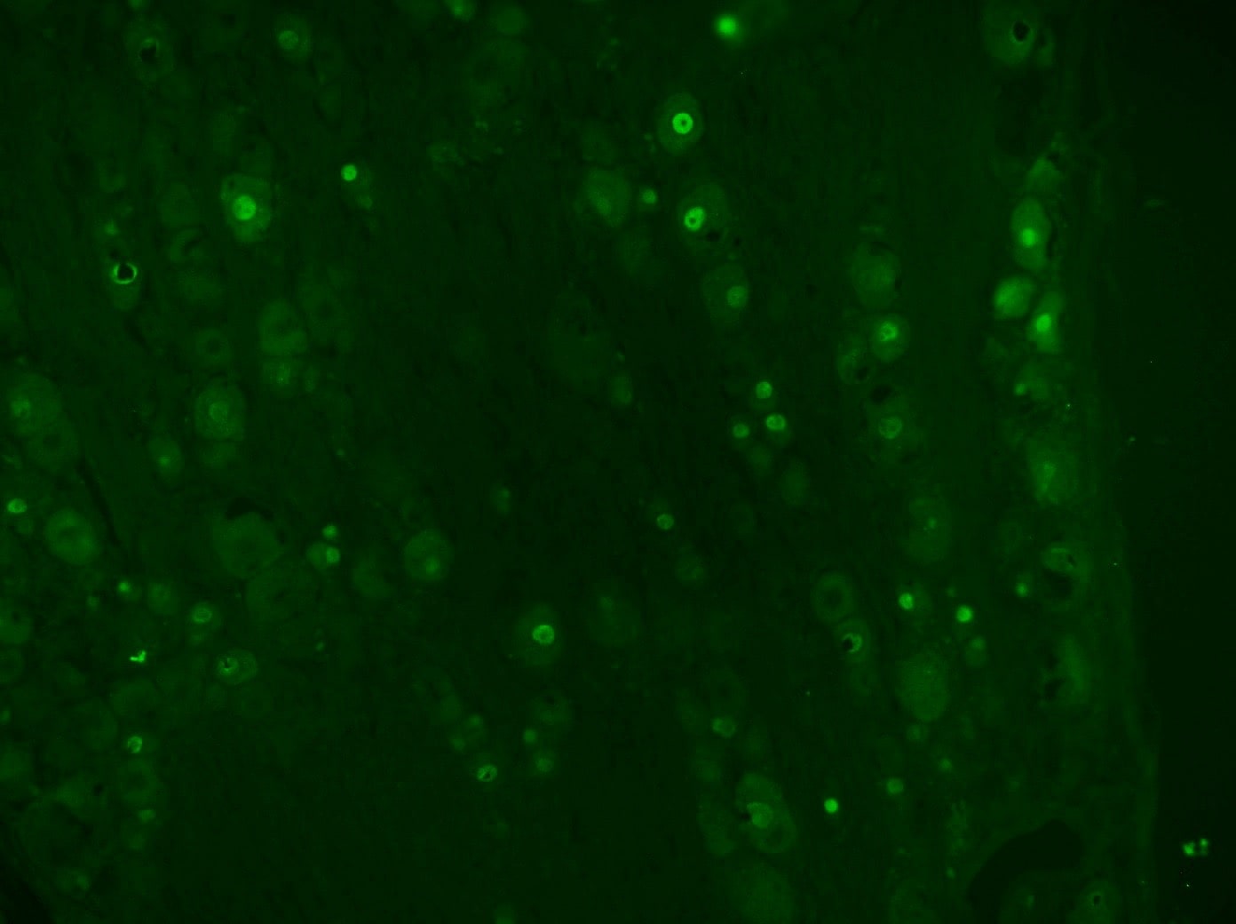
Application: Immunohistochemistry-FrozenSample Tested: Optic nerveSpecies: FelineVerified Customer | Posted 10/19/2018Image was captured under an epi-fluorescent microscope. Alexa 488 conjugated rabbit antibody was used for the secondary antibody. Immunofluorescent signal was localized in the nucleus.
-
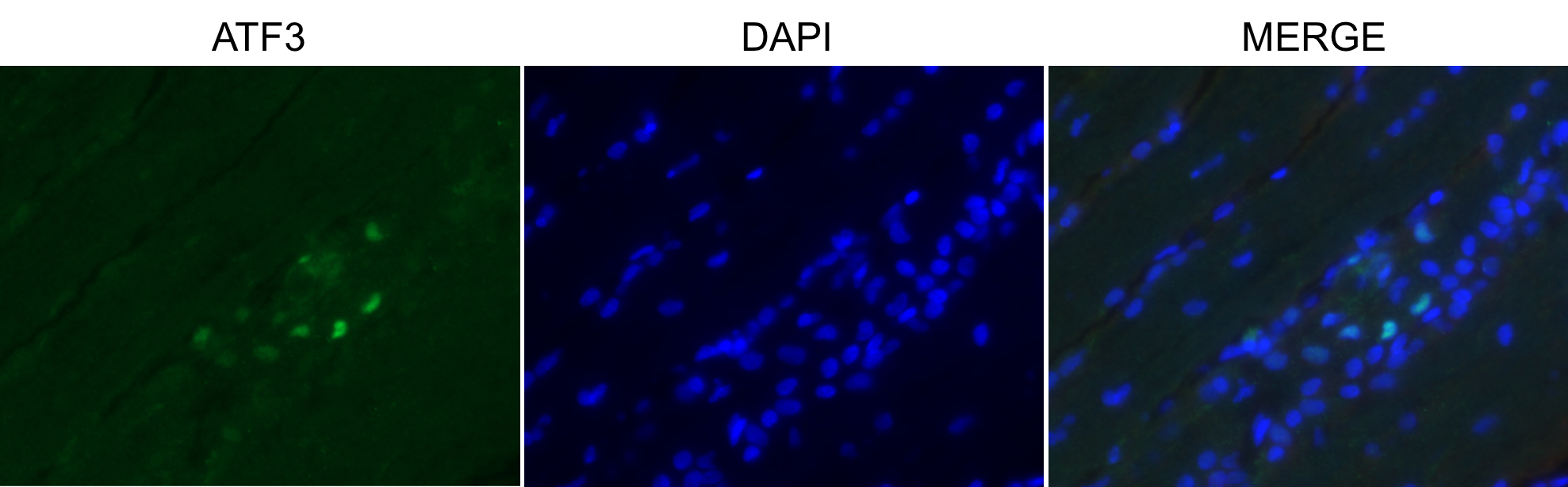
Application: Immunohistochemistry-FrozenSample Tested: Dorsal root ganglionSpecies: RatVerified Customer | Posted 09/15/2017ATF3 in rat DRG 14 days after SNIFixed frozen rat DRG sectioned at 10um and slide mounted. IHC of ATF3 (1:500) incubated overnight at RT after 1 hour blocking. Secondary incubation at RT for 1 hour using Alexa 488 goat anti-rabbit IgG at 1:1000. Snapshot taken with epifluorescent microscope at 20x. Concurrently stained contralateral image (not shown) showed no ATF3 immunoreactivity.
There are no reviews that match your criteria.
Protocols
Find general support by application which include: protocols, troubleshooting, illustrated assays, videos and webinars.
- Antigen Retrieval Protocol (PIER)
- Antigen Retrieval for Frozen Sections Protocol
- Appropriate Fixation of IHC/ICC Samples
- Cellular Response to Hypoxia Protocols
- Chromogenic IHC Staining of Formalin-Fixed Paraffin-Embedded (FFPE) Tissue Protocol
- Chromogenic Immunohistochemistry Staining of Frozen Tissue
- ClariTSA™ Fluorophore Kits
- Detection & Visualization of Antibody Binding
- Fluorescent IHC Staining of Frozen Tissue Protocol
- Graphic Protocol for Heat-induced Epitope Retrieval
- Graphic Protocol for the Preparation and Fluorescent IHC Staining of Frozen Tissue Sections
- Graphic Protocol for the Preparation and Fluorescent IHC Staining of Paraffin-embedded Tissue Sections
- Graphic Protocol for the Preparation of Gelatin-coated Slides for Histological Tissue Sections
- ICC Cell Smear Protocol for Suspension Cells
- ICC Immunocytochemistry Protocol Videos
- ICC for Adherent Cells
- IHC Sample Preparation (Frozen sections vs Paraffin)
- Immunocytochemistry (ICC) Protocol
- Immunocytochemistry Troubleshooting
- Immunofluorescence of Organoids Embedded in Cultrex Basement Membrane Extract
- Immunofluorescent IHC Staining of Formalin-Fixed Paraffin-Embedded (FFPE) Tissue Protocol
- Immunohistochemistry (IHC) and Immunocytochemistry (ICC) Protocols
- Immunohistochemistry Frozen Troubleshooting
- Immunohistochemistry Paraffin Troubleshooting
- Preparing Samples for IHC/ICC Experiments
- Preventing Non-Specific Staining (Non-Specific Binding)
- Primary Antibody Selection & Optimization
- Protocol for Heat-Induced Epitope Retrieval (HIER)
- Protocol for Making a 4% Formaldehyde Solution in PBS
- Protocol for VisUCyte™ HRP Polymer Detection Reagent
- Protocol for the Fluorescent ICC Staining of Cell Smears - Graphic
- Protocol for the Fluorescent ICC Staining of Cultured Cells on Coverslips - Graphic
- Protocol for the Preparation & Fixation of Cells on Coverslips
- Protocol for the Preparation and Chromogenic IHC Staining of Frozen Tissue Sections
- Protocol for the Preparation and Chromogenic IHC Staining of Frozen Tissue Sections - Graphic
- Protocol for the Preparation and Chromogenic IHC Staining of Paraffin-embedded Tissue Sections
- Protocol for the Preparation and Chromogenic IHC Staining of Paraffin-embedded Tissue Sections - Graphic
- Protocol for the Preparation and Fluorescent ICC Staining of Cells on Coverslips
- Protocol for the Preparation and Fluorescent ICC Staining of Non-adherent Cells
- Protocol for the Preparation and Fluorescent ICC Staining of Stem Cells on Coverslips
- Protocol for the Preparation and Fluorescent IHC Staining of Frozen Tissue Sections
- Protocol for the Preparation and Fluorescent IHC Staining of Paraffin-embedded Tissue Sections
- Protocol for the Preparation of Gelatin-coated Slides for Histological Tissue Sections
- Protocol for the Preparation of a Cell Smear for Non-adherent Cell ICC - Graphic
- TUNEL and Active Caspase-3 Detection by IHC/ICC Protocol
- The Importance of IHC/ICC Controls
- Troubleshooting Guide: Immunohistochemistry
- View all Protocols, Troubleshooting, Illustrated assays and Webinars
Loading...