ATM [p Ser1981] Antibody (10H11.E12) - BSA Free
Novus Biologicals | Catalog # NB100-306
Key Product Details
Validated by
Biological Validation
Species Reactivity
Validated:
Human, Mouse, Rat, Canine (Negative)
Cited:
Human, Mouse, Rat
Applications
Validated:
Immunohistochemistry, Immunohistochemistry-Paraffin, Western Blot, Immunocytochemistry/ Immunofluorescence, Immunoprecipitation
Cited:
Western Blot, Immunocytochemistry/ Immunofluorescence, IF/IHC
Label
Unconjugated
Antibody Source
Monoclonal Mouse IgG1 kappa Clone # 10H11.E12
Format
BSA Free
Loading...
Product Specifications
Immunogen
ATM [p Ser1981] Antibody (10H11.E12) was made to a synthetic peptide made to a region surrounding the phosphorylated Serine 1981 of human ATM. [UniProt# Q13315]
Modification
p Ser1981
Localization
Nucleus, cytoplasm.
Clonality
Monoclonal
Host
Mouse
Isotype
IgG1 kappa
Theoretical MW
351 kDa.
Disclaimer note: The observed molecular weight of the protein may vary from the listed predicted molecular weight due to post translational modifications, post translation cleavages, relative charges, and other experimental factors.
Disclaimer note: The observed molecular weight of the protein may vary from the listed predicted molecular weight due to post translational modifications, post translation cleavages, relative charges, and other experimental factors.
Scientific Data Images for ATM [p Ser1981] Antibody (10H11.E12) - BSA Free
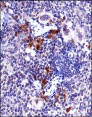
Immunohistochemistry: ATM [p Ser1981] Antibody (10H11.E12) [NB100-306] - ATM [p Ser1981] antibody (10H11.E12) [NB100-306] was tested in mouse spleen using DAB with hematoxylin counterstain.
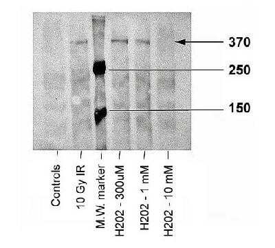
Western Blot: ATM [p Ser1981] Antibody (10H11.E12) [NB100-306] - Analysis of ATM-kinase, using ATM [p Ser1981] antibody (10H11.E12) [NB100-306]. Sample: Irradiated or peroxidated human fibroblasts. Observed molecular weight 370 kDa. Theoretical molecular weight 351 kDa.
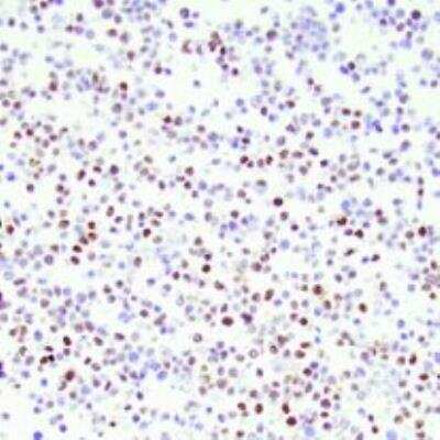
Immunocytochemistry/Immunofluorescence: ATM [p Ser1981] Antibody (10H11.E12) [NB100-306] - Human lymphoblastoid cells stained with ATM [p Ser1981] Antibody (10H11.E12). ICC/IF image submitted by a verified customer review.
![ATM [p Ser1981] Antibody (10H11.E12)](https://resources.rndsystems.com/images/products/nb100-306_mouse-monoclonal-atm-p-ser1981-antibody-10h11-e12-2552023151493.jpg "Western Blot: ATM [p Ser1981] Antibody (10H11.E12) [NB100-306] -")
![ATM [p Ser1981] Antibody (10H11.E12) - BSA Free](https://resources.rndsystems.com/images/products/nb100-306_mouse-monoclonal-atm-p-ser1981-antibody-10h11-e12-immunocytochemistry-immunofluorescence-132202617561220.jpg "Immunocytochemistry/ Immunofluorescence: ATM [p Ser1981] Antibody (10H11.E12) - BSA Free [NB100-306] -")
Immunocytochemistry/ Immunofluorescence: ATM [p Ser1981] Antibody (10H11.E12) - BSA Free [NB100-306] -
MCELNs decreased the DNA damage of H9C2 cells after radiation. (A) Immunofluorescence staining of gamma -H2A.X (green) in H9C2 cells after 48 h of culture with indicated treatment. The nucleus were stained with DAPI (blue), scale bar: 10 μm. Western blot images (B) and quantitation (C) of gamma -H2A.X in H9C2 cells after 48 h of culture with indicated treatment. (D) Immunofluorescence staining of p-ATM (green) in H9C2 cells after 48 h of culture with indicated treatment. The nucleus were stained with DAPI (blue), scale bar: 10 μm. Western blot images (E) and quantitation (F) of p-ATM and ATM in H9C2 cells after 48 h of culture with indicated treatment. IR (–/+): 0/16 Gy X-ray; MCELNs (–/+): 0/10 μg/mL. All data were represented as means +/- SD (n = 3 independent experiments). The statistical significance was evaluated by one-way ANOVA followed by the Turkey's multiple comparisons test among groups. Image collected and cropped by CiteAb from the following open publication (https://pubmed.ncbi.nlm.nih.gov/35509278), licensed under a CC-BY license. Not internally tested by Novus Biologicals.![ATM [p Ser1981] Antibody (10H11.E12) - BSA Free](https://resources.rndsystems.com/images/products/nb100-306_mouse-monoclonal-atm-p-ser1981-antibody-10h11-e12-immunocytochemistry-immunofluorescence-13220261935125.jpg "Immunocytochemistry/ Immunofluorescence: ATM [p Ser1981] Antibody (10H11.E12) - BSA Free [NB100-306] -")
Immunocytochemistry/ Immunofluorescence: ATM [p Ser1981] Antibody (10H11.E12) - BSA Free [NB100-306] -
OOEP is required for ATM activation and RAD51 recruitment to DNA damage sites Image collected and cropped by CiteAb from the following open publication (https://pubmed.ncbi.nlm.nih.gov/29955025), licensed under a CC-BY license. Not internally tested by Novus Biologicals.Applications for ATM [p Ser1981] Antibody (10H11.E12) - BSA Free
Application
Recommended Usage
Immunocytochemistry/ Immunofluorescence
1:1000
Immunohistochemistry
1:200
Immunohistochemistry-Paraffin
1:200
Immunoprecipitation
1:10-1:500
Western Blot
1:1000
Application Notes
This ATM [p Ser1981] (10H11.E12) antibody useful for Immunocytochemistry/Immunofluorescence, Immunoprecipitation, Immunohistochemistry on paraffin-embedded sections and Western blot, where a band at ~370 kDa can be seen. In IHC-P, staining was observed in the nucleus and cytoplasm of mouse spleen.
Reviewed Applications
Read 1 review rated 5 using NB100-306 in the following applications:
Formulation, Preparation, and Storage
Purification
Protein G purified
Formulation
PBS
Format
BSA Free
Preservative
0.05% Sodium Azide
Concentration
1.0 mg/ml
Shipping
The product is shipped with polar packs. Upon receipt, store it immediately at the temperature recommended below.
Stability & Storage
Aliquot and store at -20C or -80C. Avoid freeze-thaw cycles.
Background: ATM
The theoretical molecular weight of ATM is 350 kDa and it has 3 main domains: a FAT (focal adhesion targeting) domain (aa 1960-2566), a PI-3/PI-4 kinase catalytic domain (aa 2712-2962), and a C-terminal FAT domain (aa 3024-3056). ATM exists as a dimer or tetramer in its inactive state. Upon sensing DNA damage, the MRE11-RAD50-NBS1 (MRN) complex recruits ATM. The intricate process of ATM activation involves acetylation by KAT5/TIP60, autophosphorylation at Ser-1981, and dissociation into catalytically active monomers (5). Following activation, ATM phosphorylates multiple substrates such as p53/TP53 and Chk2 involved in DNA repair, checkpoint signaling, and the apoptosis pathway.
References
1. Paull TT. (2015) Mechanisms of ATM Activation. Annu Rev Biochem. 84:711-38. PMID: 25580527
2. Chaudhary MW and Al-Baradie RS. (2014) Ataxia-telangiectasia: future prospects. Appl Clin Genet. 7:159-167. PMID: 25258552
3. Stagni V, Cirotti C, and Barila D. (2018) Ataxia-Telangiectasia Mutated Kinase in the Control of Oxidative Stress, Mitochondria, and Autophagy in Cancer: A Maestro With a Large Orchestra. Front Oncol. 8:73. PMID: 29616191
4. Gumy-Pause F, Wacker P, and Sappino AP. (2004) ATM gene and lymphoid malignancies. Leukemia. 18(2):238-42. PMID: 14628072
5. Adamowicz M. (2018) Breaking up with ATM. J Immunol Sci. 2(1):26-31. PMID: 29652413
Long Name
Ataxia Telangiectasia-mutated
Alternate Names
TEL1, TELO1, TPLL
Gene Symbol
ATM
UniProt
Additional ATM Products
Product Documents for ATM [p Ser1981] Antibody (10H11.E12) - BSA Free
Certificate of Analysis
To download a Certificate of Analysis, please enter a lot or batch number in the search box below.
Product Specific Notices for ATM [p Ser1981] Antibody (10H11.E12) - BSA Free
This product is for research use only and is not approved for use in humans or in clinical diagnosis. Primary Antibodies are guaranteed for 1 year from date of receipt.
Related Research Areas
Citations for ATM [p Ser1981] Antibody (10H11.E12) - BSA Free
 Powered by Bioz
Customer Reviews for ATM [p Ser1981] Antibody (10H11.E12) - BSA Free (1)
5 out of 5
1 Customer Rating
Have you used ATM [p Ser1981] Antibody (10H11.E12) - BSA Free?
Submit a review and receive an Amazon gift card!
$25/€18/£15/$25CAN/¥2500 Yen for a review with an image
$10/€7/£6/$10CAN/¥1110 Yen for a review without an image
Submit a review
Customer Images
![ATM [p Ser1981] Antibody (10H11.E12) - BSA Free NB100-306](https://resources.rndsystems.com/images/reviews/review_nb100-306_56946_0_0.jpg)
Showing
1
-
1 of
1 review
Showing All
Filter By:
-
Application: ImmunocytochemistrySample Tested: Human lymphoblastoid cellsSpecies: HumanVerified Customer | Posted 08/27/2021Human lymphoblastoid cells
![ATM [p Ser1981] Antibody (10H11.E12) - BSA Free NB100-306](data:image/png;base64,R0lGODlhAQABAAD/ACwAAAAAAQABAAACADs=)
There are no reviews that match your criteria.
Protocols
View specific protocols for ATM [p Ser1981] Antibody (10H11.E12) - BSA Free (NB100-306):
Immunohistochemistry-Paraffin Embedded Sections
Antigen Unmasking:
Bring slides to a boil in 10 mM sodium citrate buffer (pH 6.0) then maintain at a sub-boiling temperature for 10 minutes. Cool slides on bench-top for 30 minutes (keep slides in the sodium citrate buffer at all times).
Staining:
1. Wash sections in deionized water three times for 5 minutes each.
2. Wash sections in PBS for 5 minutes.
3. Block each section with 100-400 ul blocking solution (1% BSA in PBS) for 1 hour at room temperature.
4. Remove blocking solution and add 100-400 ul diluted primary antibody. Incubate overnight at 4 C.
5. Remove antibody solution and wash sections in wash buffer three times for 5 minutes each.
6. Add 100-400 ul HRP polymer conjugated secondary antibody. Incubate 30 minutes at room temperature.
7. Wash sections three times in wash buffer for 5 minutes each.
8. Add 100-400 ul DAB substrate to each section and monitor staining closely.
9. As soon as the sections develop, immerse slides in deionized water.
10. Counterstain sections in hematoxylin.
11. Wash sections in deionized water two times for 5 minutes each.
12. Dehydrate sections.
13. Mount coverslips.
Western Blot Procedure
1) Resolve protein samples on a 6% SDS-PAGE gel at 185V for ~1.5 hours.
2) Transfer to PVDF membranes at 25V for ~1.5 hours.
3) Block the membrane with TBST+BSA and goat serum for 1 hour at RT.
4) Dilute primary antibody (NB 100-306) to 1:1,000 in blocking buffer.
5) Incubate membrane overnight at 4 degrees Celcius in diluted anti-ATM-kinase.
6) Wash 3 times ten minutes on a shaker.
7) Incubate membranes with HRP conjugated anti-mouse IgG for 1 hour (RT), diluted in blocking buffer.
8) Wash 3 times ten minutes on a shaker.
9) Add ECL reagent, as per kit directions, and expose for 1-5 seconds.
Find general support by application which include: protocols, troubleshooting, illustrated assays, videos and webinars.
- Antigen Retrieval Protocol (PIER)
- Antigen Retrieval for Frozen Sections Protocol
- Appropriate Fixation of IHC/ICC Samples
- Cellular Response to Hypoxia Protocols
- Chromogenic IHC Staining of Formalin-Fixed Paraffin-Embedded (FFPE) Tissue Protocol
- Chromogenic Immunohistochemistry Staining of Frozen Tissue
- ClariTSA™ Fluorophore Kits
- Detection & Visualization of Antibody Binding
- Fluorescent IHC Staining of Frozen Tissue Protocol
- Graphic Protocol for Heat-induced Epitope Retrieval
- Graphic Protocol for the Preparation and Fluorescent IHC Staining of Frozen Tissue Sections
- Graphic Protocol for the Preparation and Fluorescent IHC Staining of Paraffin-embedded Tissue Sections
- Graphic Protocol for the Preparation of Gelatin-coated Slides for Histological Tissue Sections
- ICC Cell Smear Protocol for Suspension Cells
- ICC Immunocytochemistry Protocol Videos
- ICC for Adherent Cells
- IHC Sample Preparation (Frozen sections vs Paraffin)
- Immunocytochemistry (ICC) Protocol
- Immunocytochemistry Troubleshooting
- Immunofluorescence of Organoids Embedded in Cultrex Basement Membrane Extract
- Immunofluorescent IHC Staining of Formalin-Fixed Paraffin-Embedded (FFPE) Tissue Protocol
- Immunohistochemistry (IHC) and Immunocytochemistry (ICC) Protocols
- Immunohistochemistry Frozen Troubleshooting
- Immunohistochemistry Paraffin Troubleshooting
- Immunoprecipitation Protocol
- Preparing Samples for IHC/ICC Experiments
- Preventing Non-Specific Staining (Non-Specific Binding)
- Primary Antibody Selection & Optimization
- Protocol for Heat-Induced Epitope Retrieval (HIER)
- Protocol for Making a 4% Formaldehyde Solution in PBS
- Protocol for VisUCyte™ HRP Polymer Detection Reagent
- Protocol for the Fluorescent ICC Staining of Cell Smears - Graphic
- Protocol for the Fluorescent ICC Staining of Cultured Cells on Coverslips - Graphic
- Protocol for the Preparation & Fixation of Cells on Coverslips
- Protocol for the Preparation and Chromogenic IHC Staining of Frozen Tissue Sections
- Protocol for the Preparation and Chromogenic IHC Staining of Frozen Tissue Sections - Graphic
- Protocol for the Preparation and Chromogenic IHC Staining of Paraffin-embedded Tissue Sections
- Protocol for the Preparation and Chromogenic IHC Staining of Paraffin-embedded Tissue Sections - Graphic
- Protocol for the Preparation and Fluorescent ICC Staining of Cells on Coverslips
- Protocol for the Preparation and Fluorescent ICC Staining of Non-adherent Cells
- Protocol for the Preparation and Fluorescent ICC Staining of Stem Cells on Coverslips
- Protocol for the Preparation and Fluorescent IHC Staining of Frozen Tissue Sections
- Protocol for the Preparation and Fluorescent IHC Staining of Paraffin-embedded Tissue Sections
- Protocol for the Preparation of Gelatin-coated Slides for Histological Tissue Sections
- Protocol for the Preparation of a Cell Smear for Non-adherent Cell ICC - Graphic
- R&D Systems Quality Control Western Blot Protocol
- TUNEL and Active Caspase-3 Detection by IHC/ICC Protocol
- The Importance of IHC/ICC Controls
- Troubleshooting Guide: Immunohistochemistry
- Troubleshooting Guide: Western Blot Figures
- Western Blot Conditions
- Western Blot Protocol
- Western Blot Protocol for Cell Lysates
- Western Blot Troubleshooting
- Western Blot Troubleshooting Guide
- View all Protocols, Troubleshooting, Illustrated assays and Webinars
FAQs for ATM [p Ser1981] Antibody (10H11.E12) - BSA Free
Showing
1
-
5 of
6 FAQs
Showing All
-
Q: Do we have any information on possible cross reactivity of NB100-306 with 53BP1?
A: We do not currently have any testing data regarding the reactivity of our ATM (pSer1981) antibody with catalogue number NB100-306 against the 53BP1 protein. An alignment of the human protein sequences reveals an overall homology of just 20% however, so it is unlikely that NB100-306 would recognise 53BP1.
-
Q: Has NB100-306 been validated on IHC-Frozen?
A: No, we haven't tested this antibody in IHC-Fr application so far, but I do not see any reason on why it should not work in this application. Technically speaking, IHC-P protocols pose the target proteins/antigens to more harsh conditions/reagents as compared to IHC-Fr as well as ICC-IF procedures, and because NB100-306 worked very well in IHC-P and ICC-IF, we strongly believe that this product should work for IHC-Fr application also!
-
Q: Is it possible to help me double confirm if these products contain any BSA? Catalog numbers: NB400-105, NB100-306, and NB100-64454.
A: No, none of these antibodies have BSA in their formulation.
-
Q: Is it possible to let me know what is the difference between NB100-307 and NB100-306 which are the same clone?
A: NB100-307 is unpurified mouse ascites, while NB100-306 is the purified version of the same antibody. They should detect similarly, with the purified one providing less background, and is tested in more applications.
-
Q: What is the observed molecular weight for this antibody?
A: The observed molecular weight is 370 kDa.
-
Q: What is the theoretical molecular weight for your ATM antibodies?
A: The theoretical molecular weight for our ATM antibodies is 351 kDa.
-
Q: Do we have any information on possible cross reactivity of NB100-306 with 53BP1?
A: We do not currently have any testing data regarding the reactivity of our ATM (pSer1981) antibody with catalogue number NB100-306 against the 53BP1 protein. An alignment of the human protein sequences reveals an overall homology of just 20% however, so it is unlikely that NB100-306 would recognise 53BP1.
-
Q: Has NB100-306 been validated on IHC-Frozen?
A: No, we haven't tested this antibody in IHC-Fr application so far, but I do not see any reason on why it should not work in this application. Technically speaking, IHC-P protocols pose the target proteins/antigens to more harsh conditions/reagents as compared to IHC-Fr as well as ICC-IF procedures, and because NB100-306 worked very well in IHC-P and ICC-IF, we strongly believe that this product should work for IHC-Fr application also!
-
Q: Is it possible to help me double confirm if these products contain any BSA? Catalog numbers: NB400-105, NB100-306, and NB100-64454.
A: No, none of these antibodies have BSA in their formulation.
-
Q: Is it possible to let me know what is the difference between NB100-307 and NB100-306 which are the same clone?
A: NB100-307 is unpurified mouse ascites, while NB100-306 is the purified version of the same antibody. They should detect similarly, with the purified one providing less background, and is tested in more applications.
-
Q: What is the observed molecular weight for this antibody?
A: The observed molecular weight is 370 kDa.
-
Q: What is the theoretical molecular weight for your ATM antibodies?
A: The theoretical molecular weight for our ATM antibodies is 351 kDa.
Loading...