![Western Blot: ATM Antibody [NB100-678]](https://resources.rndsystems.com/images/products/ATM-Antibody-Western-Blot-NB100-678-img0003.jpg "Western Blot: ATM Antibody [NB100-678]")
Key Product Details
Species Reactivity
Human
Applications
Immunohistochemistry, Immunohistochemistry-Paraffin, Western Blot, Immunoprecipitation
Label
Unconjugated
Antibody Source
Polyclonal Rabbit IgG
Format
BSA Free
Loading...
Product Specifications
Immunogen
ATM Antibody was made to a region between residues 2550 and 2600 of human ataxia telangiectasia mutated using the numbering given in SwissProt entry Q13315 (GeneID 472).
Clonality
Polyclonal
Host
Rabbit
Isotype
IgG
Theoretical MW
351 kDa.
Disclaimer note: The observed molecular weight of the protein may vary from the listed predicted molecular weight due to post translational modifications, post translation cleavages, relative charges, and other experimental factors.
Disclaimer note: The observed molecular weight of the protein may vary from the listed predicted molecular weight due to post translational modifications, post translation cleavages, relative charges, and other experimental factors.
Scientific Data Images for ATM Antibody - BSA Free
Western Blot: ATM Antibody [NB100-678]
Western Blot: ATM Antibody [NB100-678] - Samples: A) Whole cell lysate (50 ug) from human L-40 (WT) or AT-59 (AT) cells. B) Whole cell lysate (1 mg) from WT cells. Antibody: Affinity purified rabbit ATM antibody [NB100-678] used at the indicated concentrations for WB and at 0.5 ug/mg lysate for IP. Immunoprecipitated ATM was blotted with a monoclonal antibody to ATM. Detection: Chemiluminescence with an exposure time of 30 seconds. Observed molecular weight ~370 kDa. Theoretical molecular weight 351 kDa.![Western Blot: ATM Antibody [NB100-678]](https://resources.rndsystems.com/images/products/ATM-Antibody-Western-Blot-NB100-678-img0009.jpg "Western Blot: ATM Antibody [NB100-678]")
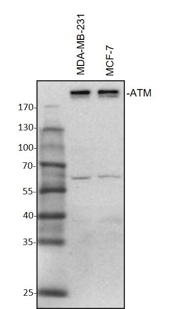
Western Blot: ATM Antibody [NB100-678]
Western Blot: ATM Antibody [NB100-678] - MDA-MB-231 and MCF-7 whole cell lysates were loaded with 50 ug/lane. 8% SDS-PAGE. ATM Antibody (NB100-678) primary antibody at 1:1000, 4C, overnight. Western blot image submitted by a verified customer review.![Immunohistochemistry: ATM Antibody [NB100-678]](https://resources.rndsystems.com/images/products/ATM-Antibody-Immunohistochemistry-NB100-678-img0007.jpg "Immunohistochemistry: ATM Antibody [NB100-678]")
Immunohistochemistry: ATM Antibody [NB100-678]
Immunohistochemistry: ATM Antibody [NB100-678] - FFPE section of human prostate-nodular hypertrophy. Affinity purified rabbit [NB100-678] used at a dilution of 1:1000 ( 1ug/mL). Detection: DAB staining using Immunohistochemistry Accessory Kit.![Western Blot: ATM Antibody [NB100-678]](https://resources.rndsystems.com/images/products/ATM-Antibody-Western-Blot-NB100-678-img0008.jpg "Western Blot: ATM Antibody [NB100-678]")
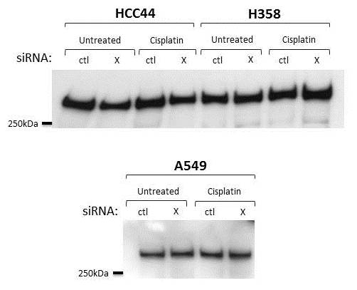
Western Blot: ATM Antibody [NB100-678]
Western Blot: ATM Antibody [NB100-678] - Analysis in whole cell lysates from human lung cancer lines with [NB100-678]. Theoretical molecular weight 351 kDa. Western blot image submitted by a verified customer review.Applications for ATM Antibody - BSA Free
Application
Recommended Usage
Immunohistochemistry
1:500 - 1:2000
Immunohistochemistry-Paraffin
1:500 - 1:2000
Immunoprecipitation
3 - 7 ug/mg lysate
Western Blot
1:5000 - 1:25000
Application Notes
Epitope retrieval is not recommended. ATM antibody validated for WB from a verified customer review.
Reviewed Applications
Read 2 reviews rated 4.5 using NB100-678 in the following applications:
Formulation, Preparation, and Storage
Purification
Immunogen affinity purified
Formulation
Tris-Citrate/Phosphate (pH 7 - 8)
Format
BSA Free
Preservative
0.09% Sodium Azide
Concentration
1.0 mg/ml
Shipping
The product is shipped with polar packs. Upon receipt, store it immediately at the temperature recommended below.
Stability & Storage
Store at 4C. Do not freeze.
Background: ATM
The theoretical molecular weight of ATM is 350 kDa and it has 3 main domains: a FAT (focal adhesion targeting) domain (aa 1960-2566), a PI-3/PI-4 kinase catalytic domain (aa 2712-2962), and a C-terminal FAT domain (aa 3024-3056). ATM exists as a dimer or tetramer in its inactive state. Upon sensing DNA damage, the MRE11-RAD50-NBS1 (MRN) complex recruits ATM. The intricate process of ATM activation involves acetylation by KAT5/TIP60, autophosphorylation at Ser-1981, and dissociation into catalytically active monomers (5). Following activation, ATM phosphorylates multiple substrates such as p53/TP53 and Chk2 involved in DNA repair, checkpoint signaling, and the apoptosis pathway.
References
1. Paull TT. (2015) Mechanisms of ATM Activation. Annu Rev Biochem. 84:711-38. PMID: 25580527
2. Chaudhary MW and Al-Baradie RS. (2014) Ataxia-telangiectasia: future prospects. Appl Clin Genet. 7:159-167. PMID: 25258552
3. Stagni V, Cirotti C, and Barila D. (2018) Ataxia-Telangiectasia Mutated Kinase in the Control of Oxidative Stress, Mitochondria, and Autophagy in Cancer: A Maestro With a Large Orchestra. Front Oncol. 8:73. PMID: 29616191
4. Gumy-Pause F, Wacker P, and Sappino AP. (2004) ATM gene and lymphoid malignancies. Leukemia. 18(2):238-42. PMID: 14628072
5. Adamowicz M. (2018) Breaking up with ATM. J Immunol Sci. 2(1):26-31. PMID: 29652413
Long Name
Ataxia Telangiectasia-mutated
Alternate Names
TEL1, TELO1, TPLL
Entrez Gene IDs
472 (Human)
Gene Symbol
ATM
UniProt
Additional ATM Products
Product Documents for ATM Antibody - BSA Free
Certificate of Analysis
To download a Certificate of Analysis, please enter a lot or batch number in the search box below.
Product Specific Notices for ATM Antibody - BSA Free
This product is for research use only and is not approved for use in humans or in clinical diagnosis. Primary Antibodies are guaranteed for 1 year from date of receipt.
Related Research Areas
Citations for ATM Antibody - BSA Free
 Powered by Bioz
Customer Reviews for ATM Antibody - BSA Free (2)
4.5 out of 5
2 Customer Ratings
Have you used ATM Antibody - BSA Free?
Submit a review and receive an Amazon gift card!
$25/€18/£15/$25CAN/¥2500 Yen for a review with an image
$10/€7/£6/$10CAN/¥1110 Yen for a review without an image
Submit a review
Customer Images
Showing
1
-
2 of
2 reviews
Showing All
Filter By:
-
Application: Western BlotSample Tested: Breast cancer cellsSpecies: HumanVerified Customer | Posted 10/28/2020Western Blot: MDA-MB-231 and MCF-7 whole cell lysates were loaded with 50 ug/lane. 8% SDS-PAGE. ATM Antibody (NB100-678) primary antibody: 1:1000, 4℃, overnight.

-
Application: Western BlotSample Tested: Non small cell lung cancer cell lines, HCC44 and Whole cell lysate from the following human lung cancer lines: H23, H358, A549, H441.Species: HumanVerified Customer | Posted 03/05/2018RIPA buffer 40ug of protein
There are no reviews that match your criteria.
Protocols
Find general support by application which include: protocols, troubleshooting, illustrated assays, videos and webinars.
- Antigen Retrieval Protocol (PIER)
- Antigen Retrieval for Frozen Sections Protocol
- Appropriate Fixation of IHC/ICC Samples
- Cellular Response to Hypoxia Protocols
- Chromogenic IHC Staining of Formalin-Fixed Paraffin-Embedded (FFPE) Tissue Protocol
- Chromogenic Immunohistochemistry Staining of Frozen Tissue
- ClariTSA™ Fluorophore Kits
- Detection & Visualization of Antibody Binding
- Fluorescent IHC Staining of Frozen Tissue Protocol
- Graphic Protocol for Heat-induced Epitope Retrieval
- Graphic Protocol for the Preparation and Fluorescent IHC Staining of Frozen Tissue Sections
- Graphic Protocol for the Preparation and Fluorescent IHC Staining of Paraffin-embedded Tissue Sections
- Graphic Protocol for the Preparation of Gelatin-coated Slides for Histological Tissue Sections
- IHC Sample Preparation (Frozen sections vs Paraffin)
- Immunofluorescent IHC Staining of Formalin-Fixed Paraffin-Embedded (FFPE) Tissue Protocol
- Immunohistochemistry (IHC) and Immunocytochemistry (ICC) Protocols
- Immunohistochemistry Frozen Troubleshooting
- Immunohistochemistry Paraffin Troubleshooting
- Immunoprecipitation Protocol
- Preparing Samples for IHC/ICC Experiments
- Preventing Non-Specific Staining (Non-Specific Binding)
- Primary Antibody Selection & Optimization
- Protocol for Heat-Induced Epitope Retrieval (HIER)
- Protocol for Making a 4% Formaldehyde Solution in PBS
- Protocol for VisUCyte™ HRP Polymer Detection Reagent
- Protocol for the Preparation & Fixation of Cells on Coverslips
- Protocol for the Preparation and Chromogenic IHC Staining of Frozen Tissue Sections
- Protocol for the Preparation and Chromogenic IHC Staining of Frozen Tissue Sections - Graphic
- Protocol for the Preparation and Chromogenic IHC Staining of Paraffin-embedded Tissue Sections
- Protocol for the Preparation and Chromogenic IHC Staining of Paraffin-embedded Tissue Sections - Graphic
- Protocol for the Preparation and Fluorescent IHC Staining of Frozen Tissue Sections
- Protocol for the Preparation and Fluorescent IHC Staining of Paraffin-embedded Tissue Sections
- Protocol for the Preparation of Gelatin-coated Slides for Histological Tissue Sections
- R&D Systems Quality Control Western Blot Protocol
- TUNEL and Active Caspase-3 Detection by IHC/ICC Protocol
- The Importance of IHC/ICC Controls
- Troubleshooting Guide: Immunohistochemistry
- Troubleshooting Guide: Western Blot Figures
- Western Blot Conditions
- Western Blot Protocol
- Western Blot Protocol for Cell Lysates
- Western Blot Troubleshooting
- Western Blot Troubleshooting Guide
- View all Protocols, Troubleshooting, Illustrated assays and Webinars
FAQs for ATM Antibody - BSA Free
Showing
1
-
2 of
2 FAQs
Showing All
-
Q: What is the isotype of this antibody?
A: The isotype for this ATM Antibody is IgG.
-
Q: What is the theoretical molecular weight for your ATM antibodies?
A: The theoretical molecular weight for our ATM antibodies is 351 kDa.
-
Q: What is the isotype of this antibody?
A: The isotype for this ATM Antibody is IgG.
-
Q: What is the theoretical molecular weight for your ATM antibodies?
A: The theoretical molecular weight for our ATM antibodies is 351 kDa.
Loading...