Bax Antibody - BSA Free
Novus Biologicals | Catalog # NB100-56095
![Western Blot: Bax Antibody [NB100-56095]](https://resources.rndsystems.com/images/products/Bax-Antibody-Western-Blot-NB100-56095-img0006.jpg "Western Blot: Bax Antibody [NB100-56095]")
Loading...
Key Product Details
Species Reactivity
Validated:
Human
Cited:
Human, Mouse
Applications
Validated:
Immunohistochemistry, Immunohistochemistry-Paraffin, Western Blot, Immunoprecipitation
Cited:
Western Blot, IF/IHC
Label
Unconjugated
Antibody Source
Polyclonal Rabbit IgG
Format
BSA Free
Loading...
Product Specifications
Immunogen
A synthetic peptide corresponding to amino acids 43-61 (PELALDPVPQDASTKKLSE) of human Bax alpha was used as immunogen, GenBank no. NP_620116.1. The immunogen sequence is 100% conserved in human Bax isoforms alpha (192 amino acids), beta (218 amino acids), epsilon (164 amino acids), sigma (179 amino acids), and psi (173 amino acids).
Clonality
Polyclonal
Host
Rabbit
Isotype
IgG
Scientific Data Images for Bax Antibody - BSA Free
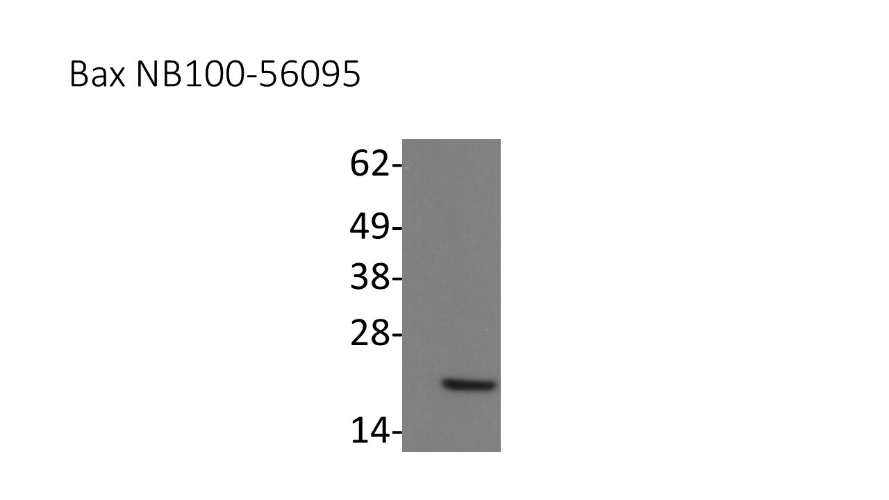
Western Blot: Bax Antibody [NB100-56095]
Western Blot: Bax Antibody [NB100-56095] - Total protein from human THP1, A431, A549 and U2OS cells was separated on a 4-15% gel by SDS-PAGE, transferred to 0.2 um PVDF membrane and blocked in 5% non-fat milk in TBST. The membrane was probed with a 1:2000 dilution of anti-BAX in 1% non-fat milk in TBST and detected with an anti-rabbit HRP secondary antibody using chemiluminescence![Immunohistochemistry-Paraffin: Bax Antibody [NB100-56095]](https://resources.rndsystems.com/images/products/Bax-Antibody-Immunohistochemistry-Paraffin-NB100-56095-img0007.jpg "Immunohistochemistry-Paraffin: Bax Antibody [NB100-56095]")
Immunohistochemistry-Paraffin: Bax Antibody [NB100-56095]
Immunohistochemistry-Paraffin: Bax Antibody [NB100-56095] - Formalin-fixed, paraffin-embedded sections of human colon cancer fom a tissue microarray stained for BAX using NB100-56095 at 1:2000. A. High Bax expression. B. Low Bax expression. Hematoxylin-eosin counterstain![Western Blot: Bax Antibody [NB100-56095]](https://resources.rndsystems.com/images/products/Bax-Antibody-Western-Blot-NB100-56095-img0004.jpg "Western Blot: Bax Antibody [NB100-56095]")
Western Blot: Bax Antibody [NB100-56095]
Western Blot: Bax Antibody [NB100-56095] - Analysis of BAX in colon tumor cell lines using this antibody. Bax is detected in the SW620 and DLD-1 cell lines. However, Bax was not detected in the cell lines (LoVo, DU145, and LS180) known to have frameshift mutations in Bax (Rampino et al. 1997). An antibody to F1ATPase was used as a protein loading control.![Western Blot: Bax Antibody [NB100-56095]](https://resources.rndsystems.com/images/products/Bax-Antibody-Western-Blot-NB100-56095-img0005.jpg "Western Blot: Bax Antibody [NB100-56095]")
Western Blot: Bax Antibody [NB100-56095]
Western Blot: Bax Antibody [NB100-56095] - Analysis of Bax in Daoy whole cell lysate using anti-Bax antibody. Image from verified customer review.
Western Blot: Bax Antibody - BSA Free [NB100-56095] -
Mitochondria dysregulation occurs due to PV administration. The 12Z cells were stimulated with PV for 18 h and then stained with TMRM or MitoSox dye. The mitochondrial membrane potential and mitochondrial reactive oxygen species were assessed using flow cytometry (A,B). Mitochondrial-dependent apoptosis signaling molecules, Bax and Bcl-2 were detected 24 h after PV treatment using western blotting (C). The specific molecular weight is represented on the left. Data are expressed as mean +/- SEM. Statistical analysis was conducted using a Dunnet one-way ANOVA (*; p < 0.05, **; p < 0.01, ***; p < 0.001). Image collected and cropped by CiteAb from the following open publication (https://pubmed.ncbi.nlm.nih.gov/35444541), licensed under a CC-BY license. Not internally tested by Novus Biologicals.Applications for Bax Antibody - BSA Free
Application
Recommended Usage
Immunohistochemistry-Paraffin
1:1000-1:5000
Western Blot
1:1000-1:2000
Application Notes
Users should optimize according to model and immunodetection system used (secondary reagents)
Reviewed Applications
Read 1 review rated 5 using NB100-56095 in the following applications:
Formulation, Preparation, and Storage
Purification
Unpurified
Formulation
Whole antisera
Format
BSA Free
Preservative
0.05% Sodium Azide
Concentration
This product is unpurified. The exact concentration of antibody is not quantifiable.
Shipping
The product is shipped with polar packs. Upon receipt, store it immediately at the temperature recommended below.
Stability & Storage
Store at 4C short term. Aliquot and store at -20C long term. Avoid freeze-thaw cycles.
Background: Bax
Long Name
Bcl Associated X Protein
Alternate Names
apoptosis regulator BAX, BCL2-associated X protein, Bcl2-L-4, BCL2L4bcl2-L-4, Bcl-2-like protein 4
Entrez Gene IDs
581 (Human)
Gene Symbol
BAX
Additional Bax Products
Product Documents for Bax Antibody - BSA Free
Certificate of Analysis
To download a Certificate of Analysis, please enter a lot or batch number in the search box below.
Product Specific Notices for Bax Antibody - BSA Free
This product is for research use only and is not approved for use in humans or in clinical diagnosis. Primary Antibodies are guaranteed for 1 year from date of receipt.
Related Research Areas
Citations for Bax Antibody - BSA Free
 Powered by Bioz
Customer Reviews for Bax Antibody - BSA Free (1)
5 out of 5
1 Customer Rating
Have you used Bax Antibody - BSA Free?
Submit a review and receive an Amazon gift card!
$25/€18/£15/$25CAN/¥2500 Yen for a review with an image
$10/€7/£6/$10CAN/¥1110 Yen for a review without an image
Submit a review
Customer Images
Showing
1
-
1 of
1 review
Showing All
Filter By:
-
Application: Western BlotSample Tested: Daoy whole cell lysate (25ug)Species: HumanVerified Customer | Posted 07/14/2016Analysis of Bax in Daoy cell lysate (25ug)

There are no reviews that match your criteria.
Protocols
Find general support by application which include: protocols, troubleshooting, illustrated assays, videos and webinars.
- Antigen Retrieval Protocol (PIER)
- Antigen Retrieval for Frozen Sections Protocol
- Appropriate Fixation of IHC/ICC Samples
- Cellular Response to Hypoxia Protocols
- Chromogenic IHC Staining of Formalin-Fixed Paraffin-Embedded (FFPE) Tissue Protocol
- Chromogenic Immunohistochemistry Staining of Frozen Tissue
- ClariTSA™ Fluorophore Kits
- Detection & Visualization of Antibody Binding
- Fluorescent IHC Staining of Frozen Tissue Protocol
- Graphic Protocol for Heat-induced Epitope Retrieval
- Graphic Protocol for the Preparation and Fluorescent IHC Staining of Frozen Tissue Sections
- Graphic Protocol for the Preparation and Fluorescent IHC Staining of Paraffin-embedded Tissue Sections
- Graphic Protocol for the Preparation of Gelatin-coated Slides for Histological Tissue Sections
- IHC Sample Preparation (Frozen sections vs Paraffin)
- Immunofluorescent IHC Staining of Formalin-Fixed Paraffin-Embedded (FFPE) Tissue Protocol
- Immunohistochemistry (IHC) and Immunocytochemistry (ICC) Protocols
- Immunohistochemistry Frozen Troubleshooting
- Immunohistochemistry Paraffin Troubleshooting
- Immunoprecipitation Protocol
- Preparing Samples for IHC/ICC Experiments
- Preventing Non-Specific Staining (Non-Specific Binding)
- Primary Antibody Selection & Optimization
- Protocol for Heat-Induced Epitope Retrieval (HIER)
- Protocol for Making a 4% Formaldehyde Solution in PBS
- Protocol for VisUCyte™ HRP Polymer Detection Reagent
- Protocol for the Preparation & Fixation of Cells on Coverslips
- Protocol for the Preparation and Chromogenic IHC Staining of Frozen Tissue Sections
- Protocol for the Preparation and Chromogenic IHC Staining of Frozen Tissue Sections - Graphic
- Protocol for the Preparation and Chromogenic IHC Staining of Paraffin-embedded Tissue Sections
- Protocol for the Preparation and Chromogenic IHC Staining of Paraffin-embedded Tissue Sections - Graphic
- Protocol for the Preparation and Fluorescent IHC Staining of Frozen Tissue Sections
- Protocol for the Preparation and Fluorescent IHC Staining of Paraffin-embedded Tissue Sections
- Protocol for the Preparation of Gelatin-coated Slides for Histological Tissue Sections
- R&D Systems Quality Control Western Blot Protocol
- TUNEL and Active Caspase-3 Detection by IHC/ICC Protocol
- The Importance of IHC/ICC Controls
- Troubleshooting Guide: Immunohistochemistry
- Troubleshooting Guide: Western Blot Figures
- Western Blot Conditions
- Western Blot Protocol
- Western Blot Protocol for Cell Lysates
- Western Blot Troubleshooting
- Western Blot Troubleshooting Guide
- View all Protocols, Troubleshooting, Illustrated assays and Webinars
Loading...
Associated Pathways

