BCAT1 Antibody (OTI3F5)
Novus Biologicals | Catalog # NBP2-01826
Key Product Details
Species Reactivity
Validated:
Human, Mouse, Canine
Cited:
Human, Mouse
Applications
Validated:
Immunohistochemistry, Immunohistochemistry-Paraffin, Western Blot
Cited:
Western Blot
Label
Unconjugated
Antibody Source
Monoclonal Mouse IgG2A Clone # OTI3F5
Loading...
Product Specifications
Immunogen
Full length human recombinant protein of human BCAT1(NP_005495) produced in HEK293T cell.
Reactivity Notes
Please note that this antibody is reactive to Mouse and derived from the same host, Mouse. Mouse-On-Mouse blocking reagent may be needed for IHC and ICC experiments to reduce high background signal. You can find these reagents under catalog numbers PK-2200-NB and MP-2400-NB. Please contact Technical Support if you have any questions.
Clonality
Monoclonal
Host
Mouse
Isotype
IgG2A
Theoretical MW
42.8 kDa.
Disclaimer note: The observed molecular weight of the protein may vary from the listed predicted molecular weight due to post translational modifications, post translation cleavages, relative charges, and other experimental factors.
Disclaimer note: The observed molecular weight of the protein may vary from the listed predicted molecular weight due to post translational modifications, post translation cleavages, relative charges, and other experimental factors.
Scientific Data Images for BCAT1 Antibody (OTI3F5)
![Western Blot: BCAT1 Antibody (OTI3F5) [NBP2-01826]](https://resources.rndsystems.com/images/products/BCAT1-Antibody-3F5-Western-Blot-NBP2-01826-img0010.jpg "Western Blot: BCAT1 Antibody (OTI3F5) [NBP2-01826]")
Western Blot: BCAT1 Antibody (OTI3F5) [NBP2-01826]
Western Blot: BCAT1 Antibody (3F5) [NBP2-01826] - Analysis of extracts (10ug) from a mouse cell line and a mouse tissue by using anti-BCAT1 monoclonal antibody. (1:200)![Immunohistochemistry-Paraffin: BCAT1 Antibody (OTI3F5) [NBP2-01826]](https://resources.rndsystems.com/images/products/BCAT1-Antibody-3F5-Immunohistochemistry-Paraffin-NBP2-01826-img0007.jpg "Immunohistochemistry-Paraffin: BCAT1 Antibody (OTI3F5) [NBP2-01826]")
Immunohistochemistry-Paraffin: BCAT1 Antibody (OTI3F5) [NBP2-01826]
Immunohistochemistry-Paraffin: BCAT1 Antibody (3F5) [NBP2-01826] - Staining of paraffin-embedded Human tonsil using anti-BCAT1 mouse monoclonal antibody.![Western Blot: BCAT1 Antibody (OTI3F5) [NBP2-01826]](https://resources.rndsystems.com/images/products/BCAT1-Antibody-3F5-Western-Blot-NBP2-01826-img0001.jpg "Western Blot: BCAT1 Antibody (OTI3F5) [NBP2-01826]")
Western Blot: BCAT1 Antibody (OTI3F5) [NBP2-01826]
Western Blot: BCAT1 Antibody (3F5) [NBP2-01826] - HEK293T cells were transfected with the pCMV6-ENTRY control (Left lane) or pCMV6-ENTRY BCAT1 (Right lane) cDNA for 48 hrs and lysed. Equivalent amounts of cell lysates (5 ug per lane) were separated by SDS-PAGE and immunoblotted with anti-BCAT1.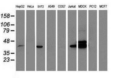
Western Blot: BCAT1 Antibody (3F5) [NBP2-01826] Analysis of extracts (35ug) from 9 different cell lines by using anti-BCAT1 monoclonal antibody.
![Western Blot: BCAT1 Antibody (OTI3F5) [NBP2-01826]](https://resources.rndsystems.com/images/products/BCAT1-Antibody-3F5-Western-Blot-NBP2-01826-img0009.jpg "Western Blot: BCAT1 Antibody (OTI3F5) [NBP2-01826]")
Western Blot: BCAT1 Antibody (OTI3F5) [NBP2-01826]
Western Blot: BCAT1 Antibody (3F5) [NBP2-01826] - Analysis of extracts (10ug) from 9 Human tissue by using anti-BCAT1 monoclonal antibody at 1:200 (1: Testis; 2: Omentum; 3: Uterus; 4: Breast; 5: Brain; 6: Liver; 7: Ovary; 8: Thyroid gland; 9: Colon)![Immunohistochemistry-Paraffin: BCAT1 Antibody (OTI3F5) [NBP2-01826]](https://resources.rndsystems.com/images/products/BCAT1-Antibody-3F5-Immunohistochemistry-Paraffin-NBP2-01826-img0002.jpg "Immunohistochemistry-Paraffin: BCAT1 Antibody (OTI3F5) [NBP2-01826]")
Immunohistochemistry-Paraffin: BCAT1 Antibody (OTI3F5) [NBP2-01826]
Immunohistochemistry-Paraffin: BCAT1 Antibody (3F5) [NBP2-01826] - Staining of paraffin-embedded Adenocarcinoma of Human endometrium tissue using anti-BCAT1 mouse monoclonal antibody.![Immunohistochemistry-Paraffin: BCAT1 Antibody (OTI3F5) [NBP2-01826]](https://resources.rndsystems.com/images/products/BCAT1-Antibody-3F5-Immunohistochemistry-Paraffin-NBP2-01826-img0003.jpg "Immunohistochemistry-Paraffin: BCAT1 Antibody (OTI3F5) [NBP2-01826]")
Immunohistochemistry-Paraffin: BCAT1 Antibody (OTI3F5) [NBP2-01826]
Immunohistochemistry-Paraffin: BCAT1 Antibody (3F5) [NBP2-01826] - Staining of paraffin-embedded Carcinoma of Human liver tissue using anti-BCAT1 mouse monoclonal antibody.![Immunohistochemistry-Paraffin: BCAT1 Antibody (OTI3F5) [NBP2-01826]](https://resources.rndsystems.com/images/products/BCAT1-Antibody-3F5-Immunohistochemistry-Paraffin-NBP2-01826-img0004.jpg "Immunohistochemistry-Paraffin: BCAT1 Antibody (OTI3F5) [NBP2-01826]")
Immunohistochemistry-Paraffin: BCAT1 Antibody (OTI3F5) [NBP2-01826]
Immunohistochemistry-Paraffin: BCAT1 Antibody (3F5) [NBP2-01826] - Staining of paraffin-embedded Human lymphoma tissue using anti-BCAT1 mouse monoclonal antibody.![Immunohistochemistry-Paraffin: BCAT1 Antibody (OTI3F5) [NBP2-01826]](https://resources.rndsystems.com/images/products/BCAT1-Antibody-3F5-Immunohistochemistry-Paraffin-NBP2-01826-img0005.jpg "Immunohistochemistry-Paraffin: BCAT1 Antibody (OTI3F5) [NBP2-01826]")
Immunohistochemistry-Paraffin: BCAT1 Antibody (OTI3F5) [NBP2-01826]
Immunohistochemistry-Paraffin: BCAT1 Antibody (3F5) [NBP2-01826] - Staining of paraffin-embedded Human pancreas tissue using anti-BCAT1 mouse monoclonal antibody.![Immunohistochemistry-Paraffin: BCAT1 Antibody (OTI3F5) [NBP2-01826]](https://resources.rndsystems.com/images/products/BCAT1-Antibody-3F5-Immunohistochemistry-Paraffin-NBP2-01826-img0006.jpg "Immunohistochemistry-Paraffin: BCAT1 Antibody (OTI3F5) [NBP2-01826]")
Immunohistochemistry-Paraffin: BCAT1 Antibody (OTI3F5) [NBP2-01826]
Immunohistochemistry-Paraffin: BCAT1 Antibody (3F5) [NBP2-01826] - Staining of paraffin-embedded Human prostate tissue using anti-BCAT1 mouse monoclonal antibody.[NBP2-01826]")
BCAT1 Antibody (OTI3F5)[NBP2-01826]
BCAT1 Antibody (OTI3F5)[NBP2-01826]Simple Western� analysis of endogenous protein IFIT3 from THP-1 lysates (0.1 mg/mL) using IFIT3 Mouse Monoclonal Antibody. The virtual lane view (left) shows the target (as indicated) at 1:50 dilution of primary antibody. The corresponding electropherogram view (right) plots chemiluminescence by molecular weight along the capillary at a 1:50 dilution of primary antibody. This experiment was performed under reducing conditions on the Jess� Simple Western instrument from ProteinSimple, a Bio-Techne brand, using the 12�230 kDa Separation Module. [NBP2-01826] -")
Western Blot: BCAT1 Antibody (OTI3F5) [NBP2-01826] -
BCAAs promote the phosphorylation of Tau(A–C) Bcat1 mRNA level is decreased in the brain tissues of diabetic, aged, or 3xTg AD mice (n=5). **P<0.01 by unpaired Student’s t-test. (D) Representative Western blot and quantitative results showing BCAT1 protein level are decreased in the brain tissues of AD mice (n=4). ***P<0.001 by unpaired Student’s t-test. (E) BCAA diet does not affect the content of amyloid beta 42 (A beta 42) in the brain tissues of AD mice (n=5). (F) Representative Western blot and quantitative results showing BCAA diet increase the level of phosphorylated Tau protein in the brain tissues of AD mice (n=4). ***p<0.001 by unpaired Student’s t-test. (G) Relative Western blot and quantitative results showing leucine increase the level of phosphorylated Tau protein in the neurons isolated from mice. The neurons were isolated from the 3xTg mice and treated with leucine (1 mM), isoleucine (1 mM), or valine (1 mM) for 24 h. The experiments were repeated for three times. ***P<0.001 by one-way ANOVA followed by Bonferroni post-hoc test. (H) Bcat1 knockdown increased the level of phosphorylated Tau protein in the neurons isolated from mice. The neurons were isolated from the 3xTg mice and infected with lentivirus carrying indicated shRNAs for 48 h. Image collected and cropped by CiteAb from the following open publication (https://pubmed.ncbi.nlm.nih.gov/29802157), licensed under a CC-BY license. Not internally tested by Novus Biologicals.Applications for BCAT1 Antibody (OTI3F5)
Application
Recommended Usage
Immunohistochemistry
1:150
Immunohistochemistry-Paraffin
1:150
Western Blot
1:500-2000
Formulation, Preparation, and Storage
Purification
Immunogen affinity purified
Formulation
PBS (pH 7.3), 1.0% BSA and 50% Glycerol
Preservative
0.02% Sodium Azide
Concentration
Please see the vial label for concentration. If unlisted please contact technical services.
Shipping
The product is shipped with polar packs. Upon receipt, store it immediately at the temperature recommended below.
Stability & Storage
Store at -20C. Avoid freeze-thaw cycles.
Background: BCAT1
Long Name
Branched Chain Amino Acid Transaminase 1
Alternate Names
BCAT-1, BCATC, BCT1, EC 2.6.1.42, ECA39, PNAS121, PP18
Entrez Gene IDs
586 (Human)
Gene Symbol
BCAT1
Additional BCAT1 Products
Product Documents for BCAT1 Antibody (OTI3F5)
Certificate of Analysis
To download a Certificate of Analysis, please enter a lot or batch number in the search box below.
Product Specific Notices for BCAT1 Antibody (OTI3F5)
This product is for research use only and is not approved for use in humans or in clinical diagnosis. Primary Antibodies are guaranteed for 1 year from date of receipt.
Citations for BCAT1 Antibody (OTI3F5)
 Powered by Bioz
Customer Reviews for BCAT1 Antibody (OTI3F5)
There are currently no reviews for this product. Be the first to review BCAT1 Antibody (OTI3F5) and earn rewards!
Have you used BCAT1 Antibody (OTI3F5)?
Submit a review and receive an Amazon gift card!
$25/€18/£15/$25CAN/¥2500 Yen for a review with an image
$10/€7/£6/$10CAN/¥1110 Yen for a review without an image
Submit a review
Protocols
Find general support by application which include: protocols, troubleshooting, illustrated assays, videos and webinars.
- Antigen Retrieval Protocol (PIER)
- Antigen Retrieval for Frozen Sections Protocol
- Appropriate Fixation of IHC/ICC Samples
- Cellular Response to Hypoxia Protocols
- Chromogenic IHC Staining of Formalin-Fixed Paraffin-Embedded (FFPE) Tissue Protocol
- Chromogenic Immunohistochemistry Staining of Frozen Tissue
- ClariTSA™ Fluorophore Kits
- Detection & Visualization of Antibody Binding
- Fluorescent IHC Staining of Frozen Tissue Protocol
- Graphic Protocol for Heat-induced Epitope Retrieval
- Graphic Protocol for the Preparation and Fluorescent IHC Staining of Frozen Tissue Sections
- Graphic Protocol for the Preparation and Fluorescent IHC Staining of Paraffin-embedded Tissue Sections
- Graphic Protocol for the Preparation of Gelatin-coated Slides for Histological Tissue Sections
- IHC Sample Preparation (Frozen sections vs Paraffin)
- Immunofluorescent IHC Staining of Formalin-Fixed Paraffin-Embedded (FFPE) Tissue Protocol
- Immunohistochemistry (IHC) and Immunocytochemistry (ICC) Protocols
- Immunohistochemistry Frozen Troubleshooting
- Immunohistochemistry Paraffin Troubleshooting
- Preparing Samples for IHC/ICC Experiments
- Preventing Non-Specific Staining (Non-Specific Binding)
- Primary Antibody Selection & Optimization
- Protocol for Heat-Induced Epitope Retrieval (HIER)
- Protocol for Making a 4% Formaldehyde Solution in PBS
- Protocol for VisUCyte™ HRP Polymer Detection Reagent
- Protocol for the Preparation & Fixation of Cells on Coverslips
- Protocol for the Preparation and Chromogenic IHC Staining of Frozen Tissue Sections
- Protocol for the Preparation and Chromogenic IHC Staining of Frozen Tissue Sections - Graphic
- Protocol for the Preparation and Chromogenic IHC Staining of Paraffin-embedded Tissue Sections
- Protocol for the Preparation and Chromogenic IHC Staining of Paraffin-embedded Tissue Sections - Graphic
- Protocol for the Preparation and Fluorescent IHC Staining of Frozen Tissue Sections
- Protocol for the Preparation and Fluorescent IHC Staining of Paraffin-embedded Tissue Sections
- Protocol for the Preparation of Gelatin-coated Slides for Histological Tissue Sections
- R&D Systems Quality Control Western Blot Protocol
- TUNEL and Active Caspase-3 Detection by IHC/ICC Protocol
- The Importance of IHC/ICC Controls
- Troubleshooting Guide: Immunohistochemistry
- Troubleshooting Guide: Western Blot Figures
- Western Blot Conditions
- Western Blot Protocol
- Western Blot Protocol for Cell Lysates
- Western Blot Troubleshooting
- Western Blot Troubleshooting Guide
- View all Protocols, Troubleshooting, Illustrated assays and Webinars
Loading...