BMAL1 Antibody - BSA Free
Novus Biologicals | Catalog # NB100-2288
![Western Blot: BMAL1 Antibody [NB100-2288]](https://resources.rndsystems.com/images/products/BMAL1-Antibody-Western-Blot-NB100-2288-img0023.jpg "Western Blot: BMAL1 Antibody [NB100-2288]")
![Western Blot: BMAL1 Antibody [NB100-2288]](https://resources.rndsystems.com/images/products/BMAL1-Antibody-Western-Blot-NB100-2288-img0021.jpg "Western Blot: BMAL1 Antibody [NB100-2288]")
Key Product Details
Validated by
Knockout/Knockdown, Biological Validation
Species Reactivity
Validated:
Human, Mouse, Rat, Amphibian, Primate
Cited:
Human, Mouse, Rat
Applications
Validated:
Knockout Validated, Immunohistochemistry, Immunohistochemistry-Paraffin, Immunohistochemistry-Frozen, Western Blot, Flow Cytometry, Flow (Intracellular), Immunocytochemistry/ Immunofluorescence, Chromatin Immunoprecipitation (ChIP)
Cited:
Immunohistochemistry, Immunohistochemistry-Paraffin, Immunohistochemistry-Frozen, Western Blot, Immunocytochemistry/ Immunofluorescence, Chemotaxis, IF/IHC, Knockdown Validated
Label
Unconjugated
Antibody Source
Polyclonal Rabbit IgG
Format
BSA Free
Loading...
Product Specifications
Immunogen
Bacterially expressed human BMAL1 (amino acids 392-626). [UniProt# O00327].
Reactivity Notes
Mouse reactivity reported in scientific literature (PMID:32732906). Amphibian reactivity reported from a verified customer review.
Localization
Nuclear
Clonality
Polyclonal
Host
Rabbit
Isotype
IgG
Theoretical MW
70 kDa.
Disclaimer note: The observed molecular weight of the protein may vary from the listed predicted molecular weight due to post translational modifications, post translation cleavages, relative charges, and other experimental factors.
Disclaimer note: The observed molecular weight of the protein may vary from the listed predicted molecular weight due to post translational modifications, post translation cleavages, relative charges, and other experimental factors.
Scientific Data Images for BMAL1 Antibody - BSA Free
Western Blot: BMAL1 Antibody [NB100-2288]
BMAL1-Antibody-Western-Blot-NB100-2288-img0021.jpg![Immunohistochemistry: BMAL1 Antibody [NB100-2288]](https://resources.rndsystems.com/images/products/BMAL1-Antibody-Chromatin-Immunoprecipitation-NB100-2288-img0018.jpg "Immunohistochemistry: BMAL1 Antibody [NB100-2288]")
Immunohistochemistry: BMAL1 Antibody [NB100-2288]
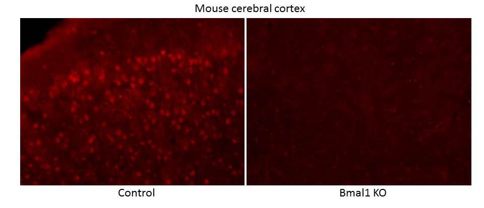
Immunohistochemistry: BMAL1 Antibody [NB100-2288] - Analysis of BMAL1 in mouse cerebral cortex (left: wt, right: Bmal1 KO) using anti-BMAL1 antibody. Image from verified customer review.![Immunohistochemistry: BMAL1 Antibody [NB100-2288]](https://resources.rndsystems.com/images/products/BMAL1-Antibody-Immunocytochemistry-Immunofluorescence-NB100-2288-img0024.jpg "Immunohistochemistry: BMAL1 Antibody [NB100-2288]")
![Immunocytochemistry/ Immunofluorescence: BMAL1 Antibody [NB100-2288]](https://resources.rndsystems.com/images/products/BMAL1-Antibody-Immunocytochemistry-Immunofluorescence-NB100-2288-img0022.jpg "Immunocytochemistry/ Immunofluorescence: BMAL1 Antibody [NB100-2288]")
![Flow (Intracellular): BMAL1 Antibody [NB100-2288]](https://resources.rndsystems.com/images/products/BMAL1-Antibody-Flow-Intracellular-NB100-2288-img0017.jpg "Flow (Intracellular): BMAL1 Antibody [NB100-2288]")
Flow (Intracellular): BMAL1 Antibody [NB100-2288]
Flow (Intracellular): BMAL1 Antibody [NB100-2288] - An intracellular stain was performed on HeLa cells with NB100-2288AF647(blue) and a matched isotype control (orange). Cells were fixed with 4% PFA and then permeabilized with 0.1% saponin. Cells were incubated in an antibody dilution of 5 ug/mL for 30 minutes at room temperature. Both antibodies were conjugated to Alexa Fluor 647.![Knockout Validated: BMAL1 Antibody [NB100-2288]](https://resources.rndsystems.com/images/products/BMAL1-Antibody-Knockout-Validated-NB100-2288-img0020.jpg "Immunohistochemistry: BMAL1 Antibody [NB100-2288]")
![Flow Cytometry: BMAL1 Antibody [NB100-2288]](https://resources.rndsystems.com/images/products/BMAL1-Antibody-Flow-Cytometry-NB100-2288-img0019.jpg "Flow Cytometry: BMAL1 Antibody [NB100-2288]")
Flow Cytometry: BMAL1 Antibody [NB100-2288]
Flow Cytometry: BMAL1 Antibody [NB100-2288] - An intracellular stain was performed on Jurkat cells with NB100-2288 and a matched isotype control. Cells were fixed with 4% PFA and then permeabilized with 0.1% saponin. Cells were incubated in an antibody dilution of 2.5 ug/mL for 30 minutes at room temperature, followed by Rabbit IgG (H+L) Cross-Adsorbed Secondary Antibody.![Western Blot: BMAL1 Antibody [NB100-2288]](https://resources.rndsystems.com/images/products/BMAL1-Antibody-Western-Blot-NB100-2288-img0014.jpg "Western Blot: BMAL1 Antibody [NB100-2288]")
Western Blot: BMAL1 Antibody [NB100-2288]
Western Blot: BMAL1 Antibody [NB100-2288] - Analysis of BMAL1 in A) MCF7, B) NIH/3T3, C) PC12.![Western Blot: BMAL1 Antibody [NB100-2288]](https://resources.rndsystems.com/images/products/BMAL1-Antibody-Western-Blot-NB100-2288-img0016.jpg "Western Blot: BMAL1 Antibody [NB100-2288]")
Western Blot: BMAL1 Antibody [NB100-2288]
Western Blot: BMAL1 Antibody [NB100-2288] - Analysis of MOP3 on 3T3/L1 cell lysates.![Immunocytochemistry/ Immunofluorescence: BMAL1 Antibody [NB100-2288]](https://resources.rndsystems.com/images/products/BMAL1-Antibody-Immunocytochemistry-Immunofluorescence-NB100-2288-img0012.jpg "Immunocytochemistry/ Immunofluorescence: BMAL1 Antibody [NB100-2288]")
Immunocytochemistry/ Immunofluorescence: BMAL1 Antibody [NB100-2288]
Immunocytochemistry/Immunofluorescence: BMAL1 Antibody [NB100-2288] - BMAL1 antibody was tested in HeLa cells at a 1:200 dilution against Dylight 488 (Green). Alpha tubulin and nuclei were counterstained against DyLight 568 (Red) and DAPI (Blue), respectively.![Immunohistochemistry: BMAL1 Antibody [NB100-2288]](https://resources.rndsystems.com/images/products/BMAL1-Antibody-Immunohistochemistry-NB100-2288-img0009.jpg "Immunohistochemistry: BMAL1 Antibody [NB100-2288]")
Immunohistochemistry: BMAL1 Antibody [NB100-2288]
Immunohistochemistry: BMAL1 Antibody [NB100-2288] - Analysis of BMAL1 in mouse brain using DAB with hematoxylin counterstain.![Immunohistochemistry-Paraffin: BMAL1 Antibody [NB100-2288]](https://resources.rndsystems.com/images/products/BMAL1-Antibody-Immunohistochemistry-Paraffin-NB100-2288-img0015.jpg "Immunohistochemistry-Paraffin: BMAL1 Antibody [NB100-2288]")
Immunohistochemistry-Paraffin: BMAL1 Antibody [NB100-2288]
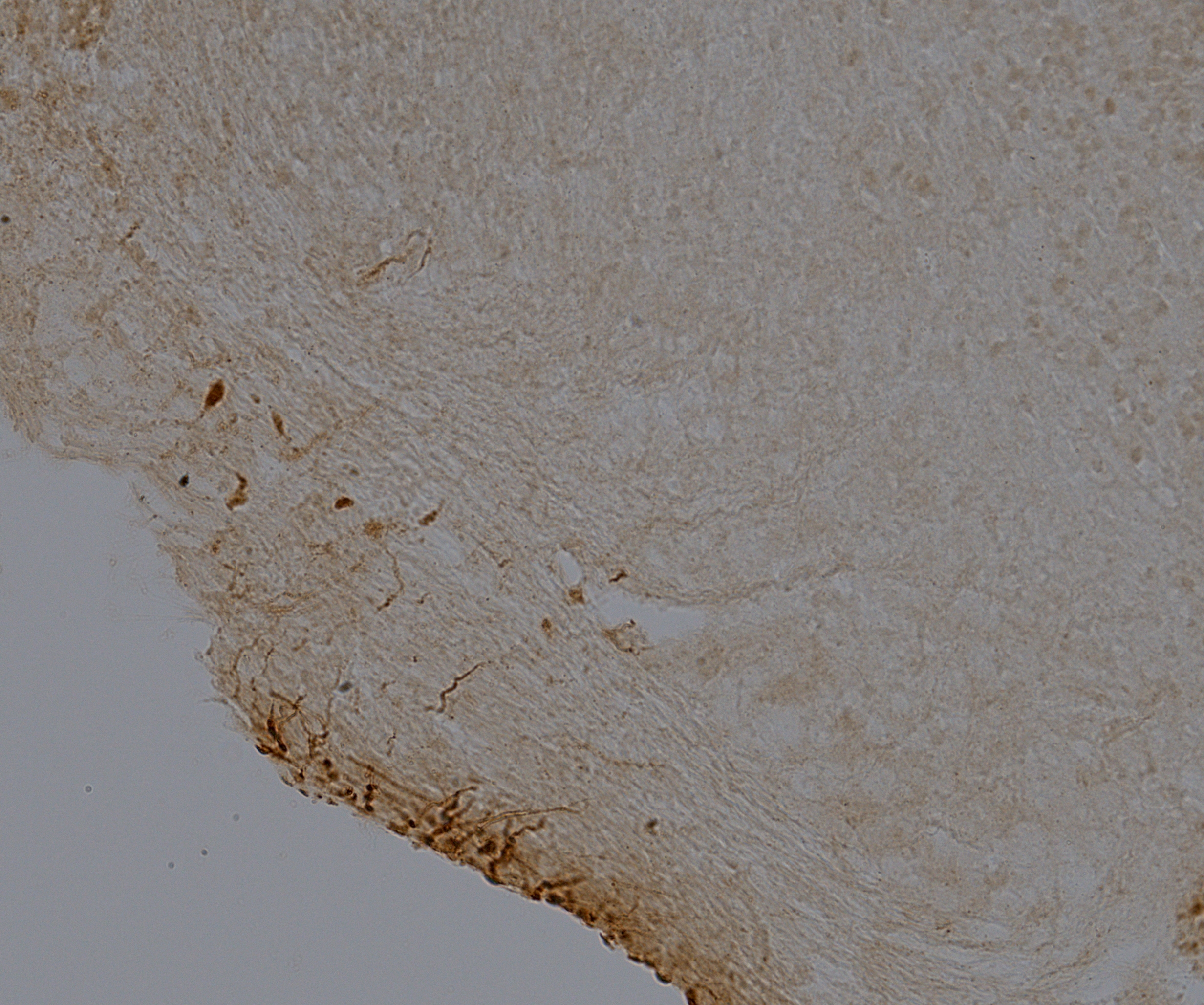
Immunohistochemistry-Paraffin: BMAL1 Antibody [NB100-2288] - Analysis of a FFPE tissue section of mouse brain using 1:200 dilution of rabbit anti-BMAL1 antibody. The staining was developed using HRP labeled anti-rabbit IgG secondary antibody and DAB reagent, and nuclei of cells were counter-stained with hematoxylin. This BMAL1 antibody generated a specific nuclear staining in most of the cells and a relatively weaker cytoplasmic signal was also observed.
Immunohistochemistry-Frozen: Rabbit Polyclonal BMAL1 Antibody [NB100-2288] -
Immunohistochemistry-Frozen: Rabbit Polyclonal BMAL1 Antibody [NB100-2288] - IHC-fixed frozen in the brain of green tree frogs. DAB staining, 1:1000 dilution. Image from a verified customer review.
BMAL1 in MCF7 Human Cell Line.
BMAL1 was detected in immersion fixed MCF7 human breast cancer cell line using Rabbit anti-BMAL1 Affinity Purified Polyclonal Antibody conjugated to Alexa Fluor® 647 (Catalog # NB100-2288AF647) (light blue) at 10 µg/mL overnight at 4C. Cells were stained counterstained with DAPI (blue). Cells were imaged using a 40X objective.
Immunocytochemistry/ Immunofluorescence: BMAL1 Antibody [NB100-2288] -
Immunocytochemistry/ Immunofluorescence: BMAL1 Antibody [NB100-2288] - Rapamycin reduces the accumulation of BMAL1 in Per2 knockout miceA. As shown in the left panel, tissue samples from Per2 knockout mice (mPER−/−) depict robust accumulation of nuclear BMAL1 (arrow) compared to control littermates (arrowhead)(*** p<0.001). Administration of Rapamycin reduces the accumulation of BMAL1 in the epidermis of mPer−/− mice (arrowhead) compared to mPer−/− mice receiving vehicle alone (** p<0.01) to levels comparable to wild-type mice receiving vehicle alone (ns: p>0.05). Image collected & cropped by CiteAb from the following publication (https://pubmed.ncbi.nlm.nih.gov/27285754), licensed under a CC-BY license. Not internally tested by Novus Biologicals.
Western Blot: BMAL1 Antibody [NB100-2288] -
Western Blot: BMAL1 Antibody [NB100-2288] - Small interference RNA targeting Raptor & Rictor disrupts BMAL1 accumulation in HNSCCTargeted disruption of Raptor (A-B) & Rictor (C-D) using siRNA results in a dose-dependent downregulation of BMAL1 in HNSCC cells. E. Disruption of PTEN by protein oxidation causes activation of mTOR signaling, resulting in accumulation of BMAL1. Notably, inhibition of mTOR signaling, particularly mTORC1 & mTORC2, results in restoration of normal BMAL1 levels in the epidermis of mice & head & neck cancer cells. These results demonstrate a novel role for mTOR in regulating nuclear levels of the core clock gene BMAL1. Image collected & cropped by CiteAb from the following publication (https://pubmed.ncbi.nlm.nih.gov/27285754), licensed under a CC-BY license. Not internally tested by Novus Biologicals.
Western Blot: BMAL1 Antibody [NB100-2288] -
Western Blot: BMAL1 Antibody [NB100-2288] - Targeted disruption of PTEN in vitro & in vivo induces activation of pS6 & BMAL1A. & B. Targeted inhibition of PTEN using siRNA results in concentration-dependent inhibition of PTEN protein in HNSCC cells. HNSCC cells show accumulation of BMAL1 & pS6 in response to PTEN inhibition. C. Immunofluorescence assay to detect BMAL1 in PTEN conditional knockout mice (K14cre PtenF/F) & control littermates. Immunofluorescences & graphic show high accumulation of nuclear BMAL1 in K14cre PtenF/F mice compared to control mice (K14cre). Scale bars represent 50 μm. Black & white images depict cells positive for BMAL1 in K14cre PtenF/F mice (arrows) compared to few BMAL-positive cells in control mice (arrowhead). Scale bars represent 10 μm. (**p<0.01). Image collected & cropped by CiteAb from the following publication (https://pubmed.ncbi.nlm.nih.gov/27285754), licensed under a CC-BY license. Not internally tested by Novus Biologicals.
Western Blot: BMAL1 Antibody [NB100-2288] -
Western Blot: BMAL1 Antibody [NB100-2288] - Small interference RNA targeting Raptor & Rictor disrupts BMAL1 accumulation in HNSCCTargeted disruption of Raptor (A-B) & Rictor (C-D) using siRNA results in a dose-dependent downregulation of BMAL1 in HNSCC cells. E. Disruption of PTEN by protein oxidation causes activation of mTOR signaling, resulting in accumulation of BMAL1. Notably, inhibition of mTOR signaling, particularly mTORC1 & mTORC2, results in restoration of normal BMAL1 levels in the epidermis of mice & head & neck cancer cells. These results demonstrate a novel role for mTOR in regulating nuclear levels of the core clock gene BMAL1. Image collected & cropped by CiteAb from the following publication (https://pubmed.ncbi.nlm.nih.gov/27285754), licensed under a CC-BY license. Not internally tested by Novus Biologicals.
Western Blot: BMAL1 Antibody [NB100-2288] -
Western Blot: BMAL1 Antibody [NB100-2288] - Targeted disruption of PTEN in vitro & in vivo induces activation of pS6 & BMAL1A. & B. Targeted inhibition of PTEN using siRNA results in concentration-dependent inhibition of PTEN protein in HNSCC cells. HNSCC cells show accumulation of BMAL1 & pS6 in response to PTEN inhibition. C. Immunofluorescence assay to detect BMAL1 in PTEN conditional knockout mice (K14cre PtenF/F) & control littermates. Immunofluorescences & graphic show high accumulation of nuclear BMAL1 in K14cre PtenF/F mice compared to control mice (K14cre). Scale bars represent 50 μm. Black & white images depict cells positive for BMAL1 in K14cre PtenF/F mice (arrows) compared to few BMAL-positive cells in control mice (arrowhead). Scale bars represent 10 μm. (**p<0.01). Image collected & cropped by CiteAb from the following publication (https://pubmed.ncbi.nlm.nih.gov/27285754), licensed under a CC-BY license. Not internally tested by Novus Biologicals.
Western Blot: BMAL1 Antibody [NB100-2288] -
Western Blot: BMAL1 Antibody [NB100-2288] - Oxidation causes accumulation of BMAL1A. Head & neck cancer cells have different expression levels of the core clock protein BMAL1. B. Immunofluorescence assay depicts accumulation of ROS (green channel) & BMAL1 (red channel) upon oxidation. C. & D. Western blot assay demonstrates time-dependent accumulation of BMAL1 upon oxidative stress in HNSCC cells (*p<0.05, **p<0.01). Image collected & cropped by CiteAb from the following publication (https://pubmed.ncbi.nlm.nih.gov/27285754), licensed under a CC-BY license. Not internally tested by Novus Biologicals.
Immunocytochemistry/ Immunofluorescence: BMAL1 Antibody [NB100-2288] -
Immunocytochemistry/ Immunofluorescence: BMAL1 Antibody [NB100-2288] - Targeted disruption of PTEN in vitro & in vivo induces activation of pS6 & BMAL1A. & B. Targeted inhibition of PTEN using siRNA results in concentration-dependent inhibition of PTEN protein in HNSCC cells. HNSCC cells show accumulation of BMAL1 & pS6 in response to PTEN inhibition. C. Immunofluorescence assay to detect BMAL1 in PTEN conditional knockout mice (K14cre PtenF/F) & control littermates. Immunofluorescences & graphic show high accumulation of nuclear BMAL1 in K14cre PtenF/F mice compared to control mice (K14cre). Scale bars represent 50 μm. Black & white images depict cells positive for BMAL1 in K14cre PtenF/F mice (arrows) compared to few BMAL-positive cells in control mice (arrowhead). Scale bars represent 10 μm. (**p<0.01). Image collected & cropped by CiteAb from the following publication (https://pubmed.ncbi.nlm.nih.gov/27285754), licensed under a CC-BY license. Not internally tested by Novus Biologicals.
Immunohistochemistry: BMAL1 Antibody [NB100-2288] -
Immunohistochemistry: BMAL1 Antibody [NB100-2288] - Astrocyte Bmal1 regulates genes with conflicting effects on A beta deposition. (A) Topro, GFAP, & BMAL1 staining in CA1 hippocampus of 4-month-old BMAL1 aKO; APP/PS1-21 mice & Cre- controls (scale bar = 100 µm). Arrows indicate astrocyte nuclei quantified as indicated by Topro nuclei surrounded by GFAP positivity. Blue circles indicate nuclei quantified as BMAL1 negative. Quantification of astrocytes counted as BMAL1- or BMAL1+ is shown on the right. n = 5 mice per group, **** = p < 0.0001 by two-way ANOVA with Sidak multiple comparisons test. (B) Heatmap of Fluidigm qPCR analysis of 20 genes involved in the circadian clock, glial activation, & Alzheimer’s Disease in cortex from Aldh1l1-CreERT2; Bmal1fl/fl mice & Cre- controls with or without APP/PS1-21 or APPNL-G-F/wt (n = 6–8 mice per group). Two-way ANOVA analysis: c = significant main effect of Cre genotype, m = main effect of A beta model, c*m = interaction effect of cre & A beta model, - = no significance (all p < 0.05). (C) Individually plotted genes from A. * = p < 0.05, ** = p < 0.005, *** = p < 0.0005 by two-way ANOVA with Sidak multiple comparisons test. Panel B was made using GraphPad Prism version 9.2 (https://www.graphpad.com). Image collected & cropped by CiteAb from the following publication (https://pubmed.ncbi.nlm.nih.gov/35110643), licensed under a CC-BY license. Not internally tested by Novus Biologicals.Applications for BMAL1 Antibody - BSA Free
Application
Recommended Usage
Immunocytochemistry/ Immunofluorescence
1:100
Immunohistochemistry
1:250. Use reported in scientific literature (PMID 33510438)
Immunohistochemistry-Frozen
reported in scientific literature (PMID 23736292)
Immunohistochemistry-Paraffin
1:250
Western Blot
0.5 ug/mL - 2 ug/mL
Application Notes
In ICC/IF, primarily nuclear staining was observed with weak cytoplasmic staining in MCF7 cells. In Western Blot, a band was observed ~70 kDa. In IHC-P, staining was observed in the nuclei of mouse brain tissue. Prior to immunostaining paraffin tissues, antigen retrieval with sodium citrate buffer (pH 6.0) is recommended. The observed molecular weight of the protein may vary from the listed predicted molecular weight due to post translational modifications, post translation cleavages, relative charges, and other experimental factors.
Reviewed Applications
Read 2 reviews rated 4 using NB100-2288 in the following applications:
Flow Cytometry Panel Builder
Bio-Techne Knows Flow Cytometry
Save time and reduce costly mistakes by quickly finding compatible reagents using the Panel Builder Tool.
Advanced Features
- Spectra Viewer - Custom analysis of spectra from multiple fluorochromes
- Spillover Popups - Visualize the spectra of individual fluorochromes
- Antigen Density Selector - Match fluorochrome brightness with antigen density

Formulation, Preparation, and Storage
Purification
Immunogen affinity purified
Formulation
PBS
Format
BSA Free
Preservative
0.02% Sodium Azide
Concentration
1.0 mg/ml
Shipping
The product is shipped with polar packs. Upon receipt, store it immediately at the temperature recommended below.
Stability & Storage
Store at 4C for up to 3 months. For longer storage, aliquot and store at -20C.
Background: BMAL1
Long Name
Brain and Muscle ARNT-Like 1
Alternate Names
ARNTL, BHLHE5, BMAL1c, JAP3, MOP3, PASD3, TIC
Gene Symbol
BMAL1
Additional BMAL1 Products
Product Documents for BMAL1 Antibody - BSA Free
Certificate of Analysis
To download a Certificate of Analysis, please enter a lot or batch number in the search box below.
Product Specific Notices for BMAL1 Antibody - BSA Free
This product is for research use only and is not approved for use in humans or in clinical diagnosis. Primary Antibodies are guaranteed for 1 year from date of receipt.
Related Research Areas
Citations for BMAL1 Antibody - BSA Free
 Powered by Bioz
Customer Reviews for BMAL1 Antibody - BSA Free (2)
4 out of 5
2 Customer Ratings
Have you used BMAL1 Antibody - BSA Free?
Submit a review and receive an Amazon gift card!
$25/€18/£15/$25CAN/¥2500 Yen for a review with an image
$10/€7/£6/$10CAN/¥1110 Yen for a review without an image
Submit a review
Customer Images
Showing
1
-
2 of
2 reviews
Showing All
Filter By:
-
Application: Immunohistochemistry-FrozenSample Tested: BrainSpecies: Hyla cinereaVerified Customer | Posted 10/20/2023IHC- fixed frozen in the brain of green tree frogs. DAB staining, 1:1000 dilution
 Bio-Techne ResponseThis review was submitted through the legacy Novus Innovators Program, reflecting a new species or application tested on a primary antibody.
 -
Application: ImmunofluorescenceSample Tested: mouse brainSpecies: MouseVerified Customer | Posted 06/29/2015Bmal1 staining in mouse cerebral cortex: wt and Bmal1 KO
There are no reviews that match your criteria.
Protocols
View specific protocols for BMAL1 Antibody - BSA Free (NB100-2288):
Immunocytochemistry Protocol
Culture cells to appropriate density in 35 mm culture dishes or 6-well plates.
1. Remove culture medium and wash the cells briefly in PBS. Add 10% formalin to the dish and fix at room temperature for 10 minutes.
2. Remove the formalin and wash the cells in PBS.
3. Permeablize the cells with 0.1% Triton X100 or other suitable detergent for 10 min.
4. Remove the permeablization buffer and wash three times for 10 minutes each in PBS. Be sure to not let the specimen dry out.
5. To block nonspecific antibody binding, incubate in 10% normal goat serum from 1 hour to overnight at room temperature.
6. Add primary antibody at appropriate dilution and incubate overnight at 4C.
7. Remove primary antibody and replace with PBS. Wash three times for 10 minutes each.
8. Add secondary antibody at appropriate dilution. Incubate for 1 hour at room temperature.
9. Remove secondary antibody and replace with PBS. Wash three times for 10 minutes each.
10. Counter stain DNA with DAPi if required.
Immunohistochemistry-Paraffin Embedded Sections
Antigen Unmasking:
Bring slides to a boil in 10 mM sodium citrate buffer (pH 6.0) then maintain at a sub-boiling temperature for 10 minutes. Cool slides on bench-top for 30 minutes (keep slides in the sodium citrate buffer at all times).
Staining:
1. Wash sections in deionized water three times for 5 minutes each.
2. Wash sections in PBS for 5 minutes.
3. Block each section with 100-400 ul blocking solution (1% BSA in PBS) for 1 hour at room temperature.
4. Remove blocking solution and add 100-400 ul diluted primary antibody. Incubate overnight at 4 C.
5. Remove antibody solution and wash sections in wash buffer three times for 5 minutes each.
6. Add 100-400 ul HRP polymer conjugated secondary antibody. Incubate 30 minutes at room temperature.
7. Wash sections three times in wash buffer for 5 minutes each.
8. Add 100-400 ul DAB substrate to each section and monitor staining closely.
9. As soon as the sections develop, immerse slides in deionized water.
10. Counterstain sections in hematoxylin.
11. Wash sections in deionized water two times for 5 minutes each.
12. Dehydrate sections.
13. Mount coverslips.
Western Blot Protocol
1. Perform SDS-PAGE on samples to be analyzed, loading 10-25 ug of total protein per lane.
2. Transfer proteins to PVDF membrane according to the instructions provided by the manufacturer of the membrane and transfer apparatus.
3. Stain the membrane with Ponceau S (or similar product) to assess transfer success, and mark molecular weight standards where appropriate.
4. Rinse the blot TBS -0.05% Tween 20 (TBST).
5. Block the membrane in 5% Non-fat milk in TBST (blocking buffer) for at least 1 hour.
6. Wash the membrane in TBST three times for 10 minutes each.
7. Dilute primary antibody in blocking buffer and incubate overnight at 4C with gentle rocking.
8. Wash the membrane in TBST three times for 10 minutes each.
9. Incubate the membrane in diluted HRP conjugated secondary antibody in blocking buffer (as per manufacturer's instructions) for 1 hour at room temperature.
10. Wash the blot in TBST three times for 10 minutes each (this step can be repeated as required to reduce background).
11. Apply the detection reagent of choice in accordance with the manufacturer's instructions.
Find general support by application which include: protocols, troubleshooting, illustrated assays, videos and webinars.
- 7-Amino Actinomycin D (7-AAD) Cell Viability Flow Cytometry Protocol
- Antigen Retrieval Protocol (PIER)
- Antigen Retrieval for Frozen Sections Protocol
- Appropriate Fixation of IHC/ICC Samples
- Cellular Response to Hypoxia Protocols
- ChIP Protocol Video
- Chromatin Immunoprecipitation (ChIP) Protocol
- Chromatin Immunoprecipitation Protocol
- Chromogenic IHC Staining of Formalin-Fixed Paraffin-Embedded (FFPE) Tissue Protocol
- Chromogenic Immunohistochemistry Staining of Frozen Tissue
- ClariTSA™ Fluorophore Kits
- Detection & Visualization of Antibody Binding
- Extracellular Membrane Flow Cytometry Protocol
- Flow Cytometry Protocol for Cell Surface Markers
- Flow Cytometry Protocol for Staining Membrane Associated Proteins
- Flow Cytometry Staining Protocols
- Flow Cytometry Troubleshooting Guide
- Fluorescent IHC Staining of Frozen Tissue Protocol
- Graphic Protocol for Heat-induced Epitope Retrieval
- Graphic Protocol for the Preparation and Fluorescent IHC Staining of Frozen Tissue Sections
- Graphic Protocol for the Preparation and Fluorescent IHC Staining of Paraffin-embedded Tissue Sections
- Graphic Protocol for the Preparation of Gelatin-coated Slides for Histological Tissue Sections
- ICC Cell Smear Protocol for Suspension Cells
- ICC Immunocytochemistry Protocol Videos
- ICC for Adherent Cells
- IHC Sample Preparation (Frozen sections vs Paraffin)
- Immunocytochemistry (ICC) Protocol
- Immunocytochemistry Troubleshooting
- Immunofluorescence of Organoids Embedded in Cultrex Basement Membrane Extract
- Immunofluorescent IHC Staining of Formalin-Fixed Paraffin-Embedded (FFPE) Tissue Protocol
- Immunohistochemistry (IHC) and Immunocytochemistry (ICC) Protocols
- Immunohistochemistry Frozen Troubleshooting
- Immunohistochemistry Paraffin Troubleshooting
- Intracellular Flow Cytometry Protocol Using Alcohol (Methanol)
- Intracellular Flow Cytometry Protocol Using Detergents
- Intracellular Nuclear Staining Flow Cytometry Protocol Using Detergents
- Intracellular Staining Flow Cytometry Protocol Using Alcohol Permeabilization
- Intracellular Staining Flow Cytometry Protocol Using Detergents to Permeabilize Cells
- Preparing Samples for IHC/ICC Experiments
- Preventing Non-Specific Staining (Non-Specific Binding)
- Primary Antibody Selection & Optimization
- Propidium Iodide Cell Viability Flow Cytometry Protocol
- Protocol for Heat-Induced Epitope Retrieval (HIER)
- Protocol for Liperfluo
- Protocol for Making a 4% Formaldehyde Solution in PBS
- Protocol for VisUCyte™ HRP Polymer Detection Reagent
- Protocol for the Characterization of Human Th22 Cells
- Protocol for the Characterization of Human Th9 Cells
- Protocol for the Fluorescent ICC Staining of Cell Smears - Graphic
- Protocol for the Fluorescent ICC Staining of Cultured Cells on Coverslips - Graphic
- Protocol for the Preparation & Fixation of Cells on Coverslips
- Protocol for the Preparation and Chromogenic IHC Staining of Frozen Tissue Sections
- Protocol for the Preparation and Chromogenic IHC Staining of Frozen Tissue Sections - Graphic
- Protocol for the Preparation and Chromogenic IHC Staining of Paraffin-embedded Tissue Sections
- Protocol for the Preparation and Chromogenic IHC Staining of Paraffin-embedded Tissue Sections - Graphic
- Protocol for the Preparation and Fluorescent ICC Staining of Cells on Coverslips
- Protocol for the Preparation and Fluorescent ICC Staining of Non-adherent Cells
- Protocol for the Preparation and Fluorescent ICC Staining of Stem Cells on Coverslips
- Protocol for the Preparation and Fluorescent IHC Staining of Frozen Tissue Sections
- Protocol for the Preparation and Fluorescent IHC Staining of Paraffin-embedded Tissue Sections
- Protocol for the Preparation of Gelatin-coated Slides for Histological Tissue Sections
- Protocol for the Preparation of a Cell Smear for Non-adherent Cell ICC - Graphic
- Protocol: Annexin V and PI Staining by Flow Cytometry
- Protocol: Annexin V and PI Staining for Apoptosis by Flow Cytometry
- R&D Systems Quality Control Western Blot Protocol
- TUNEL and Active Caspase-3 Detection by IHC/ICC Protocol
- The Importance of IHC/ICC Controls
- Troubleshooting Guide: Fluorokine Flow Cytometry Kits
- Troubleshooting Guide: Immunohistochemistry
- Troubleshooting Guide: Western Blot Figures
- Western Blot Conditions
- Western Blot Protocol
- Western Blot Protocol for Cell Lysates
- Western Blot Troubleshooting
- Western Blot Troubleshooting Guide
- View all Protocols, Troubleshooting, Illustrated assays and Webinars
Loading...