CA19-9/Sialyl Lewis A Antibody (SPM588)
Novus Biologicals | Catalog # NBP2-45199
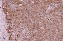
![Immunohistochemistry-Paraffin: CA19-9/Sialyl Lewis A Antibody (SPM588) [NBP2-45199]](https://resources.rndsystems.com/images/products/CA19-9-Sialyl-Lewis-A-Antibody-SPM588-Immunohistochemistry-Paraffin-NBP2-45199-img0002.jpg "Immunohistochemistry-Paraffin: CA19-9/Sialyl Lewis A Antibody (SPM588) [NBP2-45199]")
Key Product Details
Species Reactivity
Human
Applications
Immunohistochemistry, Immunohistochemistry-Paraffin, Flow Cytometry, Immunofluorescence, Immunocytochemistry/ Immunofluorescence
Label
Unconjugated
Antibody Source
Monoclonal Mouse IgM Kappa Clone # SPM588
Loading...
Product Specifications
Immunogen
Precipitin lines obtained after immuno-diffusion using MAb 116-NS-19-9 and mucins isolated from an ovarian cyst of a Lewis A+B- patient (0Le).
Localization
Cytoplasmic
Marker
GI Tumor Marker
Specificity
CA19-9, a carbohydrate epitope expressed on a high MW (400kDa) mucin glycoprotein, is a sialyl Lewisa structure which is synthesized from type 1 blood group precursor chains and is present in individuals expressing the Lewisa and/or Lewisb blood group antigens. In normal tissues, sialyl Lewisa antigen is present in ductal epithelium of the breast, kidney, salivary gland, and sweat glands. Its expression is greatly enhanced in serum as well as in the majority of tumor cells in gastrointestinal (GI) carcinomas, including adenocarcinomas of the stomach, intestine, and pancreas. Preoperative elevated CA19-9 levels in patients with stage I pancreatic carcinoma decrease to normal values following surgery. When used serially, CA19-9 can predict recurrence of disease prior to radiographic or clinical findings. This monoclonal antibody is superb for staining of formalin-fixed, paraffin-embedded tissues.
Clonality
Monoclonal
Host
Mouse
Isotype
IgM Kappa
Theoretical MW
400 kDa.
Disclaimer note: The observed molecular weight of the protein may vary from the listed predicted molecular weight due to post translational modifications, post translation cleavages, relative charges, and other experimental factors.
Disclaimer note: The observed molecular weight of the protein may vary from the listed predicted molecular weight due to post translational modifications, post translation cleavages, relative charges, and other experimental factors.
Description
200ug/ml of antibody purified from Bioreactor Concentrate. Prepared in 10 mM PBS with 0.05% BSA & 0.05% azide. Also available WITHOUT BSA at 1.0 mg/ml. (NBP3-11589)
Antibody with azide - store at 2 to 8C. Antibody without azide - store at -20 to -80C.
Antibody with azide - store at 2 to 8C. Antibody without azide - store at -20 to -80C.
Scientific Data Images for CA19-9/Sialyl Lewis A Antibody (SPM588)
Immunohistochemistry-Paraffin: CA19-9/Sialyl Lewis A Antibody (SPM588) [NBP2-45199]
Immunohistochemistry-Paraffin: CA19-9/Sialyl Lewis A Antibody (SPM588) [NBP2-45199] - Human thyroid carcinoma tissue section. Fixed with 4% formaldehyde, incubated O/N at 4C with primary antibody. IHC-P image submitted by a verified customer review.![Immunohistochemistry-Paraffin: CA19-9/Sialyl Lewis A Antibody (SPM588) [NBP2-45199]](https://resources.rndsystems.com/images/products/CA19-9-Sialyl-Lewis-A-Antibody-SPM588-Immunohistochemistry-Paraffin-NBP2-45199-img0001.jpg "Immunohistochemistry-Paraffin: CA19-9/Sialyl Lewis A Antibody (SPM588) [NBP2-45199]")
Immunohistochemistry-Paraffin: CA19-9/Sialyl Lewis A Antibody (SPM588) [NBP2-45199]
Immunohistochemistry-Paraffin: CA19-9/Sialyl Lewis A Antibody (SPM588) [NBP2-45199] - Human Colon Carcinoma stained with CA19-9 Monoclonal Antibody (SPM588)).Applications for CA19-9/Sialyl Lewis A Antibody (SPM588)
Application
Recommended Usage
Flow Cytometry
1-2 ug/million cells
Immunocytochemistry/ Immunofluorescence
1-2 ug/ml
Immunofluorescence
0.5 - 1.0 ug/ml
Immunohistochemistry-Paraffin
1-2 ug/ml
Application Notes
Immunohistochemistry (Formalin-fixed): 1 - 2 ug/mL for 30 minutes at RT. No special pretreatment is required for the immunohistochemical staining of formalin-fixed, paraffin-embedded tissues.
Optimal dilution for a specific application should be determined.
Optimal dilution for a specific application should be determined.
Reviewed Applications
Read 1 review rated 5 using NBP2-45199 in the following applications:
Flow Cytometry Panel Builder
Bio-Techne Knows Flow Cytometry
Save time and reduce costly mistakes by quickly finding compatible reagents using the Panel Builder Tool.
Advanced Features
- Spectra Viewer - Custom analysis of spectra from multiple fluorochromes
- Spillover Popups - Visualize the spectra of individual fluorochromes
- Antigen Density Selector - Match fluorochrome brightness with antigen density

Formulation, Preparation, and Storage
Purification
Protein L or PEG purified
Formulation
10 mM PBS with 0.05% BSA
Preservative
0.05% Sodium Azide
Concentration
0.2 mg/ml
Shipping
The product is shipped with polar packs. Upon receipt, store it immediately at the temperature recommended below.
Stability & Storage
Store at 4C.
Background: CA19-9
Long Name
Carbohydrate Antigen 19-9
Alternate Names
CA 19-9
Additional CA19-9 Products
Product Documents for CA19-9/Sialyl Lewis A Antibody (SPM588)
Certificate of Analysis
To download a Certificate of Analysis, please enter a lot or batch number in the search box below.
Product Specific Notices for CA19-9/Sialyl Lewis A Antibody (SPM588)
This product is for research use only and is not approved for use in humans or in clinical diagnosis. Primary Antibodies are guaranteed for 1 year from date of receipt.
Related Research Areas
Customer Reviews for CA19-9/Sialyl Lewis A Antibody (SPM588) (1)
5 out of 5
1 Customer Rating
Have you used CA19-9/Sialyl Lewis A Antibody (SPM588)?
Submit a review and receive an Amazon gift card!
$25/€18/£15/$25CAN/¥2500 Yen for a review with an image
$10/€7/£6/$10CAN/¥1110 Yen for a review without an image
Submit a review
Customer Images
Showing
1
-
1 of
1 review
Showing All
Filter By:
-
Application: Immunohistochemistry-ParaffinSample Tested: Thyroid carcinomaSpecies: HumanVerified Customer | Posted 10/14/2021Thyroid carcinoma4% formaldehyde fixed paraffin sections, incubated O/N in 4 C with primary antibody.

There are no reviews that match your criteria.
Protocols
Find general support by application which include: protocols, troubleshooting, illustrated assays, videos and webinars.
- 7-Amino Actinomycin D (7-AAD) Cell Viability Flow Cytometry Protocol
- Antigen Retrieval Protocol (PIER)
- Antigen Retrieval for Frozen Sections Protocol
- Appropriate Fixation of IHC/ICC Samples
- Cellular Response to Hypoxia Protocols
- Chromogenic IHC Staining of Formalin-Fixed Paraffin-Embedded (FFPE) Tissue Protocol
- Chromogenic Immunohistochemistry Staining of Frozen Tissue
- ClariTSA™ Fluorophore Kits
- Detection & Visualization of Antibody Binding
- Extracellular Membrane Flow Cytometry Protocol
- Flow Cytometry Protocol for Cell Surface Markers
- Flow Cytometry Protocol for Staining Membrane Associated Proteins
- Flow Cytometry Staining Protocols
- Flow Cytometry Troubleshooting Guide
- Fluorescent IHC Staining of Frozen Tissue Protocol
- Graphic Protocol for Heat-induced Epitope Retrieval
- Graphic Protocol for the Preparation and Fluorescent IHC Staining of Frozen Tissue Sections
- Graphic Protocol for the Preparation and Fluorescent IHC Staining of Paraffin-embedded Tissue Sections
- Graphic Protocol for the Preparation of Gelatin-coated Slides for Histological Tissue Sections
- ICC Cell Smear Protocol for Suspension Cells
- ICC Immunocytochemistry Protocol Videos
- ICC for Adherent Cells
- IHC Sample Preparation (Frozen sections vs Paraffin)
- Immunocytochemistry (ICC) Protocol
- Immunocytochemistry Troubleshooting
- Immunofluorescence of Organoids Embedded in Cultrex Basement Membrane Extract
- Immunofluorescent IHC Staining of Formalin-Fixed Paraffin-Embedded (FFPE) Tissue Protocol
- Immunohistochemistry (IHC) and Immunocytochemistry (ICC) Protocols
- Immunohistochemistry Frozen Troubleshooting
- Immunohistochemistry Paraffin Troubleshooting
- Intracellular Flow Cytometry Protocol Using Alcohol (Methanol)
- Intracellular Flow Cytometry Protocol Using Detergents
- Intracellular Nuclear Staining Flow Cytometry Protocol Using Detergents
- Intracellular Staining Flow Cytometry Protocol Using Alcohol Permeabilization
- Intracellular Staining Flow Cytometry Protocol Using Detergents to Permeabilize Cells
- Preparing Samples for IHC/ICC Experiments
- Preventing Non-Specific Staining (Non-Specific Binding)
- Primary Antibody Selection & Optimization
- Propidium Iodide Cell Viability Flow Cytometry Protocol
- Protocol for Heat-Induced Epitope Retrieval (HIER)
- Protocol for Liperfluo
- Protocol for Making a 4% Formaldehyde Solution in PBS
- Protocol for VisUCyte™ HRP Polymer Detection Reagent
- Protocol for the Characterization of Human Th22 Cells
- Protocol for the Characterization of Human Th9 Cells
- Protocol for the Fluorescent ICC Staining of Cell Smears - Graphic
- Protocol for the Fluorescent ICC Staining of Cultured Cells on Coverslips - Graphic
- Protocol for the Preparation & Fixation of Cells on Coverslips
- Protocol for the Preparation and Chromogenic IHC Staining of Frozen Tissue Sections
- Protocol for the Preparation and Chromogenic IHC Staining of Frozen Tissue Sections - Graphic
- Protocol for the Preparation and Chromogenic IHC Staining of Paraffin-embedded Tissue Sections
- Protocol for the Preparation and Chromogenic IHC Staining of Paraffin-embedded Tissue Sections - Graphic
- Protocol for the Preparation and Fluorescent ICC Staining of Cells on Coverslips
- Protocol for the Preparation and Fluorescent ICC Staining of Non-adherent Cells
- Protocol for the Preparation and Fluorescent ICC Staining of Stem Cells on Coverslips
- Protocol for the Preparation and Fluorescent IHC Staining of Frozen Tissue Sections
- Protocol for the Preparation and Fluorescent IHC Staining of Paraffin-embedded Tissue Sections
- Protocol for the Preparation of Gelatin-coated Slides for Histological Tissue Sections
- Protocol for the Preparation of a Cell Smear for Non-adherent Cell ICC - Graphic
- Protocol: Annexin V and PI Staining by Flow Cytometry
- Protocol: Annexin V and PI Staining for Apoptosis by Flow Cytometry
- TUNEL and Active Caspase-3 Detection by IHC/ICC Protocol
- The Importance of IHC/ICC Controls
- Troubleshooting Guide: Fluorokine Flow Cytometry Kits
- Troubleshooting Guide: Immunohistochemistry
- View all Protocols, Troubleshooting, Illustrated assays and Webinars
Loading...