CASK Antibody - BSA Free
Novus Biologicals | Catalog # NBP2-41181
![Western Blot: CASK AntibodyBSA Free [NBP2-41181]](https://resources.rndsystems.com/images/products/CASK-Antibody-Western-Blot-NBP2-41181-img0001.jpg "Western Blot: CASK AntibodyBSA Free [NBP2-41181]")
Key Product Details
Species Reactivity
Validated:
Human, Mouse, Rat
Cited:
Human
Applications
Validated:
Western Blot, ELISA, Immunocytochemistry/ Immunofluorescence, Simple Western
Cited:
Western Blot, Immunocytochemistry/ Immunofluorescence, Simple Western
Label
Unconjugated
Antibody Source
Polyclonal Rabbit IgG
Format
BSA Free
Loading...
Product Specifications
Immunogen
Antibody was raised against a 16 amino acid synthetic peptide near the amino terminus of human CASK. The immunogen is located within amino acids 760 - 810 of CASK.
Specificity
At least six alternatively spliced transcript variants have been observed.
Clonality
Polyclonal
Host
Rabbit
Isotype
IgG
Scientific Data Images for CASK Antibody - BSA Free
Western Blot: CASK AntibodyBSA Free [NBP2-41181]
Western Blot: CASK Antibody [NBP2-41181] - Western blot analysis of CASK in mouse brain tissue lysate with CASK antibody at 1 ug/ml in (A) the absence and (B) the presence of blocking peptide.![Simple Western: CASK AntibodyBSA Free [NBP2-41181]](https://resources.rndsystems.com/images/products/CASK-Antibody-Simple-Western-NBP2-41181-img0003.jpg "Simple Western: CASK AntibodyBSA Free [NBP2-41181]")
Simple Western: CASK AntibodyBSA Free [NBP2-41181]
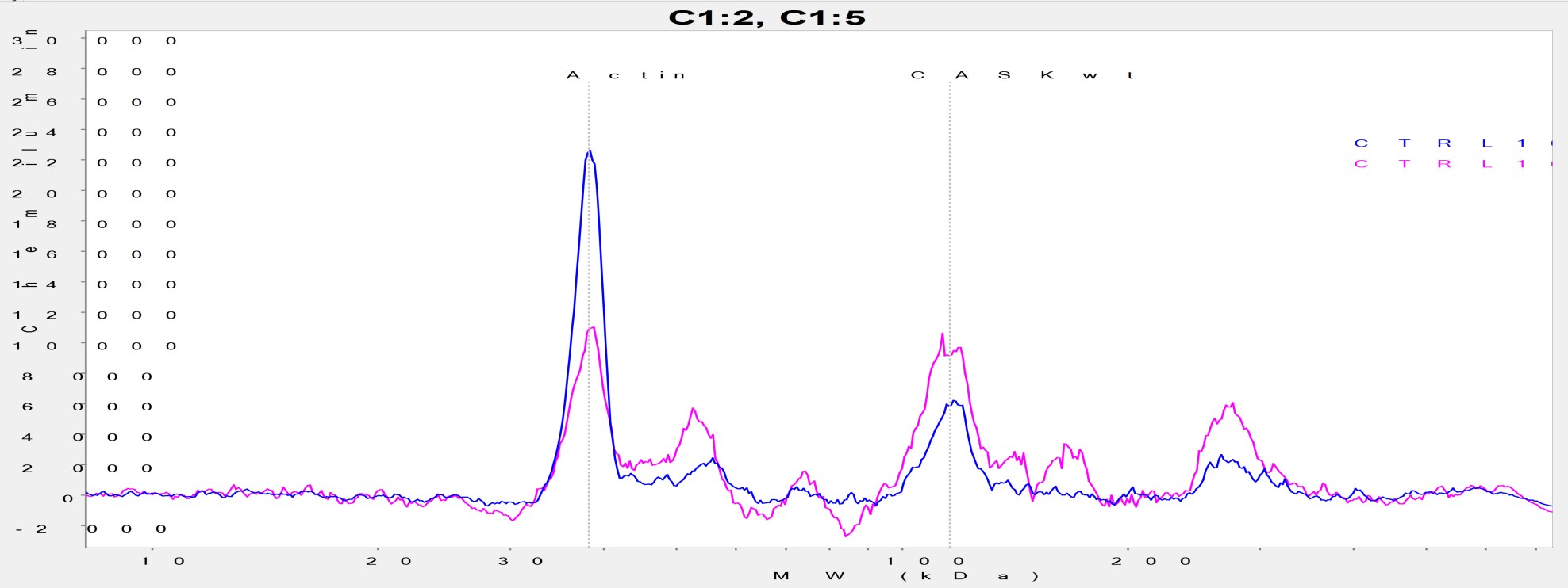
Simple Western: CASK Antibody [NBP2-41181] - 0.6 mg/ml protein lysates from differentiating human neuroepithelial stem cells run on Simple Western with 1:500 CASK.Ab (blue) and 1:1000 CASK.Ab (pink) and an actin antibody as positive control. The CASK peak is located at 116 kDa and actin at 38 kDa. Image is courtesy of customer.
Simple Western: CASK Antibody - BSA Free [NBP2-41181] -
Simple Western: CASK Antibody - BSA Free [NBP2-41181] - Presynaptic effect is limited to inhibitory VGAT presynaptic marker.a Deconvolution of cell-type populations underlying bulk RNA-sequence samples from all cell lines. b Quantification of VGlut & Homer-1 particle size (n = 4 per cell line). c Representative confocal microscopy images of neurons differentiated for 28 days & immunostained for inhibitory VGAT (green), Homer-1 (red), & Hoechst (blue). Scale = 10 µm. d Quantification of VGAT & Homer-1 particle size (n = 3 per cell line). Statistical differences between cell lines were calculated using ANOVA with post hoc Tukey HSD. *p < 0.05, **p < 0.01, ***p < 0.001. Asterisks are color-coded according to case cell lines. e In vivo concentration of GABA in the DLPFC, MFC, & putamen of ASDCASK_SS & his cotwin in relation to a typical developed control group. Image collected & cropped by CiteAb from the following publication (https://pubmed.ncbi.nlm.nih.gov/32929080), licensed under a CC-BY license. Not internally tested by Novus Biologicals.
Immunocytochemistry/ Immunofluorescence: CASK Antibody - BSA Free [NBP2-41181] -
Immunocytochemistry/ Immunofluorescence: CASK Antibody - BSA Free [NBP2-41181] - Immunofluorescence of CASK in human brain tissue with CASK antibody at 20 ug/mL.Applications for CASK Antibody - BSA Free
Application
Recommended Usage
Immunocytochemistry/ Immunofluorescence
20 ug/mL
Western Blot
1 ug/mL
Application Notes
Simple Western reported in a verified customer review.
See Simple Western Antibody Database for Simple Western validation: Tested in Differentiating human neuroepithelial stem cells, separated by Size, antibody dilution of 1:500 and 1:1000, apparent MW was 116, 38 kDa
See Simple Western Antibody Database for Simple Western validation: Tested in Differentiating human neuroepithelial stem cells, separated by Size, antibody dilution of 1:500 and 1:1000, apparent MW was 116, 38 kDa
Reviewed Applications
Read 1 review rated 5 using NBP2-41181 in the following applications:
Formulation, Preparation, and Storage
Purification
Peptide affinity purified
Formulation
PBS
Format
BSA Free
Preservative
0.02% Sodium Azide
Concentration
1 mg/ml
Shipping
The product is shipped with polar packs. Upon receipt, store it immediately at the temperature recommended below.
Stability & Storage
Store at 4C short term. Aliquot and store at -20C long term. Avoid freeze-thaw cycles.
Background: CASK
Alternate Names
Calcium/calmodulin-dependent serine protein kinase, calcium/calmodulin-dependent serine protein kinase (MAGUK family), calcium/calmodulin-dependent serine protein kinase membrane-associatedguanylate kinase, CAMGUK, CMG, EC 2.7.11, EC 2.7.11.1, FGS4CAGH39, FLJ22219, hCASK, LIN2FLJ31914, MICPCH, peripheral plasma membrane protein CASK, Protein lin-2 homolog, TNRC8, trinucleotide repeat containing 8
Gene Symbol
CASK
Additional CASK Products
Product Documents for CASK Antibody - BSA Free
Certificate of Analysis
To download a Certificate of Analysis, please enter a lot or batch number in the search box below.
Product Specific Notices for CASK Antibody - BSA Free
This product is for research use only and is not approved for use in humans or in clinical diagnosis. Primary Antibodies are guaranteed for 1 year from date of receipt.
Citations for CASK Antibody - BSA Free
 Powered by Bioz
Customer Reviews for CASK Antibody - BSA Free (1)
5 out of 5
1 Customer Rating
Have you used CASK Antibody - BSA Free?
Submit a review and receive an Amazon gift card!
$25/€18/£15/$25CAN/¥2500 Yen for a review with an image
$10/€7/£6/$10CAN/¥1110 Yen for a review without an image
Submit a review
Customer Images
Showing
1
-
1 of
1 review
Showing All
Filter By:
-
Application: Simple WesternSample Tested: Differentiated neuron progenitor cellsSpecies: HumanVerified Customer | Posted 03/13/20170.6 mg/ml protein lysates from differentiating human neuroepithelial stem cells run on Simple Western with 1:500 CASK.Ab (blue) and 1:1000 CASK.Ab (pink) and an actin antibody as positive control. The CASK peak is located at 116 kDa and actin at 38 kDa.Protein lysates have been generated using detergent free lysis buffer (TrisHCl, NaCl, EDTA, EGTA) and 5 seconds of sonication.

There are no reviews that match your criteria.
Protocols
Find general support by application which include: protocols, troubleshooting, illustrated assays, videos and webinars.
- Appropriate Fixation of IHC/ICC Samples
- Cellular Response to Hypoxia Protocols
- ClariTSA™ Fluorophore Kits
- Detection & Visualization of Antibody Binding
- ELISA Sample Preparation & Collection Guide
- ELISA Troubleshooting Guide
- How to Run an R&D Systems DuoSet ELISA
- How to Run an R&D Systems Quantikine ELISA
- How to Run an R&D Systems Quantikine™ QuicKit™ ELISA
- ICC Cell Smear Protocol for Suspension Cells
- ICC Immunocytochemistry Protocol Videos
- ICC for Adherent Cells
- Immunocytochemistry (ICC) Protocol
- Immunocytochemistry Troubleshooting
- Immunofluorescence of Organoids Embedded in Cultrex Basement Membrane Extract
- Immunohistochemistry (IHC) and Immunocytochemistry (ICC) Protocols
- Preparing Samples for IHC/ICC Experiments
- Preventing Non-Specific Staining (Non-Specific Binding)
- Primary Antibody Selection & Optimization
- Protocol for VisUCyte™ HRP Polymer Detection Reagent
- Protocol for the Fluorescent ICC Staining of Cell Smears - Graphic
- Protocol for the Fluorescent ICC Staining of Cultured Cells on Coverslips - Graphic
- Protocol for the Preparation and Fluorescent ICC Staining of Cells on Coverslips
- Protocol for the Preparation and Fluorescent ICC Staining of Non-adherent Cells
- Protocol for the Preparation and Fluorescent ICC Staining of Stem Cells on Coverslips
- Protocol for the Preparation of a Cell Smear for Non-adherent Cell ICC - Graphic
- Quantikine HS ELISA Kit Assay Principle, Alkaline Phosphatase
- Quantikine HS ELISA Kit Principle, Streptavidin-HRP Polymer
- R&D Systems Quality Control Western Blot Protocol
- Sandwich ELISA (Colorimetric) – Biotin/Streptavidin Detection Protocol
- Sandwich ELISA (Colorimetric) – Direct Detection Protocol
- TUNEL and Active Caspase-3 Detection by IHC/ICC Protocol
- The Importance of IHC/ICC Controls
- Troubleshooting Guide: ELISA
- Troubleshooting Guide: Western Blot Figures
- Western Blot Conditions
- Western Blot Protocol
- Western Blot Protocol for Cell Lysates
- Western Blot Troubleshooting
- Western Blot Troubleshooting Guide
- View all Protocols, Troubleshooting, Illustrated assays and Webinars
Loading...