Caspase-3 Antibody - (Pro and Active) - BSA Free
Novus Biologicals | Catalog # NB100-56112
![Western Blot: Caspase-3 Antibody(Pro and Active) [NB100-56112]](https://resources.rndsystems.com/images/products/Caspase-3-Antibody-Pro-and-Active-Western-Blot-NB100-56112-img0010.jpg "Western Blot: Caspase-3 Antibody(Pro and Active) [NB100-56112]")
Loading...
Key Product Details
Species Reactivity
Validated:
Human, Mouse, Rat, Canine, Gerbil
Cited:
Human, Mouse, Rat, Canine
Applications
Validated:
Immunohistochemistry, Immunohistochemistry-Paraffin, Immunohistochemistry-Frozen, Western Blot, Simple Western, Immunoprecipitation, Chromatin Immunoprecipitation, Chromatin Immunoprecipitation (ChIP)
Cited:
Immunohistochemistry-Paraffin, Western Blot, Chemotaxis
Label
Unconjugated
Antibody Source
Polyclonal Rabbit IgG
Format
BSA Free
Loading...
Product Specifications
Immunogen
This Caspase-3 Antibody - (Pro and Active) was developed against full-length human Caspase-3 protein (pro-form).
Localization
The subcellular location is primarly in the cytoplasm.
Clonality
Polyclonal
Host
Rabbit
Isotype
IgG
Theoretical MW
31.7 kDa.
Disclaimer note: The observed molecular weight of the protein may vary from the listed predicted molecular weight due to post translational modifications, post translation cleavages, relative charges, and other experimental factors.
Disclaimer note: The observed molecular weight of the protein may vary from the listed predicted molecular weight due to post translational modifications, post translation cleavages, relative charges, and other experimental factors.
Scientific Data Images for Caspase-3 Antibody - (Pro and Active) - BSA Free
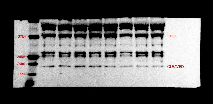
Western Blot: Caspase-3 Antibody(Pro and Active) [NB100-56112]
Western Blot: Caspase-3 Antibody - (Pro and Active) [NB100-56112] - Analysis of Caspase-3. Lysates from Jurkat cells (lane 1), normal mammary tissue (lane 2) and surgical specimens from three invasive ductal carcinomas (lanes 3-5) were normalized for total protein content (50 ug/lane) and western blotted with Caspase-3 Antibody - (Pro and Active). The ~32 kDa pro-Caspase-3 protein was detected in all samples. Active/cleaved Caspase-3 was identified in Jurkat and two ductal carcinomas (14-21 kDa large subunit).![Immunohistochemistry-Paraffin: Caspase-3 Antibody - (Pro and Active) [NB100-56112]](https://resources.rndsystems.com/images/products/Caspase-3-Antibody-Pro-and-Active-Immunohistochemistry-Paraffin-NB100-56112-img0011.jpg "Immunohistochemistry-Paraffin: Caspase-3 Antibody - (Pro and Active) [NB100-56112]")
Immunohistochemistry-Paraffin: Caspase-3 Antibody - (Pro and Active) [NB100-56112]
Immunohistochemistry-Paraffin: Caspase-3 Antibody - (Pro and Active) [NB100-56112] - Caspase-3 was detected in immersion fixed paraffin-embedded sections of human bladder tissue 1:300 dilution of rabbit polyclonal Caspase-3 Antibody - (Pro and Active)(NB100-56112), for 1 hour at room temperature followed by anti-rabbit IgG VisUCyte HRP polymer(VC003). Tissue was stained using DAB (brown) and counterstained with hematoxylin (blue).![Simple Western: Caspase-3 Antibody(Pro and Active) [NB100-56112]](https://resources.rndsystems.com/images/products/Caspase-3-Antibody-Pro-and-Active-Simple-Western-NB100-56112-img0009.jpg "Simple Western: Caspase-3 Antibody(Pro and Active) [NB100-56112]")
Simple Western: Caspase-3 Antibody(Pro and Active) [NB100-56112]
Simple Western: Caspase-3 Antibody - (Pro and Active) [NB100-56112] - Simple Western lane view shows a specific band for Caspase 3 in 0.2 mg/ml of HEK293 lysate. This experiment was performed under reducing conditions using the 12-230 kDa separation system.![Western Blot: Caspase-3 Antibody(Pro and Active) [NB100-56112]](https://resources.rndsystems.com/images/products/Caspase-3-Antibody-Pro-and-Active-Western-Blot-NB100-56112-img0012.jpg "Western Blot: Caspase-3 Antibody(Pro and Active) [NB100-56112]")
Western Blot: Caspase-3 Antibody(Pro and Active) [NB100-56112]
Caspase-3-Antibody-Pro-and-Active-Western-Blot-NB100-56112-img0012.jpg![Immunohistochemistry-Paraffin: Caspase-3 Antibody - (Pro and Active) [NB100-56112]](https://resources.rndsystems.com/images/products/Caspase-3-Antibody-Pro-and-Active-Immunohistochemistry-Paraffin-NB100-56112-img0008.jpg "Immunohistochemistry-Paraffin: Caspase-3 Antibody - (Pro and Active) [NB100-56112]")
Immunohistochemistry-Paraffin: Caspase-3 Antibody - (Pro and Active) [NB100-56112]
Immunohistochemistry-Paraffin: Caspase-3 Antibody - (Pro and Active) [NB100-56112] - Caspase-3 expression in formalin-fixed, paraffin-embedded human reactive lymph node using NB100-56112 (Caspase-3 Antibody - (Pro and Active)) at 1:2000. Staining is seen in the apoptosis-prone germinal center B lymphocytes of follicles.![Immunohistochemistry-Paraffin: Caspase-3 Antibody - (Pro and Active) [NB100-56112]](https://resources.rndsystems.com/images/products/Caspase-3-Antibody-Pro-and-Active-Immunohistochemistry-Paraffin-NB100-56112-img0007.jpg "Immunohistochemistry-Paraffin: Caspase-3 Antibody - (Pro and Active) [NB100-56112]")
Immunohistochemistry-Paraffin: Caspase-3 Antibody - (Pro and Active) [NB100-56112]
Immunohistochemistry-Paraffin: Caspase-3 Antibody - (Pro and Active) [NB100-56112] - Analysis of Caspase-3 expression in formalin-fixed, paraffin-embedded human breast ductal carcinoma in situ using this antibody at 1:2000. Staining is seen in the the cancerous ducts, but not in the normal lobulus.Applications for Caspase-3 Antibody - (Pro and Active) - BSA Free
Application
Recommended Usage
Chromatin Immunoprecipitation
1:10-1:500. Use reported in scientific literature (PMID 27735949)
Chromatin Immunoprecipitation (ChIP)
1:10-1:500
Immunohistochemistry
1:1000-1:5000
Immunohistochemistry-Frozen
1:10-1:500
Immunohistochemistry-Paraffin
1:1000-1:5000
Immunoprecipitation
1:50-1:200
Simple Western
1:200
Western Blot
1:1000-1:2000
Application Notes
Immunoprecipitation, Western Blot, Immunohistochemistry-Paraffin IHC (frozen): Users should optimize according to model and immunodetection system used (secondary reagents). In Simple Western only 10 - 15 uL of the recommended dilution is used per data point.
See Simple Western Antibody Database for Simple Western validation: Tested in Hek293 lysate 0.2 mg/mL, separated by Size, antibody dilution of 1:200, apparent MW was 40 kDa. Separated by Size-Wes, Sally Sue/Peggy Sue.
See Simple Western Antibody Database for Simple Western validation: Tested in Hek293 lysate 0.2 mg/mL, separated by Size, antibody dilution of 1:200, apparent MW was 40 kDa. Separated by Size-Wes, Sally Sue/Peggy Sue.
Reviewed Applications
Read 1 review rated 5 using NB100-56112 in the following applications:
Formulation, Preparation, and Storage
Purification
Unpurified
Formulation
Whole antisera
Format
BSA Free
Preservative
0.05% Sodium Azide
Concentration
This product is unpurified. The exact concentration of antibody is not quantifiable.
Shipping
The product is shipped with polar packs. Upon receipt, store it immediately at the temperature recommended below.
Stability & Storage
Store at -20C. Avoid freeze-thaw cycles.
Background: Caspase-3
References
1.Mu, N., Lei, Y., Wang, Y., Wang, Y., Duan, Q., Ma, G.,... Su, L. (2019). Inhibition of SIRT1/2 upregulates HSPA5 acetylation and induces pro-survival autophagy via ATF4-DDIT4-mTORC1 axis in human lung cancer cells. Apoptosis, 24(9-10), 798-811. doi:10.1007/s10495-019-01559-3
2.Sun, C. M., Enkhjargal, B., Reis, C., Zhou, K. R., Xie, Z. Y., Wu, L. Y.,... Zhang, J. H. (2019). Osteopontin attenuates early brain injury through regulating autophagy-apoptosis interaction after subarachnoid hemorrhage in rats. CNS Neurosci Ther, 25(10), 1162-1172. doi:10.1111/cns.13199
3.Louneva, N., Cohen, J. W., Han, L. Y., Talbot, K., Wilson, R. S., Bennett, D. A.,... Arnold, S. E. (2008). Caspase-3 is enriched in postsynaptic densities and increased in Alzheimer's disease. Am J Pathol, 173(5), 1488-1495. doi:10.2353/ajpath.2008.080434
Alternate Names
Apopain, CASP3, Caspase3, CPP32, LICE-1, YAMA
Gene Symbol
CASP3
Additional Caspase-3 Products
Product Documents for Caspase-3 Antibody - (Pro and Active) - BSA Free
Certificate of Analysis
To download a Certificate of Analysis, please enter a lot or batch number in the search box below.
Product Specific Notices for Caspase-3 Antibody - (Pro and Active) - BSA Free
This product is for research use only and is not approved for use in humans or in clinical diagnosis. Primary Antibodies are guaranteed for 1 year from date of receipt.
Related Research Areas
Citations for Caspase-3 Antibody - (Pro and Active) - BSA Free
 Powered by Bioz
Customer Reviews for Caspase-3 Antibody - (Pro and Active) - BSA Free (1)
5 out of 5
1 Customer Rating
Have you used Caspase-3 Antibody - (Pro and Active) - BSA Free?
Submit a review and receive an Amazon gift card!
$25/€18/£15/$25CAN/¥2500 Yen for a review with an image
$10/€7/£6/$10CAN/¥1110 Yen for a review without an image
Submit a review
Customer Images
Showing
1
-
1 of
1 review
Showing All
Filter By:
-
Application: Western BlotSample Tested: BRAIN(Substantia Nigra par compacta)Species: MouseVerified Customer | Posted 01/12/2017

There are no reviews that match your criteria.
Protocols
Find general support by application which include: protocols, troubleshooting, illustrated assays, videos and webinars.
- Antigen Retrieval Protocol (PIER)
- Antigen Retrieval for Frozen Sections Protocol
- Appropriate Fixation of IHC/ICC Samples
- Cellular Response to Hypoxia Protocols
- ChIP Protocol Video
- Chromatin Immunoprecipitation (ChIP) Protocol
- Chromatin Immunoprecipitation Protocol
- Chromogenic IHC Staining of Formalin-Fixed Paraffin-Embedded (FFPE) Tissue Protocol
- Chromogenic Immunohistochemistry Staining of Frozen Tissue
- ClariTSA™ Fluorophore Kits
- Detection & Visualization of Antibody Binding
- Fluorescent IHC Staining of Frozen Tissue Protocol
- Graphic Protocol for Heat-induced Epitope Retrieval
- Graphic Protocol for the Preparation and Fluorescent IHC Staining of Frozen Tissue Sections
- Graphic Protocol for the Preparation and Fluorescent IHC Staining of Paraffin-embedded Tissue Sections
- Graphic Protocol for the Preparation of Gelatin-coated Slides for Histological Tissue Sections
- IHC Sample Preparation (Frozen sections vs Paraffin)
- Immunofluorescent IHC Staining of Formalin-Fixed Paraffin-Embedded (FFPE) Tissue Protocol
- Immunohistochemistry (IHC) and Immunocytochemistry (ICC) Protocols
- Immunohistochemistry Frozen Troubleshooting
- Immunohistochemistry Paraffin Troubleshooting
- Immunoprecipitation Protocol
- Preparing Samples for IHC/ICC Experiments
- Preventing Non-Specific Staining (Non-Specific Binding)
- Primary Antibody Selection & Optimization
- Protocol for Heat-Induced Epitope Retrieval (HIER)
- Protocol for Making a 4% Formaldehyde Solution in PBS
- Protocol for VisUCyte™ HRP Polymer Detection Reagent
- Protocol for the Preparation & Fixation of Cells on Coverslips
- Protocol for the Preparation and Chromogenic IHC Staining of Frozen Tissue Sections
- Protocol for the Preparation and Chromogenic IHC Staining of Frozen Tissue Sections - Graphic
- Protocol for the Preparation and Chromogenic IHC Staining of Paraffin-embedded Tissue Sections
- Protocol for the Preparation and Chromogenic IHC Staining of Paraffin-embedded Tissue Sections - Graphic
- Protocol for the Preparation and Fluorescent IHC Staining of Frozen Tissue Sections
- Protocol for the Preparation and Fluorescent IHC Staining of Paraffin-embedded Tissue Sections
- Protocol for the Preparation of Gelatin-coated Slides for Histological Tissue Sections
- R&D Systems Quality Control Western Blot Protocol
- TUNEL and Active Caspase-3 Detection by IHC/ICC Protocol
- The Importance of IHC/ICC Controls
- Troubleshooting Guide: Immunohistochemistry
- Troubleshooting Guide: Western Blot Figures
- Western Blot Conditions
- Western Blot Protocol
- Western Blot Protocol for Cell Lysates
- Western Blot Troubleshooting
- Western Blot Troubleshooting Guide
- View all Protocols, Troubleshooting, Illustrated assays and Webinars
Loading...
Associated Pathways

