![Immunohistochemistry-Paraffin: Caspase-3 Antibody [NB600-1235]](https://resources.rndsystems.com/images/products/Caspase-3-Antibody-Immunohistochemistry-Paraffin-NB600-1235-img0003.jpg "Immunohistochemistry-Paraffin: Caspase-3 Antibody [NB600-1235]")
Loading...
Key Product Details
Species Reactivity
Validated:
Human, Fish
Cited:
Human, Mouse, Rat, Bovine, Canine, Fish, Insect - Drosophila melanogaster, Primate, Rabbit
Applications
Validated:
Immunohistochemistry, Immunohistochemistry-Paraffin, Immunohistochemistry-Frozen, Immunohistochemistry Free-Floating, Western Blot, Immunocytochemistry/ Immunofluorescence
Cited:
Immunohistochemistry, Immunohistochemistry-Paraffin, Immunohistochemistry Free-Floating, Western Blot, Immunocytochemistry/ Immunofluorescence, IF/IHC
Label
Unconjugated
Antibody Source
Polyclonal Rabbit IgG
Loading...
Product Specifications
Immunogen
This Caspase-3 Antibody was developed against a synthetic peptide corresponding to the cleavage site of human caspase 3 (amino acids 167-175), conjugated to KLH.
Reactivity Notes
Fish reactivity reported in scientific literature (PMID: 28675853).
Localization
Cytoplasmic and some nuclear.
Specificity
This reacts with the active form of Caspase 3 (17 kDa protein).
Clonality
Polyclonal
Host
Rabbit
Isotype
IgG
Theoretical MW
31.7 kDa.
Disclaimer note: The observed molecular weight of the protein may vary from the listed predicted molecular weight due to post translational modifications, post translation cleavages, relative charges, and other experimental factors.
Disclaimer note: The observed molecular weight of the protein may vary from the listed predicted molecular weight due to post translational modifications, post translation cleavages, relative charges, and other experimental factors.
Scientific Data Images for Caspase-3 Antibody
Immunohistochemistry-Paraffin: Caspase-3 Antibody [NB600-1235]
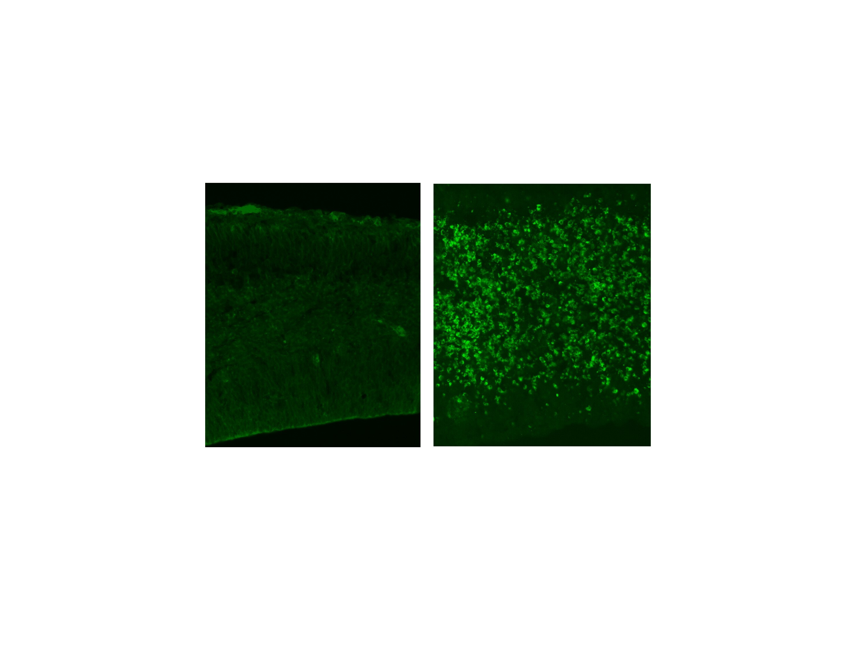
Immunohistochemistry-Paraffin: Caspase-3 Antibody [NB600-1235] - Formalin fixed paraffin embedded human tonsil stained with Caspase-3 Antibody (NB600-1235).![Immunohistochemistry-Paraffin: Caspase-3 Antibody [NB600-1235]](https://resources.rndsystems.com/images/products/Caspase-3-Antibody-Immunohistochemistry-Paraffin-NB600-1235-img0002.jpg "Immunohistochemistry-Paraffin: Caspase-3 Antibody [NB600-1235]")
Immunohistochemistry-Paraffin: Caspase-3 Antibody [NB600-1235]
Immunohistochemistry-Paraffin: Caspase-3 Antibody [NB600-1235] - Human tonsil.
Immunohistochemistry: Caspase-3 Antibody [NB600-1235] -
nb600-1235_rabbit-polyclonal-caspase-3-antibody-imgenex-img-80432-20920231721154.jpgApplications for Caspase-3 Antibody
Application
Recommended Usage
Immunocytochemistry/ Immunofluorescence
1:10-1:500
Immunohistochemistry
1:10-1:500
Immunohistochemistry-Paraffin
1:50-1:100
Application Notes
IHC-P: recommended pretreatment of citrate buffer, pH 6.0. Recommended incubation time of 30 min at RT. Use in ICC/IF was reported in the scientific literature (PMID: 23813946). Use in Western blot reported in scientific literature (PMID: 29977195). Use in IHC-FrFl reported in secitific publication PMID: 32651317. This Caspase-3 Antibody is validated for IHC-Fr from a verified customer review.
Reviewed Applications
Read 1 review rated 4 using NB600-1235 in the following applications:
Formulation, Preparation, and Storage
Purification
Affinity purified
Formulation
PBS (pH 7.4), 0.2% BSA, Tween-20
Preservative
0.05% Sodium Azide
Concentration
1.0 mg/ml
Shipping
The product is shipped with polar packs. Upon receipt, store it immediately at the temperature recommended below.
Stability & Storage
Store at 4C. Do not freeze.
Background: Caspase-3
References
1.Mu, N., Lei, Y., Wang, Y., Wang, Y., Duan, Q., Ma, G.,... Su, L. (2019). Inhibition of SIRT1/2 upregulates HSPA5 acetylation and induces pro-survival autophagy via ATF4-DDIT4-mTORC1 axis in human lung cancer cells. Apoptosis, 24(9-10), 798-811. doi:10.1007/s10495-019-01559-3
2.Sun, C. M., Enkhjargal, B., Reis, C., Zhou, K. R., Xie, Z. Y., Wu, L. Y.,... Zhang, J. H. (2019). Osteopontin attenuates early brain injury through regulating autophagy-apoptosis interaction after subarachnoid hemorrhage in rats. CNS Neurosci Ther, 25(10), 1162-1172. doi:10.1111/cns.13199
3.Louneva, N., Cohen, J. W., Han, L. Y., Talbot, K., Wilson, R. S., Bennett, D. A.,... Arnold, S. E. (2008). Caspase-3 is enriched in postsynaptic densities and increased in Alzheimer's disease. Am J Pathol, 173(5), 1488-1495. doi:10.2353/ajpath.2008.080434
Alternate Names
Apopain, CASP3, Caspase3, CPP32, LICE-1, YAMA
Gene Symbol
CASP3
UniProt
Additional Caspase-3 Products
Product Documents for Caspase-3 Antibody
Certificate of Analysis
To download a Certificate of Analysis, please enter a lot or batch number in the search box below.
Product Specific Notices for Caspase-3 Antibody
This product is for research use only and is not approved for use in humans or in clinical diagnosis. Primary Antibodies are guaranteed for 1 year from date of receipt.
Related Research Areas
Citations for Caspase-3 Antibody
 Powered by Bioz
Customer Reviews for Caspase-3 Antibody (1)
4 out of 5
1 Customer Rating
Have you used Caspase-3 Antibody?
Submit a review and receive an Amazon gift card!
$25/€18/£15/$25CAN/¥2500 Yen for a review with an image
$10/€7/£6/$10CAN/¥1110 Yen for a review without an image
Submit a review
Customer Images
Showing
1
-
1 of
1 review
Showing All
Filter By:
-
Application: Immunohistochemistry-FrozenSample Tested: Embryonic brainSpecies: MouseVerified Customer | Posted 02/19/2021mouse E14 cortex left: negative control. right: N-ethyl-N-nitrosourea was injected to induce apoptosis.dilution: 1:500
 Bio-Techne ResponseThis review was submitted through the legacy Novus Innovators Program, reflecting a new species or application tested on a primary antibody.
There are no reviews that match your criteria.
Protocols
Find general support by application which include: protocols, troubleshooting, illustrated assays, videos and webinars.
- Antigen Retrieval Protocol (PIER)
- Antigen Retrieval for Frozen Sections Protocol
- Appropriate Fixation of IHC/ICC Samples
- Cellular Response to Hypoxia Protocols
- Chromogenic IHC Staining of Formalin-Fixed Paraffin-Embedded (FFPE) Tissue Protocol
- Chromogenic Immunohistochemistry Staining of Frozen Tissue
- ClariTSA™ Fluorophore Kits
- Detection & Visualization of Antibody Binding
- Fluorescent IHC Staining of Frozen Tissue Protocol
- Graphic Protocol for Heat-induced Epitope Retrieval
- Graphic Protocol for the Preparation and Fluorescent IHC Staining of Frozen Tissue Sections
- Graphic Protocol for the Preparation and Fluorescent IHC Staining of Paraffin-embedded Tissue Sections
- Graphic Protocol for the Preparation of Gelatin-coated Slides for Histological Tissue Sections
- ICC Cell Smear Protocol for Suspension Cells
- ICC Immunocytochemistry Protocol Videos
- ICC for Adherent Cells
- IHC Sample Preparation (Frozen sections vs Paraffin)
- Immunocytochemistry (ICC) Protocol
- Immunocytochemistry Troubleshooting
- Immunofluorescence of Organoids Embedded in Cultrex Basement Membrane Extract
- Immunofluorescent IHC Staining of Formalin-Fixed Paraffin-Embedded (FFPE) Tissue Protocol
- Immunohistochemistry (IHC) and Immunocytochemistry (ICC) Protocols
- Immunohistochemistry Frozen Troubleshooting
- Immunohistochemistry Paraffin Troubleshooting
- Preparing Samples for IHC/ICC Experiments
- Preventing Non-Specific Staining (Non-Specific Binding)
- Primary Antibody Selection & Optimization
- Protocol for Heat-Induced Epitope Retrieval (HIER)
- Protocol for Making a 4% Formaldehyde Solution in PBS
- Protocol for VisUCyte™ HRP Polymer Detection Reagent
- Protocol for the Fluorescent ICC Staining of Cell Smears - Graphic
- Protocol for the Fluorescent ICC Staining of Cultured Cells on Coverslips - Graphic
- Protocol for the Preparation & Fixation of Cells on Coverslips
- Protocol for the Preparation and Chromogenic IHC Staining of Frozen Tissue Sections
- Protocol for the Preparation and Chromogenic IHC Staining of Frozen Tissue Sections - Graphic
- Protocol for the Preparation and Chromogenic IHC Staining of Paraffin-embedded Tissue Sections
- Protocol for the Preparation and Chromogenic IHC Staining of Paraffin-embedded Tissue Sections - Graphic
- Protocol for the Preparation and Fluorescent ICC Staining of Cells on Coverslips
- Protocol for the Preparation and Fluorescent ICC Staining of Non-adherent Cells
- Protocol for the Preparation and Fluorescent ICC Staining of Stem Cells on Coverslips
- Protocol for the Preparation and Fluorescent IHC Staining of Frozen Tissue Sections
- Protocol for the Preparation and Fluorescent IHC Staining of Paraffin-embedded Tissue Sections
- Protocol for the Preparation of Gelatin-coated Slides for Histological Tissue Sections
- Protocol for the Preparation of a Cell Smear for Non-adherent Cell ICC - Graphic
- R&D Systems Quality Control Western Blot Protocol
- TUNEL and Active Caspase-3 Detection by IHC/ICC Protocol
- The Importance of IHC/ICC Controls
- Troubleshooting Guide: Immunohistochemistry
- Troubleshooting Guide: Western Blot Figures
- Western Blot Conditions
- Western Blot Protocol
- Western Blot Protocol for Cell Lysates
- Western Blot Troubleshooting
- Western Blot Troubleshooting Guide
- View all Protocols, Troubleshooting, Illustrated assays and Webinars
FAQs for Caspase-3 Antibody
Showing
1
-
4 of
4 FAQs
Showing All
-
Q: Any reason for cleaved caspase 3 to be seen as purely nuclear staining or is this just an artifact?
A: Caspase-3 can translocate from the cytoplasm to the nucleus, usually during apoptosis. We do offer a 100% guarantee that this antibody will be useful as stated on the datasheet also.
-
Q: I'd like to get the information about the active/cleaved Caspase-3 (NB600-1235). The datasheet says that it reacts with the active form of Caspase-3 (17 kDa protein), but every time when I do the WB, I get two bands between 15kDa and 20kDa. The different company's cleaved caspase 3 antibody reacts with 17 and 19 kDa protein, so I wonder which band is specific to yours?
A: According to the data I could find for the active/cleaved caspase 3, the active form should appear at approximately 17kDa and also has a smaller subunit seen at approximately 12kDa. The inactive form is approximately 32kDa. These are the theoretical weights and can vary based on type of gel and samples they are run on. In most cases, it looks like most antibodies detect the active forms at 19 and 17kDa respectively. With this particular product, NB600-1235, we have not yet validated it for use in western blot, so we unfortunately do not have any information for the location of bands.
-
Q: The datasheet says that it reacts with the active form of Caspase 3 (17 kDa protein), but every time when I do the WB, I get two bands between 15kDa and 20kDa. The different company's cleaved caspase 3 antibody reacts with 17 and 19 kDa protein, so I wonder which band is specific to yours.
A: Regarding your question, NB600-1235 has been exclusively validated for IHC, not western blot and it is supposed to detect the active form of caspase3. The inactive Caspase 3 is a 31 kD protein, a heterotetramer that consists of two anti-parallel arranged heterodimers, each one formed by a 17 kDa (p17) and a 12 kDa (p12). When it is Cleaved by granzyme B, it generates the two active subunits. As a result the two bands you are observing on the gel should be correct. However, we haven't tested this antibody for western blot.
-
Q: This is a Rabbit antibody right so Do you think I can use it on rabbit tissue?
A: This antibody does react to rabbit but if you are using it with an anti-rabbit secondary, you will almost certainly see some background staining. I know that mouse-on-mouse blocking systems exist, but I am not sure if there is a rabbit equivalent at this time. The best way to use NB600-1235 on rabbit tissues would be to have it directly conjugated to biotin, HRP, or whatever detection method you are planning to use. This will eliminate the need for an anti-rabbit secondary and should give you little to no background. We do offer a custom conjugation service, so if you would like to have this antibody conjugated, please send an email to let me know what you would like to have this antibody conjugated to and I can get you a quote.
-
Q: Any reason for cleaved caspase 3 to be seen as purely nuclear staining or is this just an artifact?
A: Caspase-3 can translocate from the cytoplasm to the nucleus, usually during apoptosis. We do offer a 100% guarantee that this antibody will be useful as stated on the datasheet also.
-
Q: I'd like to get the information about the active/cleaved Caspase-3 (NB600-1235). The datasheet says that it reacts with the active form of Caspase-3 (17 kDa protein), but every time when I do the WB, I get two bands between 15kDa and 20kDa. The different company's cleaved caspase 3 antibody reacts with 17 and 19 kDa protein, so I wonder which band is specific to yours?
A: According to the data I could find for the active/cleaved caspase 3, the active form should appear at approximately 17kDa and also has a smaller subunit seen at approximately 12kDa. The inactive form is approximately 32kDa. These are the theoretical weights and can vary based on type of gel and samples they are run on. In most cases, it looks like most antibodies detect the active forms at 19 and 17kDa respectively. With this particular product, NB600-1235, we have not yet validated it for use in western blot, so we unfortunately do not have any information for the location of bands.
-
Q: The datasheet says that it reacts with the active form of Caspase 3 (17 kDa protein), but every time when I do the WB, I get two bands between 15kDa and 20kDa. The different company's cleaved caspase 3 antibody reacts with 17 and 19 kDa protein, so I wonder which band is specific to yours.
A: Regarding your question, NB600-1235 has been exclusively validated for IHC, not western blot and it is supposed to detect the active form of caspase3. The inactive Caspase 3 is a 31 kD protein, a heterotetramer that consists of two anti-parallel arranged heterodimers, each one formed by a 17 kDa (p17) and a 12 kDa (p12). When it is Cleaved by granzyme B, it generates the two active subunits. As a result the two bands you are observing on the gel should be correct. However, we haven't tested this antibody for western blot.
-
Q: This is a Rabbit antibody right so Do you think I can use it on rabbit tissue?
A: This antibody does react to rabbit but if you are using it with an anti-rabbit secondary, you will almost certainly see some background staining. I know that mouse-on-mouse blocking systems exist, but I am not sure if there is a rabbit equivalent at this time. The best way to use NB600-1235 on rabbit tissues would be to have it directly conjugated to biotin, HRP, or whatever detection method you are planning to use. This will eliminate the need for an anti-rabbit secondary and should give you little to no background. We do offer a custom conjugation service, so if you would like to have this antibody conjugated, please send an email to let me know what you would like to have this antibody conjugated to and I can get you a quote.
Loading...
Associated Pathways

