Cav1.2 Antibody
Novus Biologicals | Catalog # NBP2-76914
![Immunocytochemistry/ Immunofluorescence: Cav1.2 Antibody [NBP2-76914]](https://resources.rndsystems.com/images/products/Cav1.2-Antibody-Immunocytochemistry-Immunofluorescence-NBP2-76914-img0008.jpg "Immunocytochemistry/ Immunofluorescence: Cav1.2 Antibody [NBP2-76914]")
Loading...
Key Product Details
Species Reactivity
Validated:
Human, Mouse, Rat
Cited:
Mouse
Applications
Validated:
Immunohistochemistry, Immunohistochemistry-Paraffin, Flow Cytometry, Immunocytochemistry/ Immunofluorescence, Dot Blot
Cited:
Immunohistochemistry-Paraffin
Label
Unconjugated
Antibody Source
Polyclonal Rabbit IgG
Loading...
Product Specifications
Immunogen
Synthetic peptide within mouse Cav1.2 aa 802-851 / 2,139. (SwissProt: Q13936 Human; SwissProt: Q01815 Mouse; SwissProt: P22002 Rat)
Localization
Plasma membrane.
Clonality
Polyclonal
Host
Rabbit
Isotype
IgG
Theoretical MW
249 kDa.
Disclaimer note: The observed molecular weight of the protein may vary from the listed predicted molecular weight due to post translational modifications, post translation cleavages, relative charges, and other experimental factors.
Disclaimer note: The observed molecular weight of the protein may vary from the listed predicted molecular weight due to post translational modifications, post translation cleavages, relative charges, and other experimental factors.
Scientific Data Images for Cav1.2 Antibody
Immunocytochemistry/ Immunofluorescence: Cav1.2 Antibody [NBP2-76914]
Immunocytochemistry/Immunofluorescence: Cav1.2 Antibody [NBP2-76914] - Staining of CACNA1C in SiHa cells (green). Formalin fixed cells were permeabilized with 0.1% Triton X-100 in TBS for 10 minutes at room temperature and blocked with 10% negative goat serum for 15 minutes at room temperature. Cells were probed with the primary antibody (1/200) for 1 hour at room temperature, washed with PBS. Alexa Fluor(R)488 conjugate-Goat anti-Rabbit IgG was used as the secondary antibody at 1/1,000 dilution. The nuclear counter stain is DAPI (blue).![Immunohistochemistry-Paraffin: Cav1.2 Antibody [NBP2-76914]](https://resources.rndsystems.com/images/products/Cav1.2-Antibody-Immunohistochemistry-Paraffin-NBP2-76914-img0006.jpg "Immunohistochemistry-Paraffin: Cav1.2 Antibody [NBP2-76914]")
Immunohistochemistry-Paraffin: Cav1.2 Antibody [NBP2-76914]
Immunohistochemistry-Paraffin: Cav1.2 Antibody [NBP2-76914] - Immunohistochemical analysis of paraffin-embedded mouse heart tissue using anti-CACNA1C antibody. Counter stained with hematoxylin.![Flow Cytometry: Cav1.2 Antibody [NBP2-76914]](https://resources.rndsystems.com/images/products/Cav1.2-Antibody-Flow-Cytometry-NBP2-76914-img0007.jpg "Flow Cytometry: Cav1.2 Antibody [NBP2-76914]")
Flow Cytometry: Cav1.2 Antibody [NBP2-76914]
Flow Cytometry: Cav1.2 Antibody [NBP2-76914] - Flow cytometric analysis of SKOV-3 cells with CACNA1C antibody at 1/100 dilution (fuchsia) compared with an unlabelled control (cells without incubation with primary antibody; yellow). Alexa Fluor 488-conjugated goat anti-rabbit IgG was used as the secondary antibody.![Immunocytochemistry/ Immunofluorescence: Cav1.2 Antibody [NBP2-76914]](https://resources.rndsystems.com/images/products/Cav1.2-Antibody-Immunocytochemistry-Immunofluorescence-NBP2-76914-img0002.jpg "Immunocytochemistry/ Immunofluorescence: Cav1.2 Antibody [NBP2-76914]")
Immunocytochemistry/ Immunofluorescence: Cav1.2 Antibody [NBP2-76914]
Immunocytochemistry/Immunofluorescence: Cav1.2 Antibody [NBP2-76914] - ICC staining CACNA1C in SKOV-3 cells (green). The nuclear counter stain is DAPI (blue). Cells were fixed in paraformaldehyde, permeabilised with 0.25% Triton X100/PBS.![Immunohistochemistry-Paraffin: Cav1.2 Antibody [NBP2-76914]](https://resources.rndsystems.com/images/products/Cav1.2-Antibody-Immunohistochemistry-Paraffin-NBP2-76914-img0003.jpg "Immunohistochemistry-Paraffin: Cav1.2 Antibody [NBP2-76914]")
Immunohistochemistry-Paraffin: Cav1.2 Antibody [NBP2-76914]
Immunohistochemistry-Paraffin: Cav1.2 Antibody [NBP2-76914] - Immunohistochemical analysis of paraffin-embedded rat brain tissue using anti-CACNA1C antibody. Counter stained with hematoxylin.![Immunohistochemistry-Paraffin: Cav1.2 Antibody [NBP2-76914]](https://resources.rndsystems.com/images/products/Cav1.2-Antibody-Immunohistochemistry-Paraffin-NBP2-76914-img0004.jpg "Immunohistochemistry-Paraffin: Cav1.2 Antibody [NBP2-76914]")
Immunohistochemistry-Paraffin: Cav1.2 Antibody [NBP2-76914]
Immunohistochemistry-Paraffin: Cav1.2 Antibody [NBP2-76914] - Immunohistochemical analysis of paraffin-embedded human kidney tissue using anti-CACNA1C antibody. Counter stained with hematoxylin.![Immunohistochemistry-Paraffin: Cav1.2 Antibody [NBP2-76914]](https://resources.rndsystems.com/images/products/Cav1.2-Antibody-Immunohistochemistry-Paraffin-NBP2-76914-img0005.jpg "Immunohistochemistry-Paraffin: Cav1.2 Antibody [NBP2-76914]")
Immunohistochemistry-Paraffin: Cav1.2 Antibody [NBP2-76914]
Immunohistochemistry-Paraffin: Cav1.2 Antibody [NBP2-76914] - Immunohistochemical analysis of paraffin-embedded human uterus tissue using anti-CACNA1C antibody. Counter stained with hematoxylin.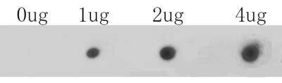
Dot Blot: Cav1.2 Antibody [NBP2-76914] - Dot blot analysis of anti-CACNA1C on PVDF. 1ug, 2ug and 4ug of immunization peptides were given in this test. Anti-CACNA1C antibody was diluted with 1/500.
Applications for Cav1.2 Antibody
Application
Recommended Usage
Dot Blot
1:500-1:1000
Flow Cytometry
1:50-1:100
Immunocytochemistry/ Immunofluorescence
1:500-1:2000
Immunohistochemistry-Paraffin
1:50-1:200
Flow Cytometry Panel Builder
Bio-Techne Knows Flow Cytometry
Save time and reduce costly mistakes by quickly finding compatible reagents using the Panel Builder Tool.
Advanced Features
- Spectra Viewer - Custom analysis of spectra from multiple fluorochromes
- Spillover Popups - Visualize the spectra of individual fluorochromes
- Antigen Density Selector - Match fluorochrome brightness with antigen density

Formulation, Preparation, and Storage
Purification
Immunogen affinity purified
Formulation
PBS (pH7.4), 0.2% BSA, 50% Glycerol
Preservative
0.05% Sodium Azide
Concentration
1 mg/ml
Shipping
The product is shipped with polar packs. Upon receipt, store it immediately at the temperature recommended below.
Stability & Storage
Store at 4C short term. Aliquot and store at -20C long term. Avoid freeze-thaw cycles.
Background: Cav1.2
Long Name
Voltage-dependent L-type calcium channel subunit alpha-1C
Alternate Names
CACH2, CACN2, CACNA1C, CACNL1A1, CCHL1A1
Gene Symbol
CACNA1C
Additional Cav1.2 Products
Product Documents for Cav1.2 Antibody
Certificate of Analysis
To download a Certificate of Analysis, please enter a lot or batch number in the search box below.
Product Specific Notices for Cav1.2 Antibody
This product is for research use only and is not approved for use in humans or in clinical diagnosis. Primary Antibodies are guaranteed for 1 year from date of receipt.
Citations for Cav1.2 Antibody
 Powered by Bioz
Customer Reviews for Cav1.2 Antibody
There are currently no reviews for this product. Be the first to review Cav1.2 Antibody and earn rewards!
Have you used Cav1.2 Antibody?
Submit a review and receive an Amazon gift card!
$25/€18/£15/$25CAN/¥2500 Yen for a review with an image
$10/€7/£6/$10CAN/¥1110 Yen for a review without an image
Submit a review
Protocols
Find general support by application which include: protocols, troubleshooting, illustrated assays, videos and webinars.
- 7-Amino Actinomycin D (7-AAD) Cell Viability Flow Cytometry Protocol
- Antigen Retrieval Protocol (PIER)
- Antigen Retrieval for Frozen Sections Protocol
- Appropriate Fixation of IHC/ICC Samples
- Cellular Response to Hypoxia Protocols
- Chromogenic IHC Staining of Formalin-Fixed Paraffin-Embedded (FFPE) Tissue Protocol
- Chromogenic Immunohistochemistry Staining of Frozen Tissue
- ClariTSA™ Fluorophore Kits
- Detection & Visualization of Antibody Binding
- Extracellular Membrane Flow Cytometry Protocol
- Flow Cytometry Protocol for Cell Surface Markers
- Flow Cytometry Protocol for Staining Membrane Associated Proteins
- Flow Cytometry Staining Protocols
- Flow Cytometry Troubleshooting Guide
- Fluorescent IHC Staining of Frozen Tissue Protocol
- Graphic Protocol for Heat-induced Epitope Retrieval
- Graphic Protocol for the Preparation and Fluorescent IHC Staining of Frozen Tissue Sections
- Graphic Protocol for the Preparation and Fluorescent IHC Staining of Paraffin-embedded Tissue Sections
- Graphic Protocol for the Preparation of Gelatin-coated Slides for Histological Tissue Sections
- ICC Cell Smear Protocol for Suspension Cells
- ICC Immunocytochemistry Protocol Videos
- ICC for Adherent Cells
- IHC Sample Preparation (Frozen sections vs Paraffin)
- Immunocytochemistry (ICC) Protocol
- Immunocytochemistry Troubleshooting
- Immunofluorescence of Organoids Embedded in Cultrex Basement Membrane Extract
- Immunofluorescent IHC Staining of Formalin-Fixed Paraffin-Embedded (FFPE) Tissue Protocol
- Immunohistochemistry (IHC) and Immunocytochemistry (ICC) Protocols
- Immunohistochemistry Frozen Troubleshooting
- Immunohistochemistry Paraffin Troubleshooting
- Intracellular Flow Cytometry Protocol Using Alcohol (Methanol)
- Intracellular Flow Cytometry Protocol Using Detergents
- Intracellular Nuclear Staining Flow Cytometry Protocol Using Detergents
- Intracellular Staining Flow Cytometry Protocol Using Alcohol Permeabilization
- Intracellular Staining Flow Cytometry Protocol Using Detergents to Permeabilize Cells
- Preparing Samples for IHC/ICC Experiments
- Preventing Non-Specific Staining (Non-Specific Binding)
- Primary Antibody Selection & Optimization
- Propidium Iodide Cell Viability Flow Cytometry Protocol
- Protocol for Heat-Induced Epitope Retrieval (HIER)
- Protocol for Liperfluo
- Protocol for Making a 4% Formaldehyde Solution in PBS
- Protocol for VisUCyte™ HRP Polymer Detection Reagent
- Protocol for the Characterization of Human Th22 Cells
- Protocol for the Characterization of Human Th9 Cells
- Protocol for the Fluorescent ICC Staining of Cell Smears - Graphic
- Protocol for the Fluorescent ICC Staining of Cultured Cells on Coverslips - Graphic
- Protocol for the Preparation & Fixation of Cells on Coverslips
- Protocol for the Preparation and Chromogenic IHC Staining of Frozen Tissue Sections
- Protocol for the Preparation and Chromogenic IHC Staining of Frozen Tissue Sections - Graphic
- Protocol for the Preparation and Chromogenic IHC Staining of Paraffin-embedded Tissue Sections
- Protocol for the Preparation and Chromogenic IHC Staining of Paraffin-embedded Tissue Sections - Graphic
- Protocol for the Preparation and Fluorescent ICC Staining of Cells on Coverslips
- Protocol for the Preparation and Fluorescent ICC Staining of Non-adherent Cells
- Protocol for the Preparation and Fluorescent ICC Staining of Stem Cells on Coverslips
- Protocol for the Preparation and Fluorescent IHC Staining of Frozen Tissue Sections
- Protocol for the Preparation and Fluorescent IHC Staining of Paraffin-embedded Tissue Sections
- Protocol for the Preparation of Gelatin-coated Slides for Histological Tissue Sections
- Protocol for the Preparation of a Cell Smear for Non-adherent Cell ICC - Graphic
- Protocol: Annexin V and PI Staining by Flow Cytometry
- Protocol: Annexin V and PI Staining for Apoptosis by Flow Cytometry
- TUNEL and Active Caspase-3 Detection by IHC/ICC Protocol
- The Importance of IHC/ICC Controls
- Troubleshooting Guide: Fluorokine Flow Cytometry Kits
- Troubleshooting Guide: Immunohistochemistry
- View all Protocols, Troubleshooting, Illustrated assays and Webinars
Loading...