CD19 Antibody (SJ25-C1) - Azide and BSA Free
Novus Biologicals | Catalog # NBP1-28375
Clone SJ25-C1 was used by HLDA to establish CD designation.
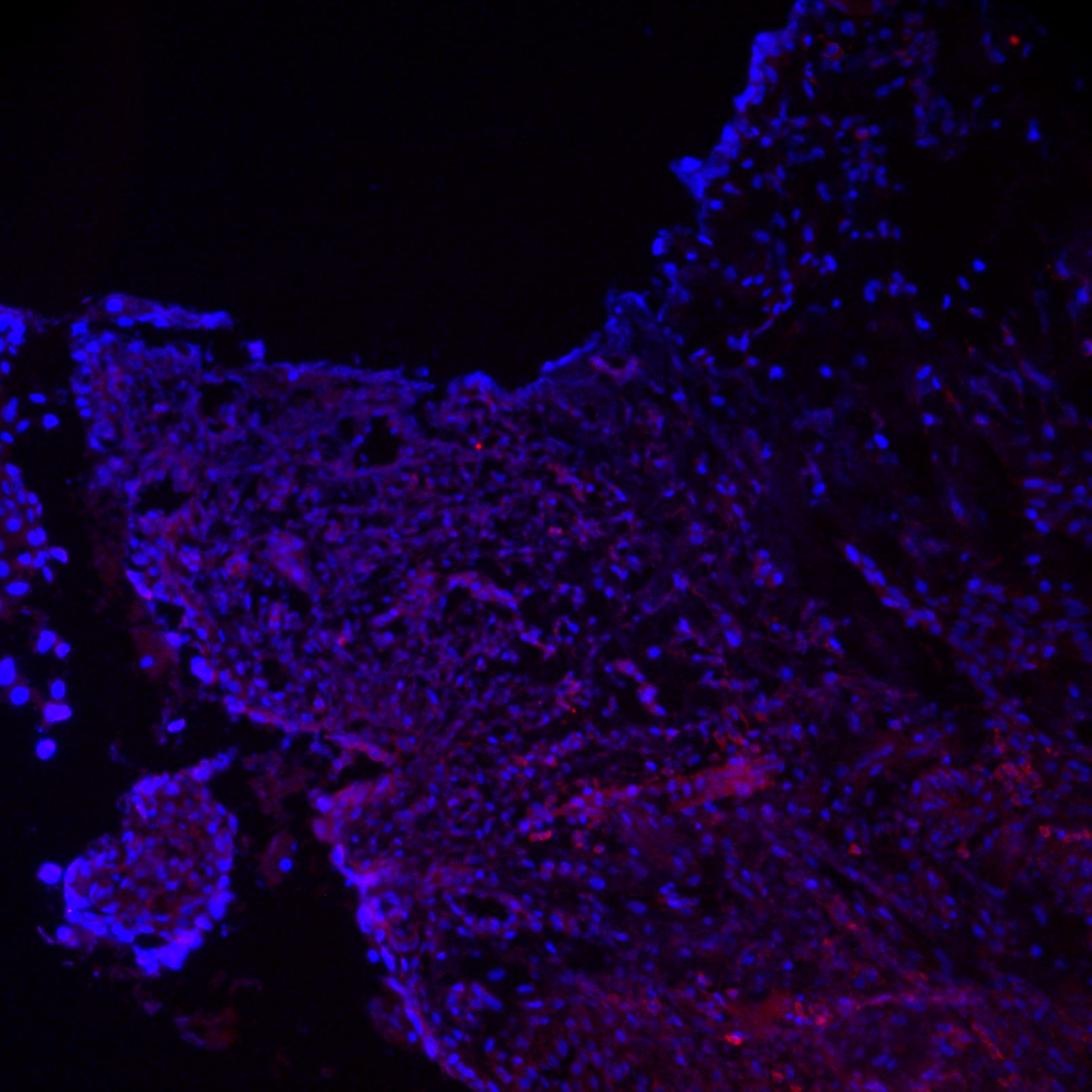
![Immunohistochemistry-Frozen: CD19 Antibody (SJ25-C1) [NBP1-28375]](https://resources.rndsystems.com/images/products/CD19-Antibody-SJ25-C1-Immunohistochemistry-Frozen-NBP1-28375-img0006.jpg "Immunohistochemistry-Frozen: CD19 Antibody (SJ25-C1) [NBP1-28375]")
Key Product Details
Species Reactivity
Human
Applications
Immunohistochemistry, Immunohistochemistry-Frozen, Flow Cytometry
Label
Unconjugated
Antibody Source
Monoclonal Mouse IgG1 kappa Clone # SJ25-C1
Format
Azide and BSA Free
Loading...
Product Specifications
Immunogen
NALM-1 and NALM-16 leukemia cell line
Clonality
Monoclonal
Host
Mouse
Isotype
IgG1 kappa
Scientific Data Images for CD19 Antibody (SJ25-C1) - Azide and BSA Free
Immunohistochemistry-Frozen: CD19 Antibody (SJ25-C1) [NBP1-28375]
Immunohistochemistry-Frozen: CD19 Antibody (SJ25-C1) [NBP1-28375] - CD19 (red) stained in human skin, counterstained with DAPI (blue) at 20x. Tissue fixed with 4% PFA, permeabilized with 0.5% Triton, and blocked with 37% Egg DI, 5% Milk DI, and 3% FSG 2% BSA TBS Ca/Azide in that order. Anti-CD19 applied at 1:500 v/v, 4C, O/N. Alexa Fluor 568 secondary antibody, with DAPI counterstain. Image from verified customer review.![Flow Cytometry: CD19 Antibody (SJ25-C1) [NBP1-28375]](https://resources.rndsystems.com/images/products/CD19-Antibody-SJ25-C1-Flow-Cytometry-NBP1-28375-img0007.jpg "Flow Cytometry: CD19 Antibody (SJ25-C1) [NBP1-28375]")
Flow Cytometry: CD19 Antibody (SJ25-C1) [NBP1-28375]
Flow Cytometry: CD19 Antibody (SJ25-C1) [NBP1-28375] - Human peripheral blood lymphocytes were stained with Mouse Anti-Human CD19-APC and Mouse Anti-Human CD3-FITC.Applications for CD19 Antibody (SJ25-C1) - Azide and BSA Free
Application
Recommended Usage
Flow Cytometry
1 ug/10^6 cells
Immunohistochemistry
1:10 - 1:500
Immunohistochemistry-Frozen
1:10 - 1:500
Application Notes
CD19 antibody validated for IHC-F from a verified customer review.
Reviewed Applications
Read 1 review rated 3 using NBP1-28375 in the following applications:
Flow Cytometry Panel Builder
Bio-Techne Knows Flow Cytometry
Save time and reduce costly mistakes by quickly finding compatible reagents using the Panel Builder Tool.
Advanced Features
- Spectra Viewer - Custom analysis of spectra from multiple fluorochromes
- Spillover Popups - Visualize the spectra of individual fluorochromes
- Antigen Density Selector - Match fluorochrome brightness with antigen density

Formulation, Preparation, and Storage
Purification
Protein A or G purified
Formulation
0.1M BBS (pH 8.2)
Format
Azide and BSA Free
Preservative
No Preservative
Concentration
0.1 mg/ml
Shipping
The product is shipped with polar packs. Upon receipt, store it immediately at the temperature recommended below.
Stability & Storage
Aliquot and store at -20C or -80C. Avoid freeze-thaw cycles.
Background: CD19
Considering the role of CD19 in BCR signaling and its expression in development from pre-B cells through plasma cells, it is understandable that CD19 dysfunction and abnormal expression is associated with numerous B cell malignancies and autoimmune disorders (1-5). CD19 expression is typically observed at relatively normal levels in B cell acute lymphoblastic leukemia (B-ALL) and chronic lymphoblastic leukemia (CLL) but is often reduced other types of lymphoma including diffuse large B cell lymphoma (DLBCL) and follicular lymphoma (FL) (1,2). On the other hand, CD19 expression is typically increased in autoimmune disorders such as systemic sclerosis (SSc) and multiple sclerosis (MS) as modeled by experimental autoimmune encephalomyelitis (EAE) (2). CD19 has become a therapeutic molecular target for the treatment of B cell lymphomas and autoimmune disorders using monoclonal antibodies (mAbs), bi-specific T cell engaging (BiTE) antibodies, and CD19-specific chimeric antigen receptor (CAR) T cells (1,2,4-6). Although anti-CD19 CAR T cell therapy has become the standard for the treatment of B cell malignancies, patients may experience relapse due to resistance mechanisms (6). Strategies to improve efficacy and limit relapse include combination of CAR T cell therapy with immune checkpoint inhibitors like anti-PD-1 (4,6).
References
1. Wang K, Wei G, Liu D. CD19: a biomarker for B cell development, lymphoma diagnosis and therapy. Exp Hematol Oncol. 2012;1(1):36. https://doi.org/10.1186/2162-3619-1-36
2. Li X, Ding Y, Zi M, et al. CD19, from bench to bedside. Immunol Lett. 2017;183:86-95. https://doi.org/10.1016/j.imlet.2017.01.010
3. Wentink MWJ, van Zelm MC, van Dongen JJM, Warnatz K, van der Burg M. Deficiencies in the CD19 complex. Clin Immunol. 2018;195:82-87. https://doi.org/10.1016/j.clim.2018.07.017
4. Frigault MJ, Maus MV. State of the art in CAR T cell therapy for CD19+ B cell malignancies. J Clin Invest. 2020;130(4):1586-1594. https://doi.org/10.1172/JCI129208
5. Penack O, Koenecke C. Complications after CD19+ CAR T-Cell Therapy. Cancers (Basel). 2020;12(11):3445. https://doi.org/10.3390/cancers12113445
6. Bouziana S, Bouzianas D. Anti-CD19 CAR-T cells: Digging in the dark side of the golden therapy. Crit Rev Oncol Hematol. 2021;157:103096. https://doi.org/10.1016/j.critrevonc.2020.103096
Alternate Names
CD19, CVID3, Leu-12
Gene Symbol
CD19
Additional CD19 Products
Product Documents for CD19 Antibody (SJ25-C1) - Azide and BSA Free
Certificate of Analysis
To download a Certificate of Analysis, please enter a lot or batch number in the search box below.
Product Specific Notices for CD19 Antibody (SJ25-C1) - Azide and BSA Free
This product is for research use only and is not approved for use in humans or in clinical diagnosis. Primary Antibodies are guaranteed for 1 year from date of receipt.
Related Research Areas
Customer Reviews for CD19 Antibody (SJ25-C1) - Azide and BSA Free (1)
3 out of 5
1 Customer Rating
Have you used CD19 Antibody (SJ25-C1) - Azide and BSA Free?
Submit a review and receive an Amazon gift card!
$25/€18/£15/$25CAN/¥2500 Yen for a review with an image
$10/€7/£6/$10CAN/¥1110 Yen for a review without an image
Submit a review
Customer Images
Showing
1
-
1 of
1 review
Showing All
Filter By:
-
Application: Immunohistochemistry-FrozenSample Tested: Frozen human skin sectionsSpecies: HumanVerified Customer | Posted 08/30/2018CD19 (red) stained in human skin, counterstained with DAPI (blue) at 20xTissue fixed with 4% PFA, permeabilized with 0.5% Triton, and blocked with 37% Egg DI, 5% Milk DI, and 3% FSG 2% BSA TBS Ca/Azide in that order. Anti-CD19 applied at 1:500 v/v, 4C, O/N. Alexa-568 secondary used, with DAPI counterstain

There are no reviews that match your criteria.
Protocols
Find general support by application which include: protocols, troubleshooting, illustrated assays, videos and webinars.
- 7-Amino Actinomycin D (7-AAD) Cell Viability Flow Cytometry Protocol
- Antigen Retrieval Protocol (PIER)
- Antigen Retrieval for Frozen Sections Protocol
- Appropriate Fixation of IHC/ICC Samples
- Cellular Response to Hypoxia Protocols
- Chromogenic IHC Staining of Formalin-Fixed Paraffin-Embedded (FFPE) Tissue Protocol
- Chromogenic Immunohistochemistry Staining of Frozen Tissue
- ClariTSA™ Fluorophore Kits
- Detection & Visualization of Antibody Binding
- Extracellular Membrane Flow Cytometry Protocol
- Flow Cytometry Protocol for Cell Surface Markers
- Flow Cytometry Protocol for Staining Membrane Associated Proteins
- Flow Cytometry Staining Protocols
- Flow Cytometry Troubleshooting Guide
- Fluorescent IHC Staining of Frozen Tissue Protocol
- Graphic Protocol for Heat-induced Epitope Retrieval
- Graphic Protocol for the Preparation and Fluorescent IHC Staining of Frozen Tissue Sections
- Graphic Protocol for the Preparation and Fluorescent IHC Staining of Paraffin-embedded Tissue Sections
- Graphic Protocol for the Preparation of Gelatin-coated Slides for Histological Tissue Sections
- IHC Sample Preparation (Frozen sections vs Paraffin)
- Immunofluorescent IHC Staining of Formalin-Fixed Paraffin-Embedded (FFPE) Tissue Protocol
- Immunohistochemistry (IHC) and Immunocytochemistry (ICC) Protocols
- Immunohistochemistry Frozen Troubleshooting
- Immunohistochemistry Paraffin Troubleshooting
- Intracellular Flow Cytometry Protocol Using Alcohol (Methanol)
- Intracellular Flow Cytometry Protocol Using Detergents
- Intracellular Nuclear Staining Flow Cytometry Protocol Using Detergents
- Intracellular Staining Flow Cytometry Protocol Using Alcohol Permeabilization
- Intracellular Staining Flow Cytometry Protocol Using Detergents to Permeabilize Cells
- Preparing Samples for IHC/ICC Experiments
- Preventing Non-Specific Staining (Non-Specific Binding)
- Primary Antibody Selection & Optimization
- Propidium Iodide Cell Viability Flow Cytometry Protocol
- Protocol for Heat-Induced Epitope Retrieval (HIER)
- Protocol for Liperfluo
- Protocol for Making a 4% Formaldehyde Solution in PBS
- Protocol for VisUCyte™ HRP Polymer Detection Reagent
- Protocol for the Characterization of Human Th22 Cells
- Protocol for the Characterization of Human Th9 Cells
- Protocol for the Preparation & Fixation of Cells on Coverslips
- Protocol for the Preparation and Chromogenic IHC Staining of Frozen Tissue Sections
- Protocol for the Preparation and Chromogenic IHC Staining of Frozen Tissue Sections - Graphic
- Protocol for the Preparation and Chromogenic IHC Staining of Paraffin-embedded Tissue Sections
- Protocol for the Preparation and Chromogenic IHC Staining of Paraffin-embedded Tissue Sections - Graphic
- Protocol for the Preparation and Fluorescent IHC Staining of Frozen Tissue Sections
- Protocol for the Preparation and Fluorescent IHC Staining of Paraffin-embedded Tissue Sections
- Protocol for the Preparation of Gelatin-coated Slides for Histological Tissue Sections
- Protocol: Annexin V and PI Staining by Flow Cytometry
- Protocol: Annexin V and PI Staining for Apoptosis by Flow Cytometry
- TUNEL and Active Caspase-3 Detection by IHC/ICC Protocol
- The Importance of IHC/ICC Controls
- Troubleshooting Guide: Fluorokine Flow Cytometry Kits
- Troubleshooting Guide: Immunohistochemistry
- View all Protocols, Troubleshooting, Illustrated assays and Webinars


