CD68/SR-D1 Antibody (SPM130)
Novus Biologicals | Catalog # NBP2-32831
Key Product Details
Species Reactivity
Validated:
Human, Canine (Negative), Chicken (Negative), Feline, Monkey, Porcine (Negative), Rabbit
Cited:
Human, Rat
Applications
Validated:
Immunohistochemistry, Immunohistochemistry-Paraffin, Immunohistochemistry-Frozen, Flow Cytometry, Immunocytochemistry/ Immunofluorescence
Cited:
Immunohistochemistry, Immunohistochemistry-Paraffin, IF/IHC
Label
Unconjugated
Antibody Source
Monoclonal Mouse IgG1 kappa Clone # SPM130
Loading...
Product Specifications
Immunogen
Subcellular fraction of human alveolar macrophages
Reactivity Notes
Does not react with Porcine, Canine or Chicken.
Localization
Cell Surface and Cytoplasmic
Marker
Macrophage Marker
Specificity
This antibody recognizes a glycoprotein of 110kDa, which is identified as CD68. It is important for identifying macrophages in tissue sections. It stains macrophages in a wide variety of human tissues, including Kupffer cells and macrophages in the red pulp of the spleen, in lamina propria of the gut, in lung alveoli, and in bone marrow. It reacts with myeloid precursors and peripheral blood granulocytes. It also reacts with plasmacytoid T cells, which are supposed to be of monocyte/macrophage origin. It shows strong granular cytoplasmic staining of chronic and acute myeloid leukemia and also reacts with rare cases of true histiocytic neoplasia. Lymphomas are negative or show few granules.
Clonality
Monoclonal
Host
Mouse
Isotype
IgG1 kappa
Theoretical MW
110 kDa.
Disclaimer note: The observed molecular weight of the protein may vary from the listed predicted molecular weight due to post translational modifications, post translation cleavages, relative charges, and other experimental factors.
Disclaimer note: The observed molecular weight of the protein may vary from the listed predicted molecular weight due to post translational modifications, post translation cleavages, relative charges, and other experimental factors.
Description
200ug/ml of antibody purified from Bioreactor Concentrate by Protein A or G. Prepared in 10 mM PBS with 0.05% BSA & 0.05% azide. Also available WITHOUT BSA & azide at 1.0 mg/ml. (NBP2-34736)
Antibody with azide - store at 2 to 8C. Antibody without azide - store at -20 to -80C.
Antibody with azide - store at 2 to 8C. Antibody without azide - store at -20 to -80C.
Scientific Data Images for CD68/SR-D1 Antibody (SPM130)
![Immunohistochemistry-Paraffin: CD68/SR-D1 Antibody (SPM130) [NBP2-32831]](https://resources.rndsystems.com/images/products/CD68-SR-D1-Antibody-SPM130-Immunohistochemistry-Paraffin-NBP2-32831-img0003.jpg "Immunohistochemistry-Paraffin: CD68/SR-D1 Antibody (SPM130) [NBP2-32831]")
Immunohistochemistry-Paraffin: CD68/SR-D1 Antibody (SPM130) [NBP2-32831]
Immunohistochemistry-Paraffin: CD68/SR-D1 Antibody (SPM130) [NBP2-32831] - Formalin-fixed, paraffin-embedded human tonsil (20X) stained with CD68 MAb (SPM130).![Immunohistochemistry-Paraffin: CD68/SR-D1 Antibody (SPM130) [NBP2-32831]](https://resources.rndsystems.com/images/products/CD68-SR-D1-Antibody-SPM130-Immunohistochemistry-Paraffin-NBP2-32831-img0004.jpg "Immunohistochemistry-Paraffin: CD68/SR-D1 Antibody (SPM130) [NBP2-32831]")
Immunohistochemistry-Paraffin: CD68/SR-D1 Antibody (SPM130) [NBP2-32831]
Immunohistochemistry-Paraffin: CD68/SR-D1 Antibody (SPM130) [NBP2-32831] - Formalin-fixed, paraffin-embedded human tonsil (10X) stained with CD68 MAb (SPM130).![Immunohistochemistry-Frozen: CD68/SR-D1 Antibody (SPM130) [NBP2-32831]](https://resources.rndsystems.com/images/products/CD68-SR-D1-Antibody-SPM130-Immunohistochemistry-Frozen-NBP2-32831-img0007.jpg "Immunohistochemistry-Frozen: CD68/SR-D1 Antibody (SPM130) [NBP2-32831]")
Immunohistochemistry-Frozen: CD68/SR-D1 Antibody (SPM130) [NBP2-32831]
Immunohistochemistry-Frozen: CD68/SR-D1 Antibody (SPM130) [NBP2-32831] - The antibody was incubated with feline spleen section at 1:100 and the slide was further stained with Alexa secondary antibody. Image was captured with epifluorescence microscope. This image was submitted via customer review.![Flow Cytometry: CD68/SR-D1 Antibody (SPM130) [NBP2-32831]](https://resources.rndsystems.com/images/products/CD68-SR-D1-Antibody-SPM130-Flow-Cytometry-NBP2-32831-img0008.jpg "Flow Cytometry: CD68/SR-D1 Antibody (SPM130) [NBP2-32831]")
Flow Cytometry: CD68/SR-D1 Antibody (SPM130) [NBP2-32831]
Flow Cytometry: CD68/SR-D1 Antibody (SPM130) [NBP2-32831] - Flow Cytometry: CD68/SR-D1 Antibody (SPM130) [Biotin] [NBP2-34736B] - Mouse peripheral blood cells were unstained (left) or stained (right) with CD68/SR-D1 antibody and anti-biotin alexa-fluor 488 secondary antibody. Image from verified customer review. [NBP2-32831] -")
Immunohistochemistry: CD68/SR-D1 Antibody (SPM130) [NBP2-32831] -
nbp2-32831_mouse-monoclonal-cd68-sr-d1-antibody-spm130-17720241044628.jpgApplications for CD68/SR-D1 Antibody (SPM130)
Application
Recommended Usage
Flow Cytometry
1-2 ug/million cells
Immunocytochemistry/ Immunofluorescence
1-2 ug/ml
Immunohistochemistry-Frozen
0.5-1.0ug/ml
Immunohistochemistry-Paraffin
1-2 ug/ml
Application Notes
Immunohistochemistry (Formalin-fixed): 1-2ug/ml for 30 minutes at RT. Staining of formalin-fixed tissues requires heating tissue sections in 10mM Tris with 1mM EDTA, pH 9.0, for 45 min at 95C followed by cooling at RT for 20 minutes.
Optimal dilution for a specific application should be determined.
Optimal dilution for a specific application should be determined.
Reviewed Applications
Read 2 reviews rated 2 using NBP2-32831 in the following applications:
Flow Cytometry Panel Builder
Bio-Techne Knows Flow Cytometry
Save time and reduce costly mistakes by quickly finding compatible reagents using the Panel Builder Tool.
Advanced Features
- Spectra Viewer - Custom analysis of spectra from multiple fluorochromes
- Spillover Popups - Visualize the spectra of individual fluorochromes
- Antigen Density Selector - Match fluorochrome brightness with antigen density

Formulation, Preparation, and Storage
Purification
Protein A or G purified
Formulation
10 mM PBS with 0.05% BSA
Preservative
0.05% Sodium Azide
Concentration
0.2 mg/ml
Shipping
The product is shipped with polar packs. Upon receipt, store it immediately at the temperature recommended below.
Stability & Storage
Store at 4C.
Background: CD68/SR-D1
CD68 is highly expressed in cells of the mononuclear phagocyte system such as macrophages, microglia, osteoclasts, and myeloid dendritic cells (DCs); and is expressed to a lesser extent in lymphoid cells (CD19+ B lymphocytes and CD4+ T lymphocytes), human umbilical cord mesenchymal stem cells (MSCs), fibroblasts, endothelial cells, multiple non-hematopoietic cancer cell lines, and human arterial intimal smooth muscle cells (SMCs). Expression has been also observed in diseased states for granulocytes and neutrophils, in particular basophils from myeloproliferative disorders and intestinal neutrophils from inflammatory bowel disease (IBD), respectively (1).
Although the function of CD68 has yet to be established, it has often been used as an immunohistochemistry (IHC) marker of inflammation and for granular cell tumors (GCTs). CD68+ tumor associated macrophages (TAMs) has been suggested to be a predictive marker for poor cancer prognosis, but a meta-analysis showed the presence of CD68 is not correlated with survival (2). In addition, a role in hepatic malaria infection has been reported based on the finding that peptide P39 binds CD68, considered a receptor for malaria sporozoite, and inhibits parasite entry into Kupffer cells. CD68 was deemed a member of the Scavenger receptor family due to its upregulation in macrophages following inflammatory stimuli, ability to bind modified LDL, phosphatidylserine, and apoptotic cells, as well as shuttling between the plasma membrane and endosomes. CD68 has been linked to atherogenesis based on binding and internalization of its ligand, oxLDL (1).
References
1. Chistiakov, DA, Killingsworth, MC, Myasoedova, VA. Orekhov AN, Bobryshev YV. (2017) CD68/macrosialin: not just a histochemical marker. Lab Invest. 97:4-13. PMID: 27869795
2. Troiano G, Caponio VCA, Adipietro I, Tepedino M, Santoro R, Laino L, Lo Russo L, Cirillo N, Lo Muzio L. (2019) Prognostic significance of CD68+ and CD163+ tumor associated macrophages in head and neck squamous cell carcinoma: A systematic review and meta-analysis. Oral Oncol. 93:66-75. PMID: 31109698.
Additional CD68/SR-D1 Products
Product Documents for CD68/SR-D1 Antibody (SPM130)
Certificate of Analysis
To download a Certificate of Analysis, please enter a lot or batch number in the search box below.
Product Specific Notices for CD68/SR-D1 Antibody (SPM130)
This product is for research use only and is not approved for use in humans or in clinical diagnosis. Primary Antibodies are guaranteed for 1 year from date of receipt.
Citations for CD68/SR-D1 Antibody (SPM130)
 Powered by Bioz
Customer Reviews for CD68/SR-D1 Antibody (SPM130) (2)
2 out of 5
2 Customer Ratings
Have you used CD68/SR-D1 Antibody (SPM130)?
Submit a review and receive an Amazon gift card!
$25/€18/£15/$25CAN/¥2500 Yen for a review with an image
$10/€7/£6/$10CAN/¥1110 Yen for a review without an image
Submit a review
Customer Images

Showing
1
-
2 of
2 reviews
Showing All
Filter By:
-
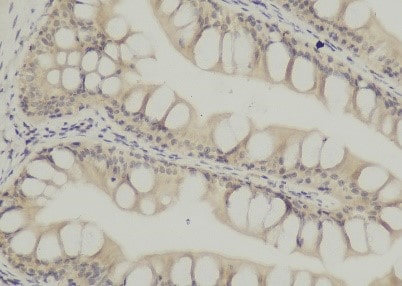
Application: Immunohistochemistry-ParaffinSample Tested: After intestinal bowelSpecies: grouperVerified Customer | Posted 10/16/2020Picture of experimental group.The experiment was repeated several times and the results were all negativeDewaxing to water: 1, paraffin sections in turn sliced into 15 min - dimethylbenzene xylene Ⅰ Ⅱ 15 min - xylene III Ⅰ 5 min - 15 min - anhydrous ethanol anhydrous ethanol Ⅱ 5 min 5 min - 75% - 85% alcohol alcohol 5 min - the distilled water to wash. 2, antigen repair: biopsy under full antigen repair citrate buffer (PH6.0) repair box in a have a certain amount of water pressure cooker, induction cooker heated to stomatal began to jet, stop heating, release the pressure, the slice on the repair box, induction cooker heated to stomatal began to jet, 5 min after close the induction cooker, the process should prevent excessive evaporation buffer, do not dry.After natural cooling, the slides were placed in PBS (PH7.4) and shaken on the decolorizing shaper for 3 times, 5min each. 3. Break endogenous peroxidase: the slides were placed in 3% hydrogen peroxide solution and incubated at room temperature and away from light for 25 min. The slides were placed in PBS (PH7.4) and shaken on a decolorizing shaper for 3 times for 5min each. Serum sealing: 3%BSA was added to cover the tissue evenly in the chemochemical ring, and the tissue was sealed at room temperature for 30min.(Primary antibody is sealed with rabbit serum from goat and BSA from other sources.) 4. Primary antibody was added: the blocking solution was gently removed, the primary antibody prepared with PBS in a certain proportion was added to the sections, and the sections were placed flat in a wet box and incubated overnight at 4°C.(Add a small amount of water in the wet box to prevent evaporation of antibodies) 5. Secondary antibody was added: the slides were placed in PBS (PH7.4) and washed by shaking on the decolorizing shaker for 3 times, 5min each.After the sections were slightly shaken and dried, the tissues were dripped with secondary antibody (HRP-labeled) of the species corresponding to the primary antibody and incubated at room temperature for 50min. 7. DAB color development: The slides were placed in PBS (PH7.4) and shaken on the decoloring shaker for 3 times, 5min each.DAB color developing solution newly prepared was added in the circle after the sections were slightly dried. The color developing time was controlled under the microscope. The positive color was brownish yellow, and the color developing was stopped after the sections were washed under tap water. 8. Restaining nuclei: restaining with hematoxylin for about 3min; washing with tap water; differentiation with hematoxylin differentiation solution for several seconds; washing with tap water; flushing with hematoxylin returning blue solution; washing with running water., dehydration and 9: the slice in turn into 5 min 5 min 75% alcohol - 85% alcohol, anhydrous ethanol Ⅰ Ⅱ 5 min - the anhydrous ethanol dehydration in 5 min - xylene Ⅰ 5 min is transparent, the slice out of xylene is a bit dry, neutral rubber sealing piece. Microscope examination, image acquisition and analysis.
 Bio-Techne ResponseThis review was submitted through the legacy Novus Innovators Program, reflecting a new species or application tested on a primary antibody.
 -
Application: Immunohistochemistry-FrozenSample Tested: spleenSpecies: FelineVerified Customer | Posted 03/09/2018The antibody was incubated at 1:100 and the slide was further stained with Alexa secondary antibody. Image was captured with epifluorescence microscope.
There are no reviews that match your criteria.
Protocols
Find general support by application which include: protocols, troubleshooting, illustrated assays, videos and webinars.
- 7-Amino Actinomycin D (7-AAD) Cell Viability Flow Cytometry Protocol
- Antigen Retrieval Protocol (PIER)
- Antigen Retrieval for Frozen Sections Protocol
- Appropriate Fixation of IHC/ICC Samples
- Cellular Response to Hypoxia Protocols
- Chromogenic IHC Staining of Formalin-Fixed Paraffin-Embedded (FFPE) Tissue Protocol
- Chromogenic Immunohistochemistry Staining of Frozen Tissue
- ClariTSA™ Fluorophore Kits
- Detection & Visualization of Antibody Binding
- Extracellular Membrane Flow Cytometry Protocol
- Flow Cytometry Protocol for Cell Surface Markers
- Flow Cytometry Protocol for Staining Membrane Associated Proteins
- Flow Cytometry Staining Protocols
- Flow Cytometry Troubleshooting Guide
- Fluorescent IHC Staining of Frozen Tissue Protocol
- Graphic Protocol for Heat-induced Epitope Retrieval
- Graphic Protocol for the Preparation and Fluorescent IHC Staining of Frozen Tissue Sections
- Graphic Protocol for the Preparation and Fluorescent IHC Staining of Paraffin-embedded Tissue Sections
- Graphic Protocol for the Preparation of Gelatin-coated Slides for Histological Tissue Sections
- ICC Cell Smear Protocol for Suspension Cells
- ICC Immunocytochemistry Protocol Videos
- ICC for Adherent Cells
- IHC Sample Preparation (Frozen sections vs Paraffin)
- Immunocytochemistry (ICC) Protocol
- Immunocytochemistry Troubleshooting
- Immunofluorescence of Organoids Embedded in Cultrex Basement Membrane Extract
- Immunofluorescent IHC Staining of Formalin-Fixed Paraffin-Embedded (FFPE) Tissue Protocol
- Immunohistochemistry (IHC) and Immunocytochemistry (ICC) Protocols
- Immunohistochemistry Frozen Troubleshooting
- Immunohistochemistry Paraffin Troubleshooting
- Intracellular Flow Cytometry Protocol Using Alcohol (Methanol)
- Intracellular Flow Cytometry Protocol Using Detergents
- Intracellular Nuclear Staining Flow Cytometry Protocol Using Detergents
- Intracellular Staining Flow Cytometry Protocol Using Alcohol Permeabilization
- Intracellular Staining Flow Cytometry Protocol Using Detergents to Permeabilize Cells
- Preparing Samples for IHC/ICC Experiments
- Preventing Non-Specific Staining (Non-Specific Binding)
- Primary Antibody Selection & Optimization
- Propidium Iodide Cell Viability Flow Cytometry Protocol
- Protocol for Heat-Induced Epitope Retrieval (HIER)
- Protocol for Liperfluo
- Protocol for Making a 4% Formaldehyde Solution in PBS
- Protocol for VisUCyte™ HRP Polymer Detection Reagent
- Protocol for the Characterization of Human Th22 Cells
- Protocol for the Characterization of Human Th9 Cells
- Protocol for the Fluorescent ICC Staining of Cell Smears - Graphic
- Protocol for the Fluorescent ICC Staining of Cultured Cells on Coverslips - Graphic
- Protocol for the Preparation & Fixation of Cells on Coverslips
- Protocol for the Preparation and Chromogenic IHC Staining of Frozen Tissue Sections
- Protocol for the Preparation and Chromogenic IHC Staining of Frozen Tissue Sections - Graphic
- Protocol for the Preparation and Chromogenic IHC Staining of Paraffin-embedded Tissue Sections
- Protocol for the Preparation and Chromogenic IHC Staining of Paraffin-embedded Tissue Sections - Graphic
- Protocol for the Preparation and Fluorescent ICC Staining of Cells on Coverslips
- Protocol for the Preparation and Fluorescent ICC Staining of Non-adherent Cells
- Protocol for the Preparation and Fluorescent ICC Staining of Stem Cells on Coverslips
- Protocol for the Preparation and Fluorescent IHC Staining of Frozen Tissue Sections
- Protocol for the Preparation and Fluorescent IHC Staining of Paraffin-embedded Tissue Sections
- Protocol for the Preparation of Gelatin-coated Slides for Histological Tissue Sections
- Protocol for the Preparation of a Cell Smear for Non-adherent Cell ICC - Graphic
- Protocol: Annexin V and PI Staining by Flow Cytometry
- Protocol: Annexin V and PI Staining for Apoptosis by Flow Cytometry
- TUNEL and Active Caspase-3 Detection by IHC/ICC Protocol
- The Importance of IHC/ICC Controls
- Troubleshooting Guide: Fluorokine Flow Cytometry Kits
- Troubleshooting Guide: Immunohistochemistry
- View all Protocols, Troubleshooting, Illustrated assays and Webinars
Loading...
Associated Pathways
