CDT1 Antibody - BSA Free
Novus Biologicals | Catalog # NB100-2567
![Western Blot: CDT1 Antibody [NB100-2567]](https://resources.rndsystems.com/images/products/CDT1-Antibody-Western-Blot-NB100-2567-img0005.jpg "Western Blot: CDT1 Antibody [NB100-2567]")
Key Product Details
Species Reactivity
Validated:
Human
Cited:
Human
Applications
Validated:
Immunohistochemistry, Immunohistochemistry-Paraffin, Western Blot, Immunoprecipitation
Cited:
Western Blot
Label
Unconjugated
Antibody Source
Polyclonal Rabbit IgG
Format
BSA Free
Loading...
Product Specifications
Immunogen
The immunogen recognized by this antibody maps to a region between residue 500 and the C-terminus (residue 546) of human Chromatin Licensing and DNA Replication Factor 1 using the numbering given in entry NP_112190.1 (GeneID 81620).
Clonality
Polyclonal
Host
Rabbit
Isotype
IgG
Theoretical MW
60.4 kDa.
Disclaimer note: The observed molecular weight of the protein may vary from the listed predicted molecular weight due to post translational modifications, post translation cleavages, relative charges, and other experimental factors.
Disclaimer note: The observed molecular weight of the protein may vary from the listed predicted molecular weight due to post translational modifications, post translation cleavages, relative charges, and other experimental factors.
Scientific Data Images for CDT1 Antibody - BSA Free
Western Blot: CDT1 Antibody [NB100-2567]
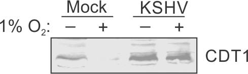
Western Blot: CDT1 Antibody [NB100-2567] - Human B cells. KSHV infection protects CDT1 from hypoxia mediated degradation. Mock or KSHV infected cells were grown under normoxic or hypoxic condition followed by measuring CDT1 levels. Western blot image submitted by a verified customer review.![Immunohistochemistry-Paraffin: CDT1 Antibody [NB100-2567]](https://resources.rndsystems.com/images/products/CDT1-Antibody-Immunohistochemistry-Paraffin-NB100-2567-img0004.jpg "Immunohistochemistry-Paraffin: CDT1 Antibody [NB100-2567]")
Immunohistochemistry-Paraffin: CDT1 Antibody [NB100-2567]
Immunohistochemistry-Paraffin: CDT1 Antibody [NB100-2567] - Human islet cell carcinoma. Antibody used at a dilution of 1:1000 (1ug/mL).![Western Blot: CDT1 Antibody [NB100-2567]](https://resources.rndsystems.com/images/products/CDT1-Antibody-Western-Blot-NB100-2567-img0003.jpg "Western Blot: CDT1 Antibody [NB100-2567]")
Western Blot: CDT1 Antibody [NB100-2567]
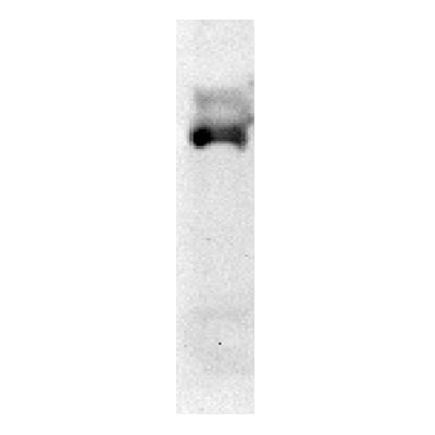
Western Blot: CDT1 Antibody [NB100-2567] - Whole cell lysate from HeLa (5, 15 and 50 ug for WB; 1 mg for IP, 20% of IP loaded) and 293T (T; 50 ug) cells. Affinity purified rabbit anti- CDT1 antibody (NB100-2567) used for WB at 1 ug/mL (A and B) and used for IP at 3 ug/mg lysate (B).Applications for CDT1 Antibody - BSA Free
Application
Recommended Usage
Immunohistochemistry
1:1000 - 1:5000
Immunohistochemistry-Paraffin
1:1000 - 1:5000
Immunoprecipitation
2-5 ug/mg lysate
Western Blot
1:1000 - 1:2500
Application Notes
Epitope retrieval with citrate buffer pH 6.0 is recommended for FFPE tissue sections.
Reviewed Applications
Read 3 reviews rated 3.3 using NB100-2567 in the following applications:
Formulation, Preparation, and Storage
Purification
Immunogen affinity purified
Formulation
Tris-Citrate/Phosphate (pH 7 - 8)
Format
BSA Free
Preservative
0.09% Sodium Azide
Concentration
1.0 mg/ml
Shipping
The product is shipped with polar packs. Upon receipt, store it immediately at the temperature recommended below.
Stability & Storage
Store at 4C. Do not freeze.
Background: CDT1
Alternate Names
chromatin licensing and DNA replication factor 1, DNA replication factor Cdt1, Double parked homolog, Double parked, Drosophila, homolog of, DUPRIS2
Entrez Gene IDs
81620 (Human)
Gene Symbol
CDT1
UniProt
Additional CDT1 Products
Product Documents for CDT1 Antibody - BSA Free
Certificate of Analysis
To download a Certificate of Analysis, please enter a lot or batch number in the search box below.
Product Specific Notices for CDT1 Antibody - BSA Free
This product is for research use only and is not approved for use in humans or in clinical diagnosis. Primary Antibodies are guaranteed for 1 year from date of receipt.
Citations for CDT1 Antibody - BSA Free
 Powered by Bioz
Customer Reviews for CDT1 Antibody - BSA Free (3)
3.3 out of 5
3 Customer Ratings
Have you used CDT1 Antibody - BSA Free?
Submit a review and receive an Amazon gift card!
$25/€18/£15/$25CAN/¥2500 Yen for a review with an image
$10/€7/£6/$10CAN/¥1110 Yen for a review without an image
Submit a review
Customer Images
Showing
1
-
3 of
3 reviews
Showing All
Filter By:
-
Application: Western BlotSample Tested: B cellsSpecies: HumanVerified Customer | Posted 10/16/2019KSHV infection protects CDT1 from hypoxia mediated degradation. Mock or KSHV infected cells were grown under normoxic or hypoxic condition followed by measuring CDT1 levels.

-
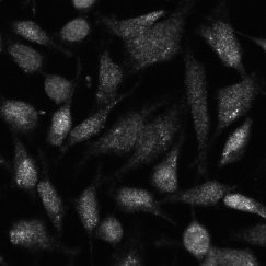
Application: ImmunocytochemistrySample Tested: hela cellSpecies: HumanVerified Customer | Posted 01/17/2018HeLa cells, fixed and stained for Cdt1. Lacking expected strong nuclear enrichment of stain.Fixed in 4% PFA for 10 min, permeablized in.2% Triton X-100 in PBS for 15 min, blocked for 1 hr in 1% BSA + 10% FBS in PBS with.1% Triton X-100. Incubated in antibody at 1:100 dilution in blocking buffer, and detected with anti-rabbit secondary antibody.
-
Application: Western BlotSample Tested: HCT116 Whole Cell Lysate, Sample Amount: 50 ugSpecies: HumanVerified Customer | Posted 02/18/2011
There are no reviews that match your criteria.
Protocols
Find general support by application which include: protocols, troubleshooting, illustrated assays, videos and webinars.
- Antigen Retrieval Protocol (PIER)
- Antigen Retrieval for Frozen Sections Protocol
- Appropriate Fixation of IHC/ICC Samples
- Cellular Response to Hypoxia Protocols
- Chromogenic IHC Staining of Formalin-Fixed Paraffin-Embedded (FFPE) Tissue Protocol
- Chromogenic Immunohistochemistry Staining of Frozen Tissue
- ClariTSA™ Fluorophore Kits
- Detection & Visualization of Antibody Binding
- Fluorescent IHC Staining of Frozen Tissue Protocol
- Graphic Protocol for Heat-induced Epitope Retrieval
- Graphic Protocol for the Preparation and Fluorescent IHC Staining of Frozen Tissue Sections
- Graphic Protocol for the Preparation and Fluorescent IHC Staining of Paraffin-embedded Tissue Sections
- Graphic Protocol for the Preparation of Gelatin-coated Slides for Histological Tissue Sections
- IHC Sample Preparation (Frozen sections vs Paraffin)
- Immunofluorescent IHC Staining of Formalin-Fixed Paraffin-Embedded (FFPE) Tissue Protocol
- Immunohistochemistry (IHC) and Immunocytochemistry (ICC) Protocols
- Immunohistochemistry Frozen Troubleshooting
- Immunohistochemistry Paraffin Troubleshooting
- Immunoprecipitation Protocol
- Preparing Samples for IHC/ICC Experiments
- Preventing Non-Specific Staining (Non-Specific Binding)
- Primary Antibody Selection & Optimization
- Protocol for Heat-Induced Epitope Retrieval (HIER)
- Protocol for Making a 4% Formaldehyde Solution in PBS
- Protocol for VisUCyte™ HRP Polymer Detection Reagent
- Protocol for the Preparation & Fixation of Cells on Coverslips
- Protocol for the Preparation and Chromogenic IHC Staining of Frozen Tissue Sections
- Protocol for the Preparation and Chromogenic IHC Staining of Frozen Tissue Sections - Graphic
- Protocol for the Preparation and Chromogenic IHC Staining of Paraffin-embedded Tissue Sections
- Protocol for the Preparation and Chromogenic IHC Staining of Paraffin-embedded Tissue Sections - Graphic
- Protocol for the Preparation and Fluorescent IHC Staining of Frozen Tissue Sections
- Protocol for the Preparation and Fluorescent IHC Staining of Paraffin-embedded Tissue Sections
- Protocol for the Preparation of Gelatin-coated Slides for Histological Tissue Sections
- R&D Systems Quality Control Western Blot Protocol
- TUNEL and Active Caspase-3 Detection by IHC/ICC Protocol
- The Importance of IHC/ICC Controls
- Troubleshooting Guide: Immunohistochemistry
- Troubleshooting Guide: Western Blot Figures
- Western Blot Conditions
- Western Blot Protocol
- Western Blot Protocol for Cell Lysates
- Western Blot Troubleshooting
- Western Blot Troubleshooting Guide
- View all Protocols, Troubleshooting, Illustrated assays and Webinars
Loading...