Chk1 [p Ser296] Antibody (SN06-50)
Novus Biologicals | Catalog # NBP2-67711
Recombinant Monoclonal Antibody
Loading...
Key Product Details
Species Reactivity
Validated:
Human, Mouse, Rat
Cited:
Human
Applications
Validated:
Immunohistochemistry, Immunohistochemistry-Paraffin, Western Blot, Immunocytochemistry/ Immunofluorescence
Cited:
Western Blot
Label
Unconjugated
Antibody Source
Recombinant Monoclonal Rabbit IgG Clone # SN06-50 expressed in HEK293
Loading...
Product Specifications
Immunogen
Synthetic phospho-peptide corresponding to residues surrounding Ser296 of human Chk1 (SwissProt: O14757 Human)
Modification
p Ser296
Localization
Chromosome, Cytoplasm, Cytoskeleton, Nucleus.
Clonality
Monoclonal
Host
Rabbit
Isotype
IgG
Scientific Data Images for Chk1 [p Ser296] Antibody (SN06-50)
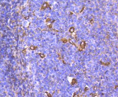
Immunohistochemistry-Paraffin: Chk1 [p Ser296] Antibody (SN06-50) [NBP2-67711] - Analysis of paraffin-embedded human tonsil tissue using anti- phospho-Chk1 (S296) antibody. Counter stained with hematoxylin.
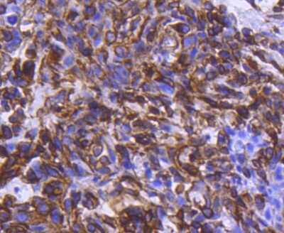
Immunohistochemistry-Paraffin: Chk1 [p Ser296] Antibody (SN06-50) [NBP2-67711] - Analysis of paraffin-embedded human breast carcinoma tissue using anti- phospho-Chk1 (S296) antibody. Counter stained with hematoxylin.
Applications for Chk1 [p Ser296] Antibody (SN06-50)
Application
Recommended Usage
Immunocytochemistry/ Immunofluorescence
1:100
Immunohistochemistry-Paraffin
1:50-1:200
Western Blot
1:500
Reviewed Applications
Read 1 review rated 5 using NBP2-67711 in the following applications:
Formulation, Preparation, and Storage
Purification
Protein A purified
Formulation
TBS (pH7.4), 0.05% BSA, 40% Glycerol
Preservative
0.05% Sodium Azide
Concentration
1 mg/ml
Shipping
The product is shipped with polar packs. Upon receipt, store it immediately at the temperature recommended below.
Stability & Storage
Store at 4C short term. Aliquot and store at -20C long term. Avoid freeze-thaw cycles.
Background: Chk1
Long Name
Checkpoint Kinase 1
Alternate Names
CHEK1
Gene Symbol
CHEK1
Additional Chk1 Products
Product Documents for Chk1 [p Ser296] Antibody (SN06-50)
Certificate of Analysis
To download a Certificate of Analysis, please enter a lot or batch number in the search box below.
Product Specific Notices for Chk1 [p Ser296] Antibody (SN06-50)
This product is for research use only and is not approved for use in humans or in clinical diagnosis. Primary Antibodies are guaranteed for 1 year from date of receipt.
Citations for Chk1 [p Ser296] Antibody (SN06-50)
 Powered by Bioz
Customer Reviews for Chk1 [p Ser296] Antibody (SN06-50) (1)
5 out of 5
1 Customer Rating
Have you used Chk1 [p Ser296] Antibody (SN06-50)?
Submit a review and receive an Amazon gift card!
$25/€18/£15/$25CAN/¥2500 Yen for a review with an image
$10/€7/£6/$10CAN/¥1110 Yen for a review without an image
Submit a review
Customer Images
![Chk1 [p Ser296] Antibody (SN06-50) NBP2-67711](https://resources.rndsystems.com/images/reviews/review_nbp2-67711_53171_0_0.jpg)
Showing
1
-
1 of
1 review
Showing All
Filter By:
-
Application: Western BlotSample Tested: Human Pancreatic Cancer Cell lineSpecies: HumanVerified Customer | Posted 08/05/2020Western Blot: Whole cell lysate from L3.6 PDAC cells prepared using RIPA buffer. Cells were treated with DMSO 48 Hr (left) or Chk1 inhibitor LY2606368 0.78nM 48 Hr (right). Total Chk1 used as loading control (lower bands)L3.6 PDAC cell line was treated 48 hours with DMSO or the Chk1 inhibitor LY2606368. Chk1 p Ser296 primary antibody dilution 1:1000 Anti rabbit HRP conjugated secondary dilution 1:2000 Blocking performed using 5% BSA on TBS-Tween solution
![Chk1 [p Ser296] Antibody (SN06-50) NBP2-67711](data:image/png;base64,R0lGODlhAQABAAD/ACwAAAAAAQABAAACADs=)
There are no reviews that match your criteria.
Protocols
Find general support by application which include: protocols, troubleshooting, illustrated assays, videos and webinars.
- Antigen Retrieval Protocol (PIER)
- Antigen Retrieval for Frozen Sections Protocol
- Appropriate Fixation of IHC/ICC Samples
- Cellular Response to Hypoxia Protocols
- Chromogenic IHC Staining of Formalin-Fixed Paraffin-Embedded (FFPE) Tissue Protocol
- Chromogenic Immunohistochemistry Staining of Frozen Tissue
- ClariTSA™ Fluorophore Kits
- Detection & Visualization of Antibody Binding
- Fluorescent IHC Staining of Frozen Tissue Protocol
- Graphic Protocol for Heat-induced Epitope Retrieval
- Graphic Protocol for the Preparation and Fluorescent IHC Staining of Frozen Tissue Sections
- Graphic Protocol for the Preparation and Fluorescent IHC Staining of Paraffin-embedded Tissue Sections
- Graphic Protocol for the Preparation of Gelatin-coated Slides for Histological Tissue Sections
- ICC Cell Smear Protocol for Suspension Cells
- ICC Immunocytochemistry Protocol Videos
- ICC for Adherent Cells
- IHC Sample Preparation (Frozen sections vs Paraffin)
- Immunocytochemistry (ICC) Protocol
- Immunocytochemistry Troubleshooting
- Immunofluorescence of Organoids Embedded in Cultrex Basement Membrane Extract
- Immunofluorescent IHC Staining of Formalin-Fixed Paraffin-Embedded (FFPE) Tissue Protocol
- Immunohistochemistry (IHC) and Immunocytochemistry (ICC) Protocols
- Immunohistochemistry Frozen Troubleshooting
- Immunohistochemistry Paraffin Troubleshooting
- Preparing Samples for IHC/ICC Experiments
- Preventing Non-Specific Staining (Non-Specific Binding)
- Primary Antibody Selection & Optimization
- Protocol for Heat-Induced Epitope Retrieval (HIER)
- Protocol for Making a 4% Formaldehyde Solution in PBS
- Protocol for VisUCyte™ HRP Polymer Detection Reagent
- Protocol for the Fluorescent ICC Staining of Cell Smears - Graphic
- Protocol for the Fluorescent ICC Staining of Cultured Cells on Coverslips - Graphic
- Protocol for the Preparation & Fixation of Cells on Coverslips
- Protocol for the Preparation and Chromogenic IHC Staining of Frozen Tissue Sections
- Protocol for the Preparation and Chromogenic IHC Staining of Frozen Tissue Sections - Graphic
- Protocol for the Preparation and Chromogenic IHC Staining of Paraffin-embedded Tissue Sections
- Protocol for the Preparation and Chromogenic IHC Staining of Paraffin-embedded Tissue Sections - Graphic
- Protocol for the Preparation and Fluorescent ICC Staining of Cells on Coverslips
- Protocol for the Preparation and Fluorescent ICC Staining of Non-adherent Cells
- Protocol for the Preparation and Fluorescent ICC Staining of Stem Cells on Coverslips
- Protocol for the Preparation and Fluorescent IHC Staining of Frozen Tissue Sections
- Protocol for the Preparation and Fluorescent IHC Staining of Paraffin-embedded Tissue Sections
- Protocol for the Preparation of Gelatin-coated Slides for Histological Tissue Sections
- Protocol for the Preparation of a Cell Smear for Non-adherent Cell ICC - Graphic
- R&D Systems Quality Control Western Blot Protocol
- TUNEL and Active Caspase-3 Detection by IHC/ICC Protocol
- The Importance of IHC/ICC Controls
- Troubleshooting Guide: Immunohistochemistry
- Troubleshooting Guide: Western Blot Figures
- Western Blot Conditions
- Western Blot Protocol
- Western Blot Protocol for Cell Lysates
- Western Blot Troubleshooting
- Western Blot Troubleshooting Guide
- View all Protocols, Troubleshooting, Illustrated assays and Webinars
Loading...