Claudin-11 Antibody - BSA Free
Novus Biologicals | Catalog # NBP1-82470
![Immunohistochemistry-Paraffin: Claudin-11 Antibody [NBP1-82470]](https://resources.rndsystems.com/images/products/Claudin-11-Antibody-Immunohistochemistry-Paraffin-NBP1-82470-img0008.jpg "Immunohistochemistry-Paraffin: Claudin-11 Antibody [NBP1-82470]")
Loading...
Key Product Details
Validated by
Orthogonal Validation
Species Reactivity
Validated:
Human
Cited:
Human, Mouse
Applications
Validated:
Immunohistochemistry, Immunohistochemistry-Paraffin
Cited:
Immunohistochemistry-Paraffin, Western Blot
Label
Unconjugated
Antibody Source
Polyclonal Rabbit IgG
Format
BSA Free
Loading...
Product Specifications
Immunogen
This antibody was developed against Recombinant Protein corresponding to amino acids: DAQAFGENRFYYTAGSSSPTHAKSAHV
Reactivity Notes
Rat (89%), Mouse (89%).
Clonality
Polyclonal
Host
Rabbit
Isotype
IgG
Scientific Data Images for Claudin-11 Antibody - BSA Free
![Western Blot: Claudin-11 Antibody [NBP1-82470]](https://resources.rndsystems.com/images/products/Claudin-11-Antibody-Western-Blot-NBP1-82470-img0013.jpg "Western Blot: Claudin-11 Antibody [NBP1-82470]")
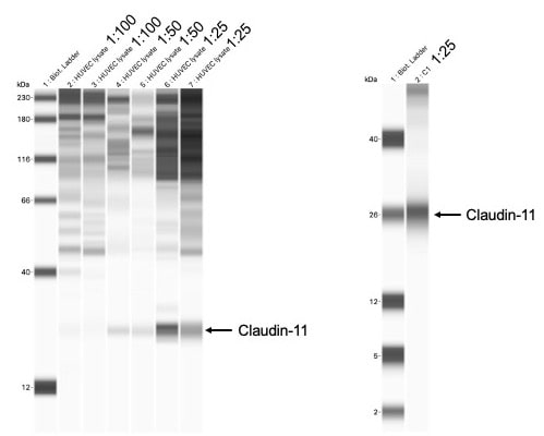
Western Blot: Claudin-11 Antibody [NBP1-82470]
Western Blot: Claudin-11 Antibody [NBP1-82470] - HUVEC (Human Umbilical Vein Endothelial Cells) lysate. Three dilutions were tested using the WES 12-230 kDa kit (left panel): Lanes 2-3, 1:100; Lanes 4-5, 1:50; and Lanes 6-7, 1:25. The 1:25 dilution was tested again using the 2-40 kDa (right panel). All test lanes utilized 0.2 mg/mL protein concentrations. The WES anti-rabbit antibody kit was used. WB image submitted by a verified customer review.![Immunohistochemistry-Paraffin: Claudin-11 Antibody [NBP1-82470]](https://resources.rndsystems.com/images/products/Claudin-11-Antibody-Immunohistochemistry-Paraffin-NBP1-82470-img0011.jpg "Immunohistochemistry-Paraffin: Claudin-11 Antibody [NBP1-82470]")
Immunohistochemistry-Paraffin: Claudin-11 Antibody [NBP1-82470]
Immunohistochemistry-Paraffin: Claudin-11 Antibody [NBP1-82470] - Staining of human Skeletal muscle shows no positivity in myocytes as expected.![Immunohistochemistry-Paraffin: Claudin-11 Antibody [NBP1-82470]](https://resources.rndsystems.com/images/products/Claudin-11-Antibody-Immunohistochemistry-Paraffin-NBP1-82470-img0009.jpg "Immunohistochemistry-Paraffin: Claudin-11 Antibody [NBP1-82470]")
Immunohistochemistry-Paraffin: Claudin-11 Antibody [NBP1-82470]
Immunohistochemistry-Paraffin: Claudin-11 Antibody [NBP1-82470] - Staining of human Cervix shows no positivity in squamous epithelial cells as expected.![Immunohistochemistry-Paraffin: Claudin-11 Antibody [NBP1-82470]](https://resources.rndsystems.com/images/products/Claudin-11-Antibody-Immunohistochemistry-Paraffin-NBP1-82470-img0010.jpg "Immunohistochemistry-Paraffin: Claudin-11 Antibody [NBP1-82470]")
Immunohistochemistry-Paraffin: Claudin-11 Antibody [NBP1-82470]
Immunohistochemistry-Paraffin: Claudin-11 Antibody [NBP1-82470] - Staining of human Pancreas shows no positivity in exocrine glandular cells as expected.![Immunohistochemistry-Paraffin: Claudin-11 Antibody [NBP1-82470]](https://resources.rndsystems.com/images/products/Claudin-11-Antibody-Immunohistochemistry-Paraffin-NBP1-82470-img0012.jpg "Immunohistochemistry-Paraffin: Claudin-11 Antibody [NBP1-82470]")
Immunohistochemistry-Paraffin: Claudin-11 Antibody [NBP1-82470]
Immunohistochemistry-Paraffin: Claudin-11 Antibody [NBP1-82470] - Staining of human Testis shows strong membranous positivity in cells in seminiferous ducts.
Western Blot: Claudin-11 Antibody [NBP1-82470] -
Western Blot: Claudin-11 Antibody [NBP1-82470] - Comparison of expression changes between young & old MRC-5 & HFF fibroblasts measured with RNA-seq & Western Blots. (a) 8 genes commonly downregulated & (b) 8 genes commonly upregulated in both cell lines. (a, b) The colors of the bars indicate the measurement technique (blue: RNA-seq; red: Western Blots/protein expression). Solid colored bars represent MRC-5 while shaded boxes represent HFF cells. The height of the bars corresponds to the logarithmic fold-change (FC) of expression between the first & the last PD investigated here (RNA-seq: log 2 RPKM FC; protein: log 2 expression ratio). Error bars indicate standard deviation from the mean. Changes statistically different comparing young & old PD (RNA-seq: DESeq; rRT-PCR/Protein: Student's t-test; n = 3) are indicated with an asterix: ∗p < 0.05, ∗∗p < 0.01, & ∗∗∗p < 0.001. (c) The blots show the protein expression levels in MRC-5 & HFF cells at young compared to old PDs. The up- or downregulation was signified by the presence or absence of bands in Western Blots. Image collected & cropped by CiteAb from the following publication (https://pubmed.ncbi.nlm.nih.gov/26339636), licensed under a CC-BY license. Not internally tested by Novus Biologicals.
Immunohistochemistry: Rabbit Polyclonal Claudin-11 Antibody [NBP1-82470]
Analysis of chicken embryo spinal cord at E9.5. Cryosections were stained using a primary antibody dilution of 1:100.White: DAPI
Red: TUBB3
Cyan: Claudin 11
Image from a verified customer review.
Applications for Claudin-11 Antibody - BSA Free
Application
Recommended Usage
Immunohistochemistry
1:50 - 1:200
Immunohistochemistry-Paraffin
1:50 - 1:200
Application Notes
IHC-Paraffin, HIER pH 6 retrieval is recommended.
Reviewed Applications
Read 2 reviews rated 4.5 using NBP1-82470 in the following applications:
Formulation, Preparation, and Storage
Purification
Affinity purified
Formulation
PBS (pH 7.2) and 40% Glycerol
Format
BSA Free
Preservative
0.02% Sodium Azide
Concentration
Concentrations vary lot to lot. See vial label for concentration. If unlisted please contact technical services.
Shipping
The product is shipped with polar packs. Upon receipt, store it immediately at the temperature recommended below.
Stability & Storage
Store at 4C short term. Aliquot and store at -20C long term. Avoid freeze-thaw cycles.
Background: Claudin-11
Alternate Names
Claudin11, CLDN11, OSP, OTM
Gene Symbol
CLDN11
Additional Claudin-11 Products
Product Documents for Claudin-11 Antibody - BSA Free
Certificate of Analysis
To download a Certificate of Analysis, please enter a lot or batch number in the search box below.
Product Specific Notices for Claudin-11 Antibody - BSA Free
This product is for research use only and is not approved for use in humans or in clinical diagnosis. Primary Antibodies are guaranteed for 1 year from date of receipt.
Related Research Areas
Citations for Claudin-11 Antibody - BSA Free
 Powered by Bioz
Customer Reviews for Claudin-11 Antibody - BSA Free (2)
4.5 out of 5
2 Customer Ratings
Have you used Claudin-11 Antibody - BSA Free?
Submit a review and receive an Amazon gift card!
$25/€18/£15/$25CAN/¥2500 Yen for a review with an image
$10/€7/£6/$10CAN/¥1110 Yen for a review without an image
Submit a review
Customer Images

Showing
1
-
2 of
2 reviews
Showing All
Filter By:
-
Application: ImmunofluorescenceSample Tested: EmbryosSpecies: Chicken and MouseVerified Customer | Posted 03/19/2026White: DAPI Red: TUBB3 Cyan: Claudin 11 Spinal cord E9.5 chicken embryo1/100 on cryosections
 Bio-Techne ResponseThis review reflects a new species or application tested on a primary antibody.
 -
Application: Simple WesternSample Tested: HUVEC (Human Umbilical Vein Endothelial Cells)Species: HumanVerified Customer | Posted 06/07/2021Three dilutions were tested using the WES 12-230 kDa kit (left panel): Lanes 2-3, 1:100; Lanes 4-5, 1:50; and Lanes 6-7, 1:25. The 1:25 dilution was tested again using the 2-40 kDa (right panel).All test lanes utilized 0.2 mg/ml protein concentrations. The WES anti-rabbit antibody kit was used.
There are no reviews that match your criteria.
Protocols
Find general support by application which include: protocols, troubleshooting, illustrated assays, videos and webinars.
- Antigen Retrieval Protocol (PIER)
- Antigen Retrieval for Frozen Sections Protocol
- Appropriate Fixation of IHC/ICC Samples
- Cellular Response to Hypoxia Protocols
- Chromogenic IHC Staining of Formalin-Fixed Paraffin-Embedded (FFPE) Tissue Protocol
- Chromogenic Immunohistochemistry Staining of Frozen Tissue
- ClariTSA™ Fluorophore Kits
- Detection & Visualization of Antibody Binding
- Fluorescent IHC Staining of Frozen Tissue Protocol
- Graphic Protocol for Heat-induced Epitope Retrieval
- Graphic Protocol for the Preparation and Fluorescent IHC Staining of Frozen Tissue Sections
- Graphic Protocol for the Preparation and Fluorescent IHC Staining of Paraffin-embedded Tissue Sections
- Graphic Protocol for the Preparation of Gelatin-coated Slides for Histological Tissue Sections
- IHC Sample Preparation (Frozen sections vs Paraffin)
- Immunofluorescent IHC Staining of Formalin-Fixed Paraffin-Embedded (FFPE) Tissue Protocol
- Immunohistochemistry (IHC) and Immunocytochemistry (ICC) Protocols
- Immunohistochemistry Frozen Troubleshooting
- Immunohistochemistry Paraffin Troubleshooting
- Preparing Samples for IHC/ICC Experiments
- Preventing Non-Specific Staining (Non-Specific Binding)
- Primary Antibody Selection & Optimization
- Protocol for Heat-Induced Epitope Retrieval (HIER)
- Protocol for Making a 4% Formaldehyde Solution in PBS
- Protocol for VisUCyte™ HRP Polymer Detection Reagent
- Protocol for the Preparation & Fixation of Cells on Coverslips
- Protocol for the Preparation and Chromogenic IHC Staining of Frozen Tissue Sections
- Protocol for the Preparation and Chromogenic IHC Staining of Frozen Tissue Sections - Graphic
- Protocol for the Preparation and Chromogenic IHC Staining of Paraffin-embedded Tissue Sections
- Protocol for the Preparation and Chromogenic IHC Staining of Paraffin-embedded Tissue Sections - Graphic
- Protocol for the Preparation and Fluorescent IHC Staining of Frozen Tissue Sections
- Protocol for the Preparation and Fluorescent IHC Staining of Paraffin-embedded Tissue Sections
- Protocol for the Preparation of Gelatin-coated Slides for Histological Tissue Sections
- TUNEL and Active Caspase-3 Detection by IHC/ICC Protocol
- The Importance of IHC/ICC Controls
- Troubleshooting Guide: Immunohistochemistry
- View all Protocols, Troubleshooting, Illustrated assays and Webinars
Loading...
Associated Pathways


