COMP/Thrombospondin-5 Antibody - BSA Free
Novus Biologicals | Catalog # NBP2-92733
![Western Blot: COMP/Thrombospondin-5 AntibodyBSA Free [NBP2-92733]](https://resources.rndsystems.com/images/products/COMP-Thrombospondin-5-Antibody-Western-Blot-NBP2-92733-img0003.jpg "Western Blot: COMP/Thrombospondin-5 AntibodyBSA Free [NBP2-92733]")
Key Product Details
Species Reactivity
Applications
Label
Antibody Source
Format
Product Specifications
Immunogen
Clonality
Host
Isotype
Scientific Data Images for COMP/Thrombospondin-5 Antibody - BSA Free
Western Blot: COMP/Thrombospondin-5 AntibodyBSA Free [NBP2-92733]
Western Blot: COMP/Thrombospondin-5 Antibody [NBP2-92733] - Analysis of extracts of various cell lines, using COMP/Thrombospondin-5 at 1:1000 dilution.Secondary antibody: HRP Goat Anti-Rabbit IgG (H+L) at 1:10000 dilution.Lysates/proteins: 25ug per lane.Blocking buffer: 3% nonfat dry milk in TBST.Detection: ECL Basic Kit.Exposure time: 1s.![Immunocytochemistry/ Immunofluorescence: COMP/Thrombospondin-5 Antibody - BSA Free [NBP2-92733]](https://resources.rndsystems.com/images/products/COMP-Thrombospondin-5-Antibody-Immunocytochemistry-Immunofluorescence-NBP2-92733-img0004.jpg "Immunocytochemistry/ Immunofluorescence: COMP/Thrombospondin-5 Antibody - BSA Free [NBP2-92733]")
Immunocytochemistry/ Immunofluorescence: COMP/Thrombospondin-5 Antibody - BSA Free [NBP2-92733]
Immunocytochemistry/Immunofluorescence: COMP/Thrombospondin-5 Antibody [NBP2-92733] - Analysis of L929 cells using COMP Rabbit pAb at dilution of 1:100 (40x lens). Blue: DAPI for nuclear staining.
Immunocytochemistry/Immunofluorescence: COMP/Thrombospondin-5 Antibody [NBP2-92733] -
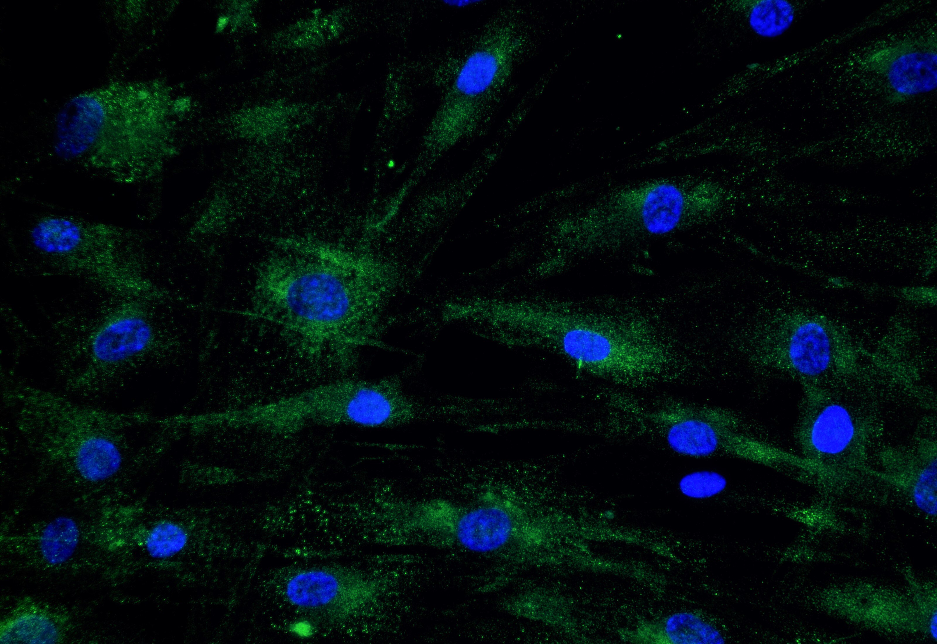
Immunocytochemistry/Immunofluorescence: COMP/Thrombospondin-5 Antibody [NBP2-92733] - Analysis of Human Dermal Fibroblasts (HDFa). Green: 1:100 COMP/Thrombospondin-5 antibody in PBS/BSA 1%, 1h room temperature Blue: DAPI. Image from verified customer review.Applications for COMP/Thrombospondin-5 Antibody - BSA Free
Immunocytochemistry/ Immunofluorescence
Western Blot
Reviewed Applications
Read 1 review rated 4 using NBP2-92733 in the following applications:
Formulation, Preparation, and Storage
Purification
Formulation
Format
Preservative
Concentration
Shipping
Stability & Storage
Background: COMP/Thrombospondin-5
Long Name
Alternate Names
Gene Symbol
Additional COMP/Thrombospondin-5 Products
Product Documents for COMP/Thrombospondin-5 Antibody - BSA Free
Certificate of Analysis
To download a Certificate of Analysis, please enter a lot or batch number in the search box below.
Product Specific Notices for COMP/Thrombospondin-5 Antibody - BSA Free
This product is for research use only and is not approved for use in humans or in clinical diagnosis. Primary Antibodies are guaranteed for 1 year from date of receipt.
Related Research Areas
Customer Reviews for COMP/Thrombospondin-5 Antibody - BSA Free (1)
Have you used COMP/Thrombospondin-5 Antibody - BSA Free?
Submit a review and receive an Amazon gift card!
$25/€18/£15/$25CAN/¥2500 Yen for a review with an image
$10/€7/£6/$10CAN/¥1110 Yen for a review without an image
Submit a review
Customer Images
-
Application: ImmunocytochemistrySample Tested: fibroblastsSpecies: HumanVerified Customer | Posted 05/25/2023Human Dermal Fibroblasts (HDFa) Green: 1:100 COMP/Thrombospondin-5 antibody in PBS/BSA 1%, 1h room temperature Blue: DAPI

There are no reviews that match your criteria.
Protocols
Find general support by application which include: protocols, troubleshooting, illustrated assays, videos and webinars.
- Appropriate Fixation of IHC/ICC Samples
- Cellular Response to Hypoxia Protocols
- ClariTSA™ Fluorophore Kits
- Detection & Visualization of Antibody Binding
- ICC Cell Smear Protocol for Suspension Cells
- ICC Immunocytochemistry Protocol Videos
- ICC for Adherent Cells
- Immunocytochemistry (ICC) Protocol
- Immunocytochemistry Troubleshooting
- Immunofluorescence of Organoids Embedded in Cultrex Basement Membrane Extract
- Immunohistochemistry (IHC) and Immunocytochemistry (ICC) Protocols
- Preparing Samples for IHC/ICC Experiments
- Preventing Non-Specific Staining (Non-Specific Binding)
- Primary Antibody Selection & Optimization
- Protocol for VisUCyte™ HRP Polymer Detection Reagent
- Protocol for the Fluorescent ICC Staining of Cell Smears - Graphic
- Protocol for the Fluorescent ICC Staining of Cultured Cells on Coverslips - Graphic
- Protocol for the Preparation and Fluorescent ICC Staining of Cells on Coverslips
- Protocol for the Preparation and Fluorescent ICC Staining of Non-adherent Cells
- Protocol for the Preparation and Fluorescent ICC Staining of Stem Cells on Coverslips
- Protocol for the Preparation of a Cell Smear for Non-adherent Cell ICC - Graphic
- R&D Systems Quality Control Western Blot Protocol
- TUNEL and Active Caspase-3 Detection by IHC/ICC Protocol
- The Importance of IHC/ICC Controls
- Troubleshooting Guide: Western Blot Figures
- Western Blot Conditions
- Western Blot Protocol
- Western Blot Protocol for Cell Lysates
- Western Blot Troubleshooting
- Western Blot Troubleshooting Guide
- View all Protocols, Troubleshooting, Illustrated assays and Webinars
FAQs for COMP/Thrombospondin-5 Antibody - BSA Free
-
Q: I have synovial fluid sample and I want to work on Western bloting can you support me,how can I prepare this sample for WB technique for assay comp(cartilage oligomatrix protein )?
A:
We currently sell four antibodies to cartilage oligomeric matrix protein (COMP), all of which are validated for Western blotting. Although we have generated Western blot data using samples from human, bovine and rabbit cartilage, we do not have any testing data derived from synovial fluid samples. You may however find the following publication useful, in which the authors sampled synovial fluid from patients with rheumatoid arthritis and prepared this fluid for Western blot detection of the proteins SP-A and SP-D: PMC 2080374
Associated Pathways
