Complement C3 Antibody (11H9) - BSA Free
Novus Biologicals | Catalog # NB200-540
Key Product Details
Validated by
Biological Validation
Species Reactivity
Validated:
Mouse, E. coli
Cited:
Mouse, Bacteria - Escherichia coli
Applications
Validated:
Immunohistochemistry, Immunohistochemistry-Paraffin, Immunohistochemistry-Frozen, Immunoassay, Flow Cytometry, Flow (Intracellular), Immunocytochemistry/ Immunofluorescence, Immunoprecipitation, CyTOF-ready
Cited:
Immunohistochemistry, Immunohistochemistry-Frozen, ELISA, Flow Cytometry, Immunocytochemistry/ Immunofluorescence, Immunoprecipitation, IF/IHC
Label
Unconjugated
Antibody Source
Monoclonal Rat IgG2A Clone # 11H9
Format
BSA Free
Loading...
Product Specifications
Immunogen
This Complement C3 Antibody (11H9) was developed against C57BL/6 thymocytes saturated with rat anti-Thy-1 monoclonal antibody of IgG2b subclass (RmT1).
Reactivity Notes
Use in Mouse reported in scientific literature (PMID:34433493). Use in E. coli reported in scientific literature (PMID:32422907).
Localization
Secreted
Specificity
Mouse Complement C3 and its activation products, C3b, iC3b, C3d and C3dg
Clonality
Monoclonal
Host
Rat
Isotype
IgG2A
Theoretical MW
187 kDa.
Disclaimer note: The observed molecular weight of the protein may vary from the listed predicted molecular weight due to post translational modifications, post translation cleavages, relative charges, and other experimental factors.
Disclaimer note: The observed molecular weight of the protein may vary from the listed predicted molecular weight due to post translational modifications, post translation cleavages, relative charges, and other experimental factors.
Scientific Data Images for Complement C3 Antibody (11H9) - BSA Free
![Flow (Intracellular): Complement C3 Antibody (11H9) - BSA Free [NB200-540]](https://resources.rndsystems.com/images/products/Complement-C3-Antibody-11H9-Flow-Intracellular-NB200-540-img0005.jpg "Flow (Intracellular): Complement C3 Antibody (11H9) - BSA Free [NB200-540]")
Flow (Intracellular): Complement C3 Antibody (11H9) - BSA Free [NB200-540]
Flow (Intracellular): Complement C3 Antibody (11H9) [NB200-540] - An intracellular stain was performed on RAW 246.7 cells with Complement C3 (11H9-3-2) antibody NB200-540 (blue) and a matched isotype control NBP2-31382 (orange). Cells were either treated with 3uM Monensin for 3 hours to block the secretion of Complement C3 (B) or grown in normal media (A). Cells were fixed with 4% PFA and then permeablized with 0.1% saponin. Cells were incubated in an antibody dilution of 2 ug/mL for 30 minutes at room temperature, followed by mouse F(ab)2 IgG (H+L) PE-conjugated secondary antibody (F0102B, R&D Systems).![Immunocytochemistry/ Immunofluorescence: Complement C3 Antibody (11H9) - BSA Free [NB200-540]](https://resources.rndsystems.com/images/products/Complement-C3-Antibody-11H9-Immunocytochemistry-Immunofluorescence-NB200-540-img0002.jpg "Immunocytochemistry/ Immunofluorescence: Complement C3 Antibody (11H9) - BSA Free [NB200-540]")
Immunocytochemistry/ Immunofluorescence: Complement C3 Antibody (11H9) - BSA Free [NB200-540]
Immunocytochemistry/Immunofluorescence: Complement C3 Antibody (11H9) [NB200-540] - C3 protein fragments deposited on kidney cells of MPL-lpr mouse. Staining with antibody 11H9. Glomerular staining pattern. Fixation in 4% paraformaldehyde in PBS pH 7.4. Vibratome sections of 4 um. Pretreated with 3% hydrogen peroxide for 20 min to quench endogenous peroxidases. Microwaved in antigen unmasking solution for 2-5 minutes as antigen retrieval.![Immunohistochemistry-Paraffin: Complement C3 Antibody (11H9) - BSA Free [NB200-540]](https://resources.rndsystems.com/images/products/Complement-C3-Antibody-11H9-Immunohistochemistry-Paraffin-NB200-540-img0004.jpg "Immunohistochemistry-Paraffin: Complement C3 Antibody (11H9) - BSA Free [NB200-540]")
Immunohistochemistry-Paraffin: Complement C3 Antibody (11H9) - BSA Free [NB200-540]
Immunohistochemistry-Paraffin: Complement C3 Antibody (11H9) [NB200-540] - Complement C3 protein in a FFPE tissue section of mouse liver using 1:100 dilution of Complement C3 antibody (clone 11H9) NB200-540. Weak but distinct membrane-cytoplasmic immunopositivity was observed in hepatocytes and few cells developed punctate membrane staining.![Flow Cytometry: Complement C3 Antibody (11H9) - BSA Free [NB200-540]](https://resources.rndsystems.com/images/products/Complement-C3-Antibody-11H9-Flow-Cytometry-NB200-540-img0006.jpg "Flow Cytometry: Complement C3 Antibody (11H9) - BSA Free [NB200-540]")
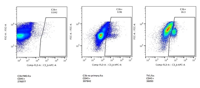
Flow Cytometry: Complement C3 Antibody (11H9) - BSA Free [NB200-540]
Flow Cytometry: Complement C3 Antibody (11H9) [NB200-540] - Left panel: FMO, Middle panel: No primary antibody control, Right panel: sample. Day 6 murine mammary tumors processed and stained for analysis with flow cytometry. The C3b+ population of CD45+ cells is what the gate in each sample is exhibiting. WB image submitted by a verified customer review.![Immunohistochemistry-Paraffin: Complement C3 Antibody (11H9) - BSA Free [NB200-540]](https://resources.rndsystems.com/images/products/Complement-C3-Antibody-11H9-Immunohistochemistry-Paraffin-NB200-540-img0003.jpg "Immunohistochemistry-Paraffin: Complement C3 Antibody (11H9) - BSA Free [NB200-540]")
Immunohistochemistry-Paraffin: Complement C3 Antibody (11H9) - BSA Free [NB200-540]
Immunohistochemistry-Paraffin: Complement C3 Antibody (11H9) [NB200-540] - Complement C3 protein in a FFPE tissue section of mouse lymph node using 1:100 dilution of Complement C3 antibody (clone 11H9) NB200-540. This representative photomicrograph shows a membrane-cytoplasmic immunopositivity in non-germinal center cells, and few cells developed an intense staining for this target protein.Applications for Complement C3 Antibody (11H9) - BSA Free
Application
Recommended Usage
Flow (Intracellular)
1 ug/ml
Flow Cytometry
1 ug/ml
Immunoassay
0.5 ug/well in PBS
Immunocytochemistry/ Immunofluorescence
1:10-1:500
Immunohistochemistry
1:10-1:500
Immunohistochemistry-Frozen
1:10-1:500
Immunohistochemistry-Paraffin
1:200
Immunoprecipitation
1:10-1:500
Reviewed Applications
Read 2 reviews rated 4.5 using NB200-540 in the following applications:
Flow Cytometry Panel Builder
Bio-Techne Knows Flow Cytometry
Save time and reduce costly mistakes by quickly finding compatible reagents using the Panel Builder Tool.
Advanced Features
- Spectra Viewer - Custom analysis of spectra from multiple fluorochromes
- Spillover Popups - Visualize the spectra of individual fluorochromes
- Antigen Density Selector - Match fluorochrome brightness with antigen density

Formulation, Preparation, and Storage
Purification
Protein G purified
Formulation
PBS
Format
BSA Free
Preservative
0.02% Sodium Azide
Concentration
1.0 mg/ml
Shipping
The product is shipped with polar packs. Upon receipt, store it immediately at the temperature recommended below.
Stability & Storage
Store at 4C short term. Aliquot and store at -20C long term. Avoid freeze-thaw cycles.
Background: Complement C3
Both elevated levels and reduced levels of Complement C3 has been implicated in diseases pathologies (6). Deficiency in Complement proteins can result in autoimmune disorders including systemic lupus erythematosus, which is more often associated with C1 or C4 deficiency and only rarely with C3 deficiency (6). However, C3 deficiency typically results in increased risk of recurrent bacterial infections and glomerulonephritis, characterized by inflammation of the filtering glomeruli in the kidneys (6). Additionally, elevated levels of C3a and C4a is seen in patients with antiphospholipid antibody syndrome (6). Serum levels of C3 are also higher in rheumatoid arthritis cases (6). The complement system has become a target for drugs and therapeutics aimed at modulating innate immunity (7). For instance, compstatin is a peptide that binds to C3, inhibiting convertase activity and cleavage and can be used to treat diseases associated with uncontrolled C3 activation (7). C3-inhibitors and other complement inhibitors are a promising drug candidate for treatment of many diseases (7).
References
1. Mathern, D. R., & Heeger, P. S. (2015). Molecules Great and Small: The Complement System. Clinical Journal of the American Society of Nephrology: CJASN. https://doi.org/10.2215/CJN.06230614
2. Merle, N. S., Church, S. E., Fremeaux-Bacchi, V., & Roumenina, L. T. (2015). Complement System Part I - Molecular Mechanisms of Activation and Regulation. Frontiers in Immunology. https://doi.org/10.3389/fimmu.2015.00262
3. Ricklin, D., Reis, E. S., Mastellos, D. C., Gros, P., & Lambris, J. D. (2016). Complement component C3 - The "Swiss Army Knife" of innate immunity and host defense. Immunological Reviews. https://doi.org/10.1111/imr.12500
4. Merle, N. S., Noe, R., Halbwachs-Mecarelli, L., Fremeaux-Bacchi, V., & Roumenina, L. T. (2015). Complement System Part II: Role in Immunity. Frontiers in Immunology. https://doi.org/10.3389/fimmu.2015.00257
5. Sahu, A., & Lambris, J. D. (2001). Structure and biology of complement protein C3, a connecting link between innate and acquired immunity. Immunological Reviews. https://doi.org/10.1034/j.1600-065x.2001.1800103.x
6. Vignesh, P., Rawat, A., Sharma, M., & Singh, S. (2017). Complement in autoimmune diseases. Clinica Chimica Acta; International Journal of Clinical Chemistry. https://doi.org/10.1016/j.cca.2016.12.017
7. Mastellos, D. C., Yancopoulou, D., Kokkinos, P., Huber-Lang, M., Hajishengallis, G., Biglarnia, A. R., Lupu, F., Nilsson, B., Risitano, A. M., Ricklin, D., & Lambris, J. D. (2015). Compstatin: a C3-targeted complement inhibitor reaching its prime for bedside intervention. European Journal of Clinical Investigation. https://doi.org/10.1111/eci.12419
Alternate Names
Acylation Stimulating Protein, acylation-stimulating protein cleavage product, AHUS5, ARMD9, ASP, C3, C3 and PZP-like alpha-2-macroglobulin domain-containing protein 1, C3a, C3a anaphylatoxin, C3adesArg, C3b, C3bc, C3-beta-c, complement C3, Complement C3 alpha chain, Complement C3 beta chain, Complement C3b alpha' chain, Complement C3c alpha' chain fragment 1, Complement C3c alpha' chain fragment 2, Complement C3d fragment, Complement C3dg fragment, Complement C3f fragment, Complement C3g fragment, complement component 3, complement component C3, complement component C3a, complement component C3b, CPAMD1, EC 3.4.21.43, epididymis secretory sperm binding protein Li 62p, HEL-S-62p, prepro-C3
Entrez Gene IDs
12266 (Mouse)
Gene Symbol
C3
UniProt
Additional Complement C3 Products
Product Documents for Complement C3 Antibody (11H9) - BSA Free
Certificate of Analysis
To download a Certificate of Analysis, please enter a lot or batch number in the search box below.
Product Specific Notices for Complement C3 Antibody (11H9) - BSA Free
This product is for research use only and is not approved for use in humans or in clinical diagnosis. Primary Antibodies are guaranteed for 1 year from date of receipt.
Citations for Complement C3 Antibody (11H9) - BSA Free
 Powered by Bioz
Customer Reviews for Complement C3 Antibody (11H9) - BSA Free (2)
4.5 out of 5
2 Customer Ratings
Have you used Complement C3 Antibody (11H9) - BSA Free?
Submit a review and receive an Amazon gift card!
$25/€18/£15/$25CAN/¥2500 Yen for a review with an image
$10/€7/£6/$10CAN/¥1110 Yen for a review without an image
Submit a review
Customer Images
Showing
1
-
2 of
2 reviews
Showing All
Filter By:
-
Application: Flow CytometrySample Tested: Breast tumorSpecies: MouseVerified Customer | Posted 05/16/2019Flow cytometry gatingDespite some issues with our secondary antibody, this antibody worked well for flow cytometry analysis of mouse tumor tissue.

-
Application: ImmunocytochemistrySample Tested: brain and spinal cordSpecies: MouseVerified Customer | Posted 04/01/2017
There are no reviews that match your criteria.
Protocols
View specific protocols for Complement C3 Antibody (11H9) - BSA Free (NB200-540):
Protocol for Flow Cytometry Intracellular Staining
Sample Preparation.
1. Grow cells to 60-85% confluency. Flow cytometry requires between 2 x 10⁵ and 1 x 10⁶ cells for optimal performance.
2. If cells are adherent, harvest gently by washing once with staining buffer and then scraping. Avoid using trypsin as this can disrupt certain epitopes of interest. If enzymatic harvest is required, use Accutase, Collagenase, or TrypLE Express for a less damaging option.
3. Reserve 100 uL for counting, then transfer cell volume into a 50 mL conical tube and centrifuge for 8 minutes at 400 RCF.
a. Count cells using a hemocytometer and a 1:1 trypan blue exclusion stain to determine cell viability before starting the flow protocol. If cells appear blue, do not proceed.
4. Re-suspend cells to a concentration of 1 x 10⁶ cells/mL in staining buffer (NBP2-26247).
5. Aliquot out 100 uL samples in accordance with your experimental samples.
Tip: When cell surface and intracellular staining are required in the same sample, it is advisable that the cell surface staining be performed first since the fixation and permeabilization steps might reduce the availability of surface antigens.
Intracellular Staining.
Tip: When performing intracellular staining, it is important to use appropriate fixation and permeabilization reagents based upon the target and its subcellular location. Generally, our Intracellular Flow Assay Kit (NBP2-29450) is a good place to start as it contains an optimized combination of reagents for intracellular staining as well as an inhibitor of intracellular protein transport (necessary if staining secreted proteins). Certain targets may require more gentle or transient permeabilization protocols such as the commonly employed methanol or saponin-based methods.
Protocol for Cytoplasmic Targets:
1. Fix the cells by adding 100 uL fixation solution (such as 4% PFA) to each sample for 10-15 minutes.
2. Permeabilize cells by adding 100 uL of a permeabilization buffer to every 1 x 10⁶ cells present in the sample. Mix well and incubate at room temperature for 15 minutes.
a. For cytoplasmic targets, use a gentle permeabilization solution such as 1X PBS + 0.5% Saponin or 1X PBS + 0.5% Tween-20.
b. To maintain the permeabilized state throughout your experiment, use staining buffer + 0.1% of the permeabilization reagent (i.e. 0.1% Tween-20 or 0.1% Saponin).
3. Following the 15 minute incubation, add 2 mL of the staining buffer + 0.1% permeabilizer to each sample.
4. Centrifuge for 1 minute at 400 RCF.
5. Discard supernatant and re-suspend in 100 uL of staining buffer + 0.1% permeabilizer.
6. Add appropriate amount of each antibody (eg. 1 test or 1 ug per sample, as experimentally determined).
7. Mix well and incubate at room temperature for 30 minutes- 1 hour. Gently mix samples every 10-15 minutes.
8. Following the primary/conjugate incubation, add 1-2 mL/sample of staining buffer +0.1% permeabilizer and centrifuge for 1 minute at 400 RCF.
9. Wash twice by re-suspending cells in staining buffer (2 mL for tubes or 200 uL for wells) and centrifuging at 400 RCF for 5 minutes. Discard supernatant.
10. Add appropriate amount of secondary antibody (as experimentally determined) to each sample.
11. Incubate at room temperature in dark for 20 minutes.
12. Add 1-2 mL of staining buffer and centrifuge at 400 RCF for 1 minute and discard supernatant.
13. Wash twice by re-suspending cells in staining buffer (2 mL for tubes or 200 uL for wells) and centrifuging at 400 RCF for 5 minutes. Discard supernatant.
14. Resuspend in an appropriate volume of staining buffer (usually 500 uL per sample) and proceed with analysis on your flow cytometer.
Immunohistochemistry-Paraffin Embedded Sections
Antigen Unmasking:
Bring slides to a boil in 10 mM sodium citrate buffer (pH 6.0) then maintain at a sub-boiling temperature for 10 minutes. Cool slides on bench-top for 30 minutes (keep slides in the sodium citrate buffer at all times).
Staining:
1. Wash sections in deionized water three times for 5 minutes each.
2. Wash sections in PBS for 5 minutes.
3. Block each section with 100-400 ul blocking solution (1% BSA in PBS) for 1 hour at room temperature.
4. Remove blocking solution and add 100-400 ul diluted primary antibody. Incubate overnight at 4 C.
5. Remove antibody solution and wash sections in wash buffer three times for 5 minutes each.
6. Add 100-400 ul HRP polymer conjugated secondary antibody. Incubate 30 minutes at room temperature.
7. Wash sections three times in wash buffer for 5 minutes each.
8. Add 100-400 ul DAB substrate to each section and monitor staining closely.
9. As soon as the sections develop, immerse slides in deionized water.
10. Counterstain sections in hematoxylin.
11. Wash sections in deionized water two times for 5 minutes each.
12. Dehydrate sections.
13. Mount coverslips.
Find general support by application which include: protocols, troubleshooting, illustrated assays, videos and webinars.
- 7-Amino Actinomycin D (7-AAD) Cell Viability Flow Cytometry Protocol
- Antigen Retrieval Protocol (PIER)
- Antigen Retrieval for Frozen Sections Protocol
- Appropriate Fixation of IHC/ICC Samples
- Cellular Response to Hypoxia Protocols
- Chromogenic IHC Staining of Formalin-Fixed Paraffin-Embedded (FFPE) Tissue Protocol
- Chromogenic Immunohistochemistry Staining of Frozen Tissue
- ClariTSA™ Fluorophore Kits
- Detection & Visualization of Antibody Binding
- ELISA Sample Preparation & Collection Guide
- ELISA Troubleshooting Guide
- Extracellular Membrane Flow Cytometry Protocol
- Flow Cytometry Protocol for Cell Surface Markers
- Flow Cytometry Protocol for Staining Membrane Associated Proteins
- Flow Cytometry Staining Protocols
- Flow Cytometry Troubleshooting Guide
- Fluorescent IHC Staining of Frozen Tissue Protocol
- Graphic Protocol for Heat-induced Epitope Retrieval
- Graphic Protocol for the Preparation and Fluorescent IHC Staining of Frozen Tissue Sections
- Graphic Protocol for the Preparation and Fluorescent IHC Staining of Paraffin-embedded Tissue Sections
- Graphic Protocol for the Preparation of Gelatin-coated Slides for Histological Tissue Sections
- How to Run an R&D Systems DuoSet ELISA
- How to Run an R&D Systems Quantikine ELISA
- How to Run an R&D Systems Quantikine™ QuicKit™ ELISA
- ICC Cell Smear Protocol for Suspension Cells
- ICC Immunocytochemistry Protocol Videos
- ICC for Adherent Cells
- IHC Sample Preparation (Frozen sections vs Paraffin)
- Immunocytochemistry (ICC) Protocol
- Immunocytochemistry Troubleshooting
- Immunofluorescence of Organoids Embedded in Cultrex Basement Membrane Extract
- Immunofluorescent IHC Staining of Formalin-Fixed Paraffin-Embedded (FFPE) Tissue Protocol
- Immunohistochemistry (IHC) and Immunocytochemistry (ICC) Protocols
- Immunohistochemistry Frozen Troubleshooting
- Immunohistochemistry Paraffin Troubleshooting
- Immunoprecipitation Protocol
- Intracellular Flow Cytometry Protocol Using Alcohol (Methanol)
- Intracellular Flow Cytometry Protocol Using Detergents
- Intracellular Nuclear Staining Flow Cytometry Protocol Using Detergents
- Intracellular Staining Flow Cytometry Protocol Using Alcohol Permeabilization
- Intracellular Staining Flow Cytometry Protocol Using Detergents to Permeabilize Cells
- Preparing Samples for IHC/ICC Experiments
- Preventing Non-Specific Staining (Non-Specific Binding)
- Primary Antibody Selection & Optimization
- Propidium Iodide Cell Viability Flow Cytometry Protocol
- Protocol for Heat-Induced Epitope Retrieval (HIER)
- Protocol for Liperfluo
- Protocol for Making a 4% Formaldehyde Solution in PBS
- Protocol for VisUCyte™ HRP Polymer Detection Reagent
- Protocol for the Characterization of Human Th22 Cells
- Protocol for the Characterization of Human Th9 Cells
- Protocol for the Fluorescent ICC Staining of Cell Smears - Graphic
- Protocol for the Fluorescent ICC Staining of Cultured Cells on Coverslips - Graphic
- Protocol for the Preparation & Fixation of Cells on Coverslips
- Protocol for the Preparation and Chromogenic IHC Staining of Frozen Tissue Sections
- Protocol for the Preparation and Chromogenic IHC Staining of Frozen Tissue Sections - Graphic
- Protocol for the Preparation and Chromogenic IHC Staining of Paraffin-embedded Tissue Sections
- Protocol for the Preparation and Chromogenic IHC Staining of Paraffin-embedded Tissue Sections - Graphic
- Protocol for the Preparation and Fluorescent ICC Staining of Cells on Coverslips
- Protocol for the Preparation and Fluorescent ICC Staining of Non-adherent Cells
- Protocol for the Preparation and Fluorescent ICC Staining of Stem Cells on Coverslips
- Protocol for the Preparation and Fluorescent IHC Staining of Frozen Tissue Sections
- Protocol for the Preparation and Fluorescent IHC Staining of Paraffin-embedded Tissue Sections
- Protocol for the Preparation of Gelatin-coated Slides for Histological Tissue Sections
- Protocol for the Preparation of a Cell Smear for Non-adherent Cell ICC - Graphic
- Protocol: Annexin V and PI Staining by Flow Cytometry
- Protocol: Annexin V and PI Staining for Apoptosis by Flow Cytometry
- Quantikine HS ELISA Kit Assay Principle, Alkaline Phosphatase
- Quantikine HS ELISA Kit Principle, Streptavidin-HRP Polymer
- Sandwich ELISA (Colorimetric) – Biotin/Streptavidin Detection Protocol
- Sandwich ELISA (Colorimetric) – Direct Detection Protocol
- TUNEL and Active Caspase-3 Detection by IHC/ICC Protocol
- The Importance of IHC/ICC Controls
- Troubleshooting Guide: ELISA
- Troubleshooting Guide: Fluorokine Flow Cytometry Kits
- Troubleshooting Guide: Immunohistochemistry
- View all Protocols, Troubleshooting, Illustrated assays and Webinars
FAQs for Complement C3 Antibody (11H9) - BSA Free
Showing
1
-
1 of
1 FAQ
Showing All
-
Q: I am trying to establish a method to measure mice C3 levels by nephelometry. I would be most grateful if you could provide me with some help, regarding the choice of the Ab. I totally understand that since such a method has never been tried, I do not expect any guaranties.
A: We have never performed nephelometry in our lab, and do not have a protocol or advice to provide about this application. However, it seems to me that you should choose an antibody that is capable of recognizing its target in its folded conformation. Therefore, I would suggest trying an antibody that has been validated for ICC or IHC.
Loading...