CSE1L/CAS/Exportin-2 Antibody (3D8) - Azide and BSA Free
Novus Biologicals | Catalog # H00001434-M08
![Western Blot: CSE1L/CAS/Exportin-2 Antibody (3D8) [H00001434-M08]](https://resources.rndsystems.com/images/products/CSE1L-CAS-Exportin-2-Antibody-3D8-Western-Blot-H00001434-M08-img0013.jpg "Western Blot: CSE1L/CAS/Exportin-2 Antibody (3D8) [H00001434-M08]")
Loading...
Key Product Details
Species Reactivity
Human, Mouse
Applications
Immunohistochemistry, Immunohistochemistry-Paraffin, Western Blot, ELISA, Immunocytochemistry/ Immunofluorescence
Label
Unconjugated
Antibody Source
Monoclonal Mouse IgG1 kappa Clone # 3D8
Format
Azide and BSA Free
Loading...
Product Specifications
Immunogen
CSE1L (NP_001307, 872 a.a. ~ 971 a.a) partial recombinant protein with GST tag. MW of the GST tag alone is 26 KDa. LIGLFELPEDDTIPDEEHFIDIEDTPGYQTAFSQLAFAGKKEHDPVGQMVNNPKIHLAQSLHKLSTACPGRVPSMVSTSLNAEALQYLQGYLQAARVTLL
Specificity
CSE1L - CSE1 chromosome segregation 1-like (yeast)
Clonality
Monoclonal
Host
Mouse
Isotype
IgG1 kappa
Description
Quality control test: Antibody Reactive Against Recombinant Protein.
Scientific Data Images for CSE1L/CAS/Exportin-2 Antibody (3D8) - Azide and BSA Free
Western Blot: CSE1L/CAS/Exportin-2 Antibody (3D8) [H00001434-M08]
Western Blot: CSE1L/CAS/Exportin-2 Antibody (3D8) [H00001434-M08] - Analysis of CSE1L expression in Hela S3 NE (Cat # L013V3).![Immunocytochemistry/ Immunofluorescence: CSE1L/CAS/Exportin-2 Antibody (3D8) [H00001434-M08]](https://resources.rndsystems.com/images/products/CSE1L-CAS-Exportin-2-Antibody-3D8-Immunocytochemistry-Immunofluorescence-H00001434-M08-img0010.jpg "Immunocytochemistry/ Immunofluorescence: CSE1L/CAS/Exportin-2 Antibody (3D8) [H00001434-M08]")
Immunocytochemistry/ Immunofluorescence: CSE1L/CAS/Exportin-2 Antibody (3D8) [H00001434-M08]
Immunocytochemistry/Immunofluorescence: CSE1L/CAS/Exportin-2 Antibody (3D8) [H00001434-M08] - Analysis of monoclonal antibody to CSE1L on HeLa cell. Antibody concentration 10 ug/mL.![Immunohistochemistry-Paraffin: CSE1L/CAS/Exportin-2 Antibody (3D8) [H00001434-M08]](https://resources.rndsystems.com/images/products/CSE1L-CAS-Exportin-2-Antibody-3D8-Immunohistochemistry-Paraffin-H00001434-M08-img0014.jpg "Immunohistochemistry-Paraffin: CSE1L/CAS/Exportin-2 Antibody (3D8) [H00001434-M08]")
Immunohistochemistry-Paraffin: CSE1L/CAS/Exportin-2 Antibody (3D8) [H00001434-M08]
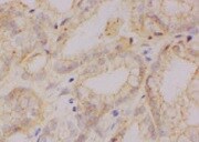
Immunohistochemistry-Paraffin: CSE1L/CAS/Exportin-2 Antibody (3D8) [H00001434-M08] - Mouse prostate tissue section stained with primary antibody for 10 hrs. IHC-P image submitted by a verified customer review.![Immunohistochemistry-Paraffin: CSE1L/CAS/Exportin-2 Antibody (3D8) [H00001434-M08]](https://resources.rndsystems.com/images/products/CSE1L-CAS-Exportin-2-Antibody-3D8-Immunohistochemistry-Paraffin-H00001434-M08-img0012.jpg "Immunohistochemistry-Paraffin: CSE1L/CAS/Exportin-2 Antibody (3D8) [H00001434-M08]")
Immunohistochemistry-Paraffin: CSE1L/CAS/Exportin-2 Antibody (3D8) [H00001434-M08]
Immunohistochemistry-Paraffin: CSE1L/CAS/Exportin-2 Antibody (3D8) [H00001434-M08] - Analysis of monoclonal antibody to CSE1L on FFPE human prostate. Antibody concentration 3 ug/mL.![ELISA: CSE1L/CAS/Exportin-2 Antibody (3D8) [H00001434-M08]](https://resources.rndsystems.com/images/products/CSE1L-CAS-Exportin-2-Antibody-3D8-ELISA-H00001434-M08-img0005.jpg "ELISA: CSE1L/CAS/Exportin-2 Antibody (3D8) [H00001434-M08]")
ELISA: CSE1L/CAS/Exportin-2 Antibody (3D8) [H00001434-M08]
ELISA: CSE1L/CAS/Exportin-2 Antibody (3D8) [H00001434-M08] - Detection limit for recombinant GST tagged CSE1L is approximately 3ng/ml as a capture antibody. [H00001434-M08] -")
Western Blot: CSE1L/CAS/Exportin-2 Antibody (3D8) [H00001434-M08] -
Western Blot: CSE1L/CAS/Exportin-2 Antibody (3D8) [H00001434-M08] - Vemurafenib treatment inhibits the phosphorylation of CSE1L & ERK1/2. a Vemurafenib treatment inhibited the phosphorylation of ERK1/2 & CSE1L. The levels of hyper-phosphorylated CSE1L, hypo-phosphorylated CSE1L, & phospho-ERK1/2 in A375 melanoma cells treated with or without 1 μM vemurafenib for 24 h were subjected to immunoblotting with anti-CSE1L (clone 3D8), anti-phospho-CSE1L, & anti-phospho-ERK1/2 antibodies. beta -actin levels were assayed as a control. b A representative image shows vemurafenib-induced apoptotic body formation in A375 melanoma cells. Cells were treated with or without 1 μM vemurafenib for 72 h. c DNA fragmentation induced by vemurafenib in A375 melanoma cells treated with or without 1 μM vemurafenib for 72 h. Each immunoblot was repeated at least three times & showed similar results. The data shown here are the representative immunoblots. Image collected & cropped by CiteAb from the following publication (http://www.translational-medicine.com/content/13/1/191), licensed under a CC-BY license. Not internally tested by Novus Biologicals. [H00001434-M08] -")
Western Blot: CSE1L/CAS/Exportin-2 Antibody (3D8) [H00001434-M08] -
Western Blot: CSE1L/CAS/Exportin-2 Antibody (3D8) [H00001434-M08] - Presence of hyper-, hypo-, & non-phosphorylated CSE1L in tumor cells as analyzed by antibodies against CSE1L & phosphorylated CSE1L. a The levels of phosphorylated & non-phosphorylated CSE1L in B16-dEV, B16-Ras, & PD98059-treated B16-Ras cells were analyzed with anti-CSE1L antibody (clone 3D8) as indicated. b The levels of phospho-CSE1L & phospho-ERK1/2 in B16-dEV, B16-CSE1L, & B16-Ras cells were analyzed with anti-phospho-CSE1L & anti-phospho-ERK1/2 antibodies as indicated. c Presence of hyper- & hypo-phosphorylated CSE1L in B16-Ras cells analyzed using lambda protein phosphatase. B16-Ras cell lysates treated with or without lambda protein phosphatase were subjected to immunoblotting with anti-CSE1L (3D8), anti-phospho-CSE1L, & anti-phospho-ERK1/2 antibodies as indicated. d Presence of non-phosphorylated & phosphorylated CSE1L as analyzed using serum-starved & serum re-fed non-cancerous cell lines. The phosphorylation of CSE1L in cell lysates from serum starved or serum starved & serum retreated HT-29 colorectal cancer cells, human foreskin fibroblast cells, NIH3T3 cells, & B16F10 melanoma cells were analyzed using the anti-CSE1L (clone 24) antibody. e Distribution of secretory phospho-CSE1L in the extracellular secretion vesicles (arrowhead) surrounding B16-Ras cells was analyzed by immunofluorescence with anti-phospho-CSE1L antibodies. Each immunoblot was repeated at least three times & showed similar results. The data shown here are the representative immunoblots. beta -actin levels were assayed as a control. pp-CSE1L hyper-phosphorylated CSE1L, p-CSE1L hypo-phosphorylated CSE1L, non-p-CSE1L non-phosphorylated CSE1L. Image collected & cropped by CiteAb from the following publication (http://www.translational-medicine.com/content/13/1/191), licensed under a CC-BY license. Not internally tested by Novus Biologicals. [H00001434-M08] -")
Western Blot: CSE1L/CAS/Exportin-2 Antibody (3D8) [H00001434-M08] -
Western Blot: CSE1L/CAS/Exportin-2 Antibody (3D8) [H00001434-M08] - Sorafenib treatment inhibits the phosphorylation of CSE1L & ERK1/2. a Sorafenib treatment inhibited the phosphorylation of ERK1/2 & CSE1L. The levels of hyper-phosphorylated CSE1L, hypo-phosphorylated CSE1L, & phospho-ERK1/2 in HT-29 colorectal cancer cells treated with or without 6 μM sorafenib for 24 h were subjected to immunoblotting with anti-CSE1L (clone 3D8), anti-phospho-CSE1L, & anti-phospho-ERK1/2 antibodies. beta -actin levels were assayed as a control. Each immunoblot was repeated at least three times & showed similar results. The data shown here are the representative immunoblots. b The cell numbers of HT-29 colorectal cancer cells treated with or without 6 μM sorafenib for 96 h were counted using trypan blue exclusion assays. The graph summarizes the results of three independent assays. Image collected & cropped by CiteAb from the following publication (http://www.translational-medicine.com/content/13/1/191), licensed under a CC-BY license. Not internally tested by Novus Biologicals.Applications for CSE1L/CAS/Exportin-2 Antibody (3D8) - Azide and BSA Free
Application
Recommended Usage
Western Blot
1:500
Application Notes
Antibody reactivity against cell lysate and recombinant protein for WB.
Reviewed Applications
Read 1 review rated 5 using H00001434-M08 in the following applications:
Formulation, Preparation, and Storage
Purification
IgG purified
Formulation
PBS, pH 7.4
Format
Azide and BSA Free
Preservative
No Preservative
Concentration
Concentrations vary lot to lot. See vial label for concentration. If unlisted please contact technical services.
Shipping
The product is shipped with polar packs. Upon receipt, store it immediately at the temperature recommended below.
Stability & Storage
Aliquot and store at -20C or -80C. Avoid freeze-thaw cycles.
Background: CSE1L/CAS
Long Name
Chromosome segregation 1-like protein
Alternate Names
CAS, CSE1, XPO2
Entrez Gene IDs
1434 (Human)
Gene Symbol
CSE1L
OMIM
601342 (Human)
UniProt
Additional CSE1L/CAS Products
Product Documents for CSE1L/CAS/Exportin-2 Antibody (3D8) - Azide and BSA Free
Certificate of Analysis
To download a Certificate of Analysis, please enter a lot or batch number in the search box below.
Product Specific Notices for CSE1L/CAS/Exportin-2 Antibody (3D8) - Azide and BSA Free
This product is produced by and distributed for Abnova, a company based in Taiwan.
This product is for research use only and is not approved for use in humans or in clinical diagnosis. Primary Antibodies are guaranteed for 1 year from date of receipt.
Citations for CSE1L/CAS/Exportin-2 Antibody (3D8) - Azide and BSA Free
 Powered by Bioz
Customer Reviews for CSE1L/CAS/Exportin-2 Antibody (3D8) - Azide and BSA Free (1)
5 out of 5
1 Customer Rating
Have you used CSE1L/CAS/Exportin-2 Antibody (3D8) - Azide and BSA Free?
Submit a review and receive an Amazon gift card!
$25/€18/£15/$25CAN/¥2500 Yen for a review with an image
$10/€7/£6/$10CAN/¥1110 Yen for a review without an image
Submit a review
Customer Images
Showing
1
-
1 of
1 review
Showing All
Filter By:
-
Application: Immunohistochemistry-ParaffinSample Tested: Prostate tissueSpecies: MouseVerified Customer | Posted 12/09/2021Prostate tissueIncubated samples with the primary antibody for 10 hours

There are no reviews that match your criteria.
Protocols
Find general support by application which include: protocols, troubleshooting, illustrated assays, videos and webinars.
- Antigen Retrieval Protocol (PIER)
- Antigen Retrieval for Frozen Sections Protocol
- Appropriate Fixation of IHC/ICC Samples
- Cellular Response to Hypoxia Protocols
- Chromogenic IHC Staining of Formalin-Fixed Paraffin-Embedded (FFPE) Tissue Protocol
- Chromogenic Immunohistochemistry Staining of Frozen Tissue
- ClariTSA™ Fluorophore Kits
- Detection & Visualization of Antibody Binding
- ELISA Sample Preparation & Collection Guide
- ELISA Troubleshooting Guide
- Fluorescent IHC Staining of Frozen Tissue Protocol
- Graphic Protocol for Heat-induced Epitope Retrieval
- Graphic Protocol for the Preparation and Fluorescent IHC Staining of Frozen Tissue Sections
- Graphic Protocol for the Preparation and Fluorescent IHC Staining of Paraffin-embedded Tissue Sections
- Graphic Protocol for the Preparation of Gelatin-coated Slides for Histological Tissue Sections
- How to Run an R&D Systems DuoSet ELISA
- How to Run an R&D Systems Quantikine ELISA
- How to Run an R&D Systems Quantikine™ QuicKit™ ELISA
- ICC Cell Smear Protocol for Suspension Cells
- ICC Immunocytochemistry Protocol Videos
- ICC for Adherent Cells
- IHC Sample Preparation (Frozen sections vs Paraffin)
- Immunocytochemistry (ICC) Protocol
- Immunocytochemistry Troubleshooting
- Immunofluorescence of Organoids Embedded in Cultrex Basement Membrane Extract
- Immunofluorescent IHC Staining of Formalin-Fixed Paraffin-Embedded (FFPE) Tissue Protocol
- Immunohistochemistry (IHC) and Immunocytochemistry (ICC) Protocols
- Immunohistochemistry Frozen Troubleshooting
- Immunohistochemistry Paraffin Troubleshooting
- Preparing Samples for IHC/ICC Experiments
- Preventing Non-Specific Staining (Non-Specific Binding)
- Primary Antibody Selection & Optimization
- Protocol for Heat-Induced Epitope Retrieval (HIER)
- Protocol for Making a 4% Formaldehyde Solution in PBS
- Protocol for VisUCyte™ HRP Polymer Detection Reagent
- Protocol for the Fluorescent ICC Staining of Cell Smears - Graphic
- Protocol for the Fluorescent ICC Staining of Cultured Cells on Coverslips - Graphic
- Protocol for the Preparation & Fixation of Cells on Coverslips
- Protocol for the Preparation and Chromogenic IHC Staining of Frozen Tissue Sections
- Protocol for the Preparation and Chromogenic IHC Staining of Frozen Tissue Sections - Graphic
- Protocol for the Preparation and Chromogenic IHC Staining of Paraffin-embedded Tissue Sections
- Protocol for the Preparation and Chromogenic IHC Staining of Paraffin-embedded Tissue Sections - Graphic
- Protocol for the Preparation and Fluorescent ICC Staining of Cells on Coverslips
- Protocol for the Preparation and Fluorescent ICC Staining of Non-adherent Cells
- Protocol for the Preparation and Fluorescent ICC Staining of Stem Cells on Coverslips
- Protocol for the Preparation and Fluorescent IHC Staining of Frozen Tissue Sections
- Protocol for the Preparation and Fluorescent IHC Staining of Paraffin-embedded Tissue Sections
- Protocol for the Preparation of Gelatin-coated Slides for Histological Tissue Sections
- Protocol for the Preparation of a Cell Smear for Non-adherent Cell ICC - Graphic
- Quantikine HS ELISA Kit Assay Principle, Alkaline Phosphatase
- Quantikine HS ELISA Kit Principle, Streptavidin-HRP Polymer
- R&D Systems Quality Control Western Blot Protocol
- Sandwich ELISA (Colorimetric) – Biotin/Streptavidin Detection Protocol
- Sandwich ELISA (Colorimetric) – Direct Detection Protocol
- TUNEL and Active Caspase-3 Detection by IHC/ICC Protocol
- The Importance of IHC/ICC Controls
- Troubleshooting Guide: ELISA
- Troubleshooting Guide: Immunohistochemistry
- Troubleshooting Guide: Western Blot Figures
- Western Blot Conditions
- Western Blot Protocol
- Western Blot Protocol for Cell Lysates
- Western Blot Troubleshooting
- Western Blot Troubleshooting Guide
- View all Protocols, Troubleshooting, Illustrated assays and Webinars
Loading...