D4-GDI/RhoGDI2 Antibody (97A1015) - (cleavage specific) - BSA Free
Novus Biologicals | Catalog # NB100-56725
Key Product Details
Validated by
Biological Validation
Species Reactivity
Validated:
Human, Mouse
Cited:
Mouse
Applications
Validated:
Immunohistochemistry, Immunohistochemistry-Paraffin, Western Blot, Flow Cytometry, Flow (Intracellular), Immunoprecipitation
Cited:
Immunohistochemistry-Paraffin, Western Blot
Label
Unconjugated
Antibody Source
Monoclonal Mouse IgG2b Kappa Clone # 97A1015
Format
BSA Free
Loading...
Product Specifications
Immunogen
A synthetic peptide corresponding to the region of the Fas-induced cleavage site of human D4-GDI/RhoGDI2 was used as the immunogen.
Clonality
Monoclonal
Host
Mouse
Isotype
IgG2b Kappa
Scientific Data Images for D4-GDI/RhoGDI2 Antibody (97A1015) - (cleavage specific) - BSA Free
![Western Blot: D4-GDI/RhoGDI2 Antibody (97A1015)(cleavage specific)BSA Free [NB100-56725]](https://resources.rndsystems.com/images/products/D4-GDI-RhoGDI2-Antibody-97A1015-cleavage-specific-Western-Blot-NB100-56725-img0001.jpg "Western Blot: D4-GDI/RhoGDI2 Antibody (97A1015)(cleavage specific)BSA Free [NB100-56725]")
Western Blot: D4-GDI/RhoGDI2 Antibody (97A1015)(cleavage specific)BSA Free [NB100-56725]
Western Blot: D4-GDI/RhoGDI2 Antibody (97A1015) - (cleavage specific) [NB100-56725] - WB analysis: HL60 cells were treated for different time periods with 10 ng/ml of anti-Fas mAb (NB100-56725, Clone DX2). Harvested cells were lysed and resolved by SDS-PAGE and transferred onto membrane. D4-GDI/RhoGDI2 cleavage was detected.![Immunohistochemistry-Paraffin: D4-GDI/RhoGDI2 Antibody (97A1015) - (cleavage specific) - BSA Free [NB100-56725]](https://resources.rndsystems.com/images/products/D4-GDI-RhoGDI2-Antibody-97A1015-cleavage-specific-Immunohistochemistry-Paraffin-NB100-56725-img0002.jpg "Immunohistochemistry-Paraffin: D4-GDI/RhoGDI2 Antibody (97A1015) - (cleavage specific) - BSA Free [NB100-56725]")
Immunohistochemistry-Paraffin: D4-GDI/RhoGDI2 Antibody (97A1015) - (cleavage specific) - BSA Free [NB100-56725]
Immunohistochemistry-Paraffin: D4-GDI/RhoGDI2 Antibody (97A1015) - (cleavage specific) [NB100-56725] - analysis of D4GD1-cv in formalin-fixed, paraffin-embedded human breast tumor tissue using an isotype control (top) and this antibody.![Flow (Intracellular): D4-GDI/RhoGDI2 Antibody (97A1015) - (cleavage specific) - BSA Free [NB100-56725]](https://resources.rndsystems.com/images/products/D4-GDI-RhoGDI2-Antibody-97A1015-cleavage-specific-Flow-Intracellular-NB100-56725-img0011.jpg "Flow (Intracellular): D4-GDI/RhoGDI2 Antibody (97A1015) - (cleavage specific) - BSA Free [NB100-56725]")
Flow (Intracellular): D4-GDI/RhoGDI2 Antibody (97A1015) - (cleavage specific) - BSA Free [NB100-56725]
Flow (Intracellular): D4-GDI/RhoGDI2 Antibody (97A1015) - (cleavage specific) [NB100-56725] - Intracellular analysis of cleaved D4-GDI in 10^6 untreated (left) and 3 hour staurosporine-treated (right) Jurkat cells using 0.5 ug of this antibody was used for this test. Green histogram represents isotype control; red represents anti-D4-GDI antibody. Image using the Alexa Fluor 647 format of this antibody.![Western Blot: D4-GDI/RhoGDI2 Antibody (97A1015)(cleavage specific)BSA Free [NB100-56725]](https://resources.rndsystems.com/images/products/D4-GDI-RhoGDI2-Antibody-97A1015-cleavage-specific-Western-Blot-NB100-56725-img0005.jpg "Western Blot: D4-GDI/RhoGDI2 Antibody (97A1015)(cleavage specific)BSA Free [NB100-56725]")
Western Blot: D4-GDI/RhoGDI2 Antibody (97A1015)(cleavage specific)BSA Free [NB100-56725]
Western Blot: D4-GDI/RhoGDI2 Antibody (97A1015) - (cleavage specific) [NB100-56725] - Western blot analysis of D4GDI in Jurkat cells using NB100-56556 (IMG-396A) at 2 ug/ml and NB100-56725 (IMG-160A) at 0.1 ug/ml. Cells were treated with 2 uM staurosporine for different time periods. NB100-56556 (IMG-396A) detects both the full-length and cleaved forms of D4GDI. While (NB100-56725) IMG-160A specifically detects only the cleaved form of the protein.![Flow (Intracellular): D4-GDI/RhoGDI2 Antibody (97A1015) - (cleavage specific) - BSA Free [NB100-56725]](https://resources.rndsystems.com/images/products/D4-GDI-RhoGDI2-Antibody-97A1015-cleavage-specific-Flow-Intracellular-NB100-56725-img0004.jpg "Flow (Intracellular): D4-GDI/RhoGDI2 Antibody (97A1015) - (cleavage specific) - BSA Free [NB100-56725]")
Flow (Intracellular): D4-GDI/RhoGDI2 Antibody (97A1015) - (cleavage specific) - BSA Free [NB100-56725]
Flow (Intracellular): D4-GDI/RhoGDI2 Antibody (97A1015) - (cleavage specific) [NB100-56725] - HL60 cells were left untreated (green) or treated with camptothecin to induce apoptosis (red). Cells were analyzed by intracellular flow cytometry using either the (0.5 ug antibody/test/1 million cells) and ((as seen in B). this is because (cells not undergoing apoptosis) and cleaved (apoptotic cells) forms of D4-GDI.![Flow (Intracellular): D4-GDI/RhoGDI2 Antibody (97A1015) - (cleavage specific) - BSA Free [NB100-56725]](https://resources.rndsystems.com/images/products/D4-GDI-RhoGDI2-Antibody-97A1015-cleavage-specific-Flow-Intracellular-NB100-56725-img0009.jpg "Flow (Intracellular): D4-GDI/RhoGDI2 Antibody (97A1015) - (cleavage specific) - BSA Free [NB100-56725]")
Flow (Intracellular): D4-GDI/RhoGDI2 Antibody (97A1015) - (cleavage specific) - BSA Free [NB100-56725]
Flow (Intracellular): D4-GDI/RhoGDI2 Antibody (97A1015) - (cleavage specific) [NB100-56725] - Analysis using the FITC conjugate of NB100-56725. Staining of cleaved D4-GDI in 10^6 camptothecin-treated (red) and untreated (green) HL60 cells using 0.5 ug of NB100-56060. The shaded histogram represents cells without antibody.![Flow (Intracellular): D4-GDI/RhoGDI2 Antibody (97A1015) - (cleavage specific) - BSA Free [NB100-56725]](https://resources.rndsystems.com/images/products/D4-GDI-RhoGDI2-Antibody-97A1015-cleavage-specific-Flow-Intracellular-NB100-56725-img0010.jpg "Flow (Intracellular): D4-GDI/RhoGDI2 Antibody (97A1015) - (cleavage specific) - BSA Free [NB100-56725]")
Flow (Intracellular): D4-GDI/RhoGDI2 Antibody (97A1015) - (cleavage specific) - BSA Free [NB100-56725]
Flow (Intracellular): D4-GDI/RhoGDI2 Antibody (97A1015) - (cleavage specific) [NB100-56725] - Analysis using the FITC conjugate of NB100-56725. Staining of cleaved D4-GDI in untreated (left) and staurosporine-treated (right) Ramos cells using 0.5 ug of was used for this test.Applications for D4-GDI/RhoGDI2 Antibody (97A1015) - (cleavage specific) - BSA Free
Application
Recommended Usage
Flow (Intracellular)
0.5-1 ug/10^6 cells
Flow Cytometry
0.5-1 ug/10^6 cells
Immunohistochemistry
1:10-1:500
Immunohistochemistry-Paraffin
1:10-1:500
Immunoprecipitation
1-2 ug/ml
Western Blot
2 ug/ml
Application Notes
In anti-Fas treated Jurkat, an approx. 23 kDa band should be observed.
Reviewed Applications
Read 1 review rated 4 using NB100-56725 in the following applications:
Flow Cytometry Panel Builder
Bio-Techne Knows Flow Cytometry
Save time and reduce costly mistakes by quickly finding compatible reagents using the Panel Builder Tool.
Advanced Features
- Spectra Viewer - Custom analysis of spectra from multiple fluorochromes
- Spillover Popups - Visualize the spectra of individual fluorochromes
- Antigen Density Selector - Match fluorochrome brightness with antigen density

Formulation, Preparation, and Storage
Purification
Protein G purified
Formulation
PBS
Format
BSA Free
Preservative
0.05% Sodium Azide
Concentration
1.0 mg/ml
Shipping
The product is shipped with polar packs. Upon receipt, store it immediately at the temperature recommended below.
Stability & Storage
Store at 4C short term. Aliquot and store at -20C long term. Avoid freeze-thaw cycles.
Background: D4-GDI/RhoGDI2
Alternate Names
ARHGDIB Rho GDP dissociation inhibitor (GDI) beta, D4, GDIA2, GDID4, LYGDI, Ly-GDI, RAP1GN1, RhoGDI2
Gene Symbol
ARHGDIB
Additional D4-GDI/RhoGDI2 Products
Product Documents for D4-GDI/RhoGDI2 Antibody (97A1015) - (cleavage specific) - BSA Free
Certificate of Analysis
To download a Certificate of Analysis, please enter a lot or batch number in the search box below.
Product Specific Notices for D4-GDI/RhoGDI2 Antibody (97A1015) - (cleavage specific) - BSA Free
This product is for research use only and is not approved for use in humans or in clinical diagnosis. Primary Antibodies are guaranteed for 1 year from date of receipt.
Citations for D4-GDI/RhoGDI2 Antibody (97A1015) - (cleavage specific) - BSA Free
 Powered by Bioz
Customer Reviews for D4-GDI/RhoGDI2 Antibody (97A1015) - (cleavage specific) - BSA Free (1)
4 out of 5
1 Customer Rating
Have you used D4-GDI/RhoGDI2 Antibody (97A1015) - (cleavage specific) - BSA Free?
Submit a review and receive an Amazon gift card!
$25/€18/£15/$25CAN/¥2500 Yen for a review with an image
$10/€7/£6/$10CAN/¥1110 Yen for a review without an image
Submit a review
Customer Images
Showing
1
-
1 of
1 review
Showing All
Filter By:
-
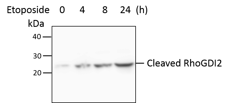
Application: Western BlotSample Tested: HT-29 human colon adenocarcinoma cell lineSpecies: HumanVerified Customer | Posted 07/18/2018HT-29 cells were treated with etoposide (100 ug/ml) for indicated time points. Total cell lysates were subjected to western blot. PVDF membrane were probed with 2 um/ml D4-GDI/RhoGDI2 (cleavage specific) Antibody (NB100-56725). A specific band was detected for cleaved RhoGDI2 at approximately 23 kDa. This experiment was conducted under reducing conditions.

There are no reviews that match your criteria.
Protocols
Find general support by application which include: protocols, troubleshooting, illustrated assays, videos and webinars.
- 7-Amino Actinomycin D (7-AAD) Cell Viability Flow Cytometry Protocol
- Antigen Retrieval Protocol (PIER)
- Antigen Retrieval for Frozen Sections Protocol
- Appropriate Fixation of IHC/ICC Samples
- Cellular Response to Hypoxia Protocols
- Chromogenic IHC Staining of Formalin-Fixed Paraffin-Embedded (FFPE) Tissue Protocol
- Chromogenic Immunohistochemistry Staining of Frozen Tissue
- ClariTSA™ Fluorophore Kits
- Detection & Visualization of Antibody Binding
- Extracellular Membrane Flow Cytometry Protocol
- Flow Cytometry Protocol for Cell Surface Markers
- Flow Cytometry Protocol for Staining Membrane Associated Proteins
- Flow Cytometry Staining Protocols
- Flow Cytometry Troubleshooting Guide
- Fluorescent IHC Staining of Frozen Tissue Protocol
- Graphic Protocol for Heat-induced Epitope Retrieval
- Graphic Protocol for the Preparation and Fluorescent IHC Staining of Frozen Tissue Sections
- Graphic Protocol for the Preparation and Fluorescent IHC Staining of Paraffin-embedded Tissue Sections
- Graphic Protocol for the Preparation of Gelatin-coated Slides for Histological Tissue Sections
- IHC Sample Preparation (Frozen sections vs Paraffin)
- Immunofluorescent IHC Staining of Formalin-Fixed Paraffin-Embedded (FFPE) Tissue Protocol
- Immunohistochemistry (IHC) and Immunocytochemistry (ICC) Protocols
- Immunohistochemistry Frozen Troubleshooting
- Immunohistochemistry Paraffin Troubleshooting
- Immunoprecipitation Protocol
- Intracellular Flow Cytometry Protocol Using Alcohol (Methanol)
- Intracellular Flow Cytometry Protocol Using Detergents
- Intracellular Nuclear Staining Flow Cytometry Protocol Using Detergents
- Intracellular Staining Flow Cytometry Protocol Using Alcohol Permeabilization
- Intracellular Staining Flow Cytometry Protocol Using Detergents to Permeabilize Cells
- Preparing Samples for IHC/ICC Experiments
- Preventing Non-Specific Staining (Non-Specific Binding)
- Primary Antibody Selection & Optimization
- Propidium Iodide Cell Viability Flow Cytometry Protocol
- Protocol for Heat-Induced Epitope Retrieval (HIER)
- Protocol for Liperfluo
- Protocol for Making a 4% Formaldehyde Solution in PBS
- Protocol for VisUCyte™ HRP Polymer Detection Reagent
- Protocol for the Characterization of Human Th22 Cells
- Protocol for the Characterization of Human Th9 Cells
- Protocol for the Preparation & Fixation of Cells on Coverslips
- Protocol for the Preparation and Chromogenic IHC Staining of Frozen Tissue Sections
- Protocol for the Preparation and Chromogenic IHC Staining of Frozen Tissue Sections - Graphic
- Protocol for the Preparation and Chromogenic IHC Staining of Paraffin-embedded Tissue Sections
- Protocol for the Preparation and Chromogenic IHC Staining of Paraffin-embedded Tissue Sections - Graphic
- Protocol for the Preparation and Fluorescent IHC Staining of Frozen Tissue Sections
- Protocol for the Preparation and Fluorescent IHC Staining of Paraffin-embedded Tissue Sections
- Protocol for the Preparation of Gelatin-coated Slides for Histological Tissue Sections
- Protocol: Annexin V and PI Staining by Flow Cytometry
- Protocol: Annexin V and PI Staining for Apoptosis by Flow Cytometry
- R&D Systems Quality Control Western Blot Protocol
- TUNEL and Active Caspase-3 Detection by IHC/ICC Protocol
- The Importance of IHC/ICC Controls
- Troubleshooting Guide: Fluorokine Flow Cytometry Kits
- Troubleshooting Guide: Immunohistochemistry
- Troubleshooting Guide: Western Blot Figures
- Western Blot Conditions
- Western Blot Protocol
- Western Blot Protocol for Cell Lysates
- Western Blot Troubleshooting
- Western Blot Troubleshooting Guide
- View all Protocols, Troubleshooting, Illustrated assays and Webinars
Loading...